
Zorotypus novobritannicus, Terry, Matthew D. & Whiting, Michael F., 2012
|
publication ID |
https://doi.org/ 10.5281/zenodo.215428 |
|
DOI |
https://doi.org/10.5281/zenodo.6167758 |
|
persistent identifier |
https://treatment.plazi.org/id/0392C77B-FF9C-FF9D-FF7C-7B68CFEAFA5E |
|
treatment provided by |
Plazi |
|
scientific name |
Zorotypus novobritannicus |
| status |
sp. nov. |
Zorotypus novobritannicus n. sp.
Figures 1–16 View FIGURES 1 – 4 View FIGURES 5 – 8 View FIGURES 9 – 11 View FIGURE 12 View FIGURES 13 – 15 View FIGURE 16
Diagnosis. The new species can be diagnosed by a combination of unique characteristics: metafemur of both sexes with 3 spines along the lateral ventral margin and 7 spines along the medial ventral margin, cerci of both sexes with dense fields of palmate setae along lateral and ventral sides, female sternum 8 (S8) with distinct medial posterior notch, male tergum 9 (T9) with large bicornuate medial notch.
Apterous female (holotype). Body length (without antennae) 2.6 mm; antennae 1.2 mm. Integument color varying slightly from light to darker brown with head and margin of the tergites darkest. Head subtriangular with posterior margins slightly wider than anterior of pronotum; no external evidence of eyes, ocelli absent; short to medium-length setae dispersed across dorsal side of head ( Fig. 1 View FIGURES 1 – 4 ). Antennae ( Fig. 1 View FIGURES 1 – 4 ) with 9 flagellomeres; first flagellomere large and goblet shaped; second flagellomere half the length of first; third through ninth flagellomere of various lengths; all flagellomeres with moderately dispersed fine setae of short to medium length; cuticle of antennae becoming progressively paler in more distal flagellomeres, particularly pronounced in flagellomeres 7–9. Labrum with fine, dense setation along anterior margin ( Fig. 1 View FIGURES 1 – 4 ). Mandible with 4 apical teeth and well-developed molar region ( Fig. 2 View FIGURES 1 – 4 ), heavily sclerotized with much darker cuticle along the molar region and at each condyle.
Maxilla ( Fig.3 View FIGURES 1 – 4 ) with 2 distinct, large setae along lateral margin; elongate lacinia and galea, lacinia with medial groove allowing for a tight fit of the lateral margin of the galea; galea with very fine, dense setation at the apex; maxillary palp five-segmented with first palpomere (P1) short, P2 elongate and slightly longer than P3, P4 short, P5 the longest and twice as wide as other palpomeres, all palpomeres except the first covered with dispersed setae of moderate length. Labium ( Fig. 4 View FIGURES 1 – 4 ) with well-defined paraglossa with very dense, fine setation at distal end; glossa small, obscure even at high stereoscopic magnification; labial palp 3-segmented with P1 narrow and elongate, P2 very short, and P3 nearly as long as P1 and twice as wide.
Pronotum ( Fig. 5 View FIGURES 5 – 8 ) subrectangular with anterior margin rounded, moderate setation across entire dorsal surface with pronounced row of larger setae along the anterior and lateral margins; mesonotum ( Fig. 5 View FIGURES 5 – 8 ) subrectangular, same length as pronotum, straight anterior margin oriented along the transverse plane of specimen, posterior margin with pronounced triangular edge slightly overlapping metanotum, setation slightly less dense than that of pronotum with most pronounced distribution along the lateral margins; metanotum ( Fig. 5 View FIGURES 5 – 8 ) roughly oval and approximately two-thirds the length of pronotum with sparse setation becoming denser along the lateral margins.
All legs ( Fig. 6–8 View FIGURES 5 – 8 ) with moderate to dense setae of medium length; two tarsal segments, segment one much shorter than elongated second segment, especially pronounced in the metatarsus, and two tarsal claws. Profemur ( Fig. 6 View FIGURES 5 – 8 ) 2.5 times longer than broad (as measured at maxima); mesofemur ( Fig. 7 View FIGURES 5 – 8 ) of same length but less broad (three times longer than broad); both protibia and mesotibia lacking spines, but setae becoming slightly stouter near the distal ends. Metafemur ( Fig. 8 View FIGURES 5 – 8 ) much larger than other femora with denser and longer setae; proximal end stout with distal end tapering markedly; posterior dorsal margin with 3 pronounced stiff spines of roughly equal length and posterior ventral margin with 7 spines increasing in size toward the distal end; metatibia approximately equal in length to metafemur, but much narrower and with 2 paired, dark brown subapical spines.
Abdominal T1 ( Fig. 9 View FIGURES 9 – 11 ) covered by posterior margin of metanotum, T2-6 non-overlapping and with transverse row of 6–8 setae along posterior margin and patches of setae along lateral margins; T7–8 slightly broader than anterior terga, also with transverse row of setae along lateral and posterior margins and slightly overlapping posterior tergites; T9 with pronounced quadrate lobe medially along the anterior margin, covered by T8, but still visible through the integument ( Fig. 9 View FIGURES 9 – 11 ), 6 medium-length setae along posterior margin, lateral margins thin and wrapped around the cerci. T10 non-overlapping and much smaller than other abdominal tergites, with 4 setae. Cerci ( Fig. 9 View FIGURES 9 – 11 ) single-segmented with 5–6 elongate setae that become progressively longer distally; dense fields of much smaller spiniform and palmate setae located along lateral and ventral sides of cerci (fig. 12), these small setae not readily discerned with a stereomicroscope; elongate lobes of the palmate setae with 2–12 elongate lobes.
All abdominal sterna (S1-8) with sparse setae across surface and pronounced transverse row of setae along the posterior margin; S8 ( Fig. 10 View FIGURES 9 – 11 ) much broader than other sterna and with distinct medial notch. Large, membranous region containing openings to the alimentary and reproductive tracts between S8 and S9 (partially illustrated in Fig. 10 View FIGURES 9 – 11 ); S9 with moderately dispersed setae of various lengths ( Fig. 11 View FIGURES 9 – 11 ); 2 small sclerites with 3 small setae each (possibly remnants of S10, at the posterior tip of the abdomen (fig. 11).
Apterous male. As described for the female except as follows: abdominal T9 with large bicornuate notch beginning medially at the posterior margin with two transverse extensions approximately half way to the lateral edge ( Fig. 13 View FIGURES 13 – 15 ); abdominal T10 with pronounced mating hook ( Figs. 13, 15 View FIGURES 13 – 15 ). Abdominal S8 roughly oval, twice as broad as S7; S9 narrow, approximately two-thirds the width of S8 with only very fine setation along posterior margin ( Fig. 14 View FIGURES 13 – 15 ); S9 articulated with sclerotized portions of terminalia. Everted male terminalia with large membranous area with three distinct lobes in dorsal aspect ( Fig. 13 View FIGURES 13 – 15 ); sclerotized portions of terminalia located along the ventral side of membranous area, consisting of an M-shaped base articulated with S9 and two posteriorly projecting arms curving anteromedially near the distal end ( Figs. 14–15 View FIGURES 13 – 15 ); thin, sclerotized medial projection emerging from posterior portion of the membranous area, curving around on the ventral surface of the everted terminalia to end in a lobed tip with several small spines ( Fig. 15 View FIGURES 13 – 15 ); sclerotized portions of the terminalia likely articulating with each other, but obscured by the membranous portions in all observed specimens.
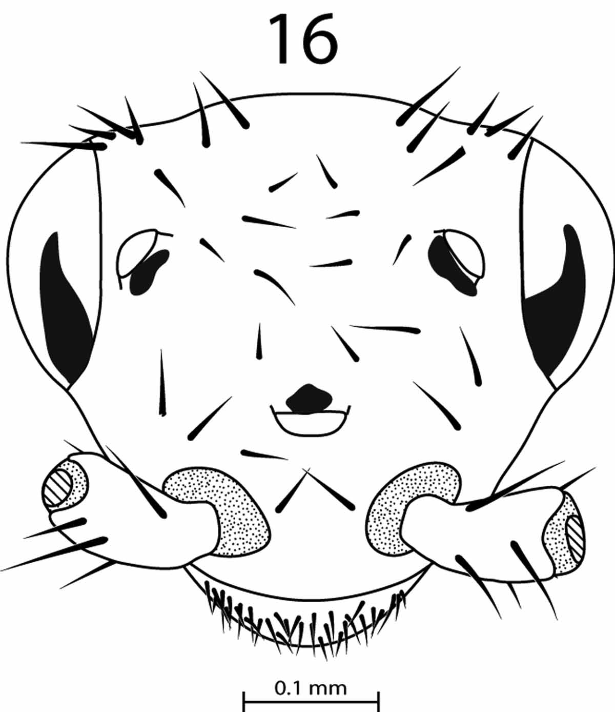
Dealated female. As described for the holotype except as follows: integument slightly darker and more opaque (dark to very dark brown); head slightly broader with well-developed eyes that have both clear, transparent areas and mottled black pigmentation ( Fig. 16 View FIGURE 16 ); one medial ocellus and two lateral ocelli, all with irregular, dark pigmentation near the base ( Fig. 16 View FIGURE 16 ). Thorax noticeably more heavily sclerotized with prominent scars on T2 and T3 as well as remnants of basal wing elements.
Dealated Male. Integument, head and thorax as described for dealate female, but with posterior abdominal features as described for apterous male.
Alate juvenile. One late-stage juvenile alate collected: light, non-sclerotized integument of non-alate juveniles, but with prominent wing pads on the mesonotum and metanotum that extend beyond the margin of the next posterior segments.
Type material. Apterous female holotype, Papua New Guinea, East New Britain Province, Kerevat, 4° 22.13’ S 152° 2.47’ E, 23 July 2008, M. Whiting et al., colls. Paratypes: 24 apterous females, 8 apterous males, 67 apterous juveniles of all stages, 16 dealated females, 19 dealated males, 1 late-stage juvenile with well-developed wing pads, same locality information as the holotype.
Etymology. The specific name refers to the province of New Britain in which the specimens were collected.


No known copyright restrictions apply. See Agosti, D., Egloff, W., 2009. Taxonomic information exchange and copyright: the Plazi approach. BMC Research Notes 2009, 2:53 for further explanation.
|
Kingdom |
|
|
Phylum |
|
|
Class |
|
|
Order |
|
|
Family |
|
|
Genus |