
Kinnecaris barrambie, Karanovic & Cooper, 2011
|
publication ID |
https://doi.org/ 10.11646/zootaxa.3026.1.1 |
|
persistent identifier |
https://treatment.plazi.org/id/03924C3A-FFB6-A606-FF41-FC7AAD64FD5E |
|
treatment provided by |
Felipe |
|
scientific name |
Kinnecaris barrambie |
| status |
sp. nov. |
Kinnecaris barrambie sp. nov.
( Figs. 5 View FIGURE 5 & 6 View FIGURE 6 )
Type locality. Australia, Western Australia, Yilgarn region, Barambie , bore B3M, 27.213178° S 118.919514° E.
Type material. Holotype male and allotype female on one SEM stub in toto coated with carbon ( WAM C47183), both collected at type locality, leg. E. Thomas & V. Campagna, 26 November 2008, oeRBV055 . Paratype: one female dissected on one slide ( WAM C47184), Australia, Western Australia, Yilgarn region, Barambie , bore B14M, 26.832544° S 118.886097° E, leg. E. Thomas & V. Campagna, 26 November 2008, oeRBV0585 .
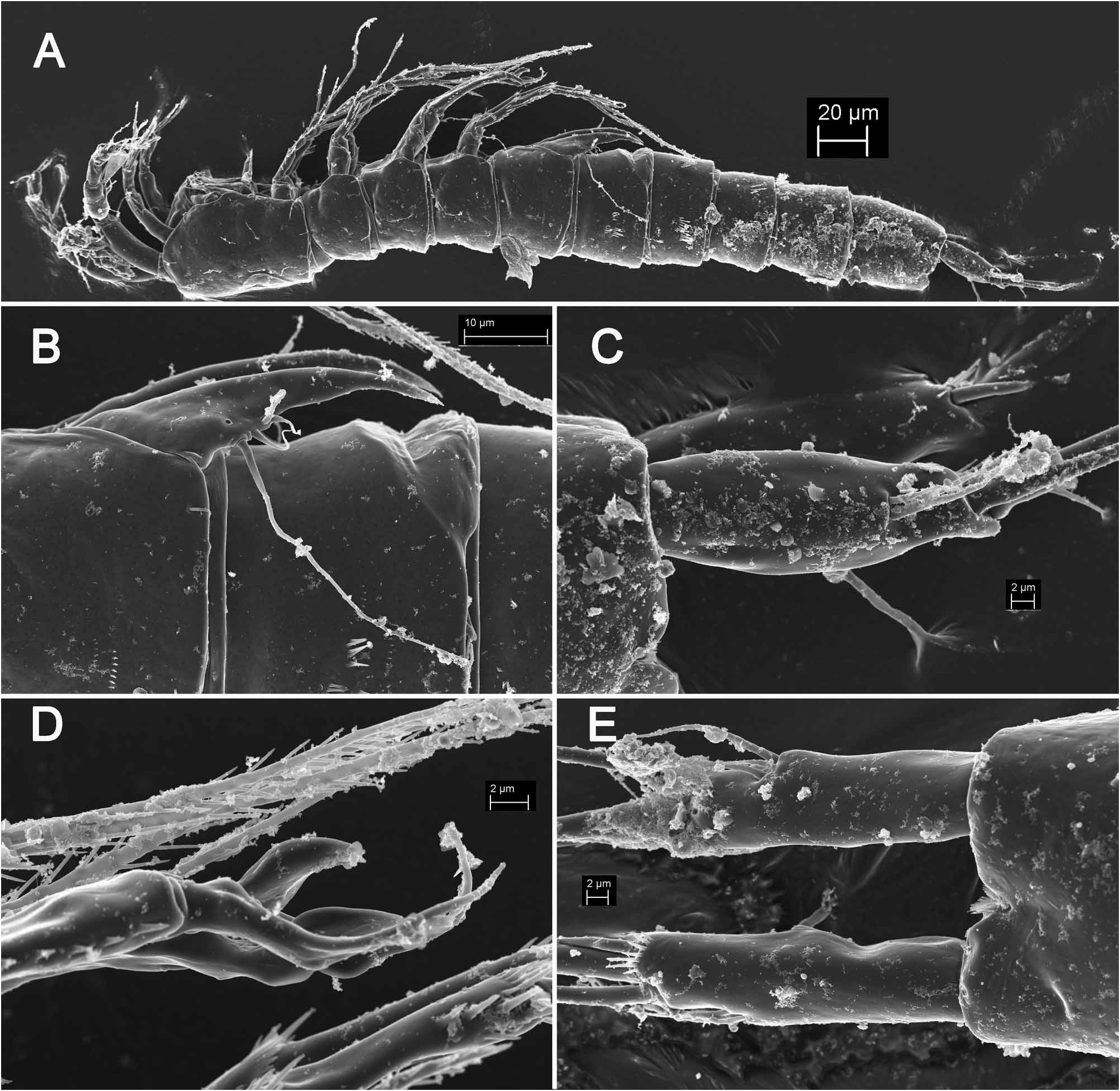
Description. Male (based on holotype). Total body length, measured in the same way as in Kinnecaris lakewayi (see above), 328 µm. Surface of integument of all somites with sparse and shallow cuticular pits. Colour, naupliar eye, rostrum, body segmentation, and pore and sensilla pattern of all somites as in K. lakewayi . Habitus ( Fig. 5A View FIGURE 5 ) cylindrical and very slender, without any dorsal demarcation between prosome and urosome, but urosome wider in lateral view; prosome/urosome ratio about 0.7; greatest width from dorsal view at posterior end of cephalothorax. Body length/width ratio about 7.5; cephalothorax 1.1 times as wide as genital somite. Integument relatively weakly sclerotized, ornamented with several very short dorsal and lateral rows of minute spinules on all urosomites, and some additional larger spinules ventrally or laterally on second (genital), third and fourth urosomites ( Fig. 5A, B View FIGURE 5 ). Cephalothorax with clearly visible ( Fig. 5A View FIGURE 5 ) posterior double dorsal cuticular window; fourth and fifth urosomites with pair of lateral circular windows each, although these harder to distinguish than in K. lakewayi .
Cephalothorax ( Fig. 5A View FIGURE 5 ) about 1.5 times as long as wide in dorsal view; representing about 18% of total body length. Surface of cephalic shield, tergites and pleuras of free pedigerous somites ornamented as in K. lakewayi , except for presence of sparse and shallow cuticular pits and fewer minute spinules on fifth pedigerous (first urosomal) somite ( Fig. 5A, B View FIGURE 5 ).
Genital somite ( Fig. 5A, B View FIGURE 5 ) with three short lateral rows of spinules (from five to seven) at midlength, three pairs of sensilla on posterior margin (one dorsal, one dorso-lateral, and one ventro-lateral), one pair of very small cuticular pores in anterior part ventro-laterally, and several dorsal and lateral rows of minute spinules (pore and sensilla pattern being same as in K. lakewayi ); spermatophore not visible inside.
Third urosomite ( Fig. 5A View FIGURE 5 ) with dorso-lateral large spinules (two parallel short rows of eight spinules each), ventro-lateral rows of large spinules, six posterior sensilla, and several dorsal rows of minute spinules.
Fourth urosomite ( Fig. 5A View FIGURE 5 ) ornamented with two or three large spinules laterally (covered with dirt in Fig. 5A View FIGURE 5 ), and four midventral groups of four large spinules; lateral oval cuticular window not easy to observe (more visible with light compound microscope).
Fifth (preanal) urosomite ( Fig. 5A View FIGURE 5 ) with similar windows to those observed in fourth urosomite but without any large spinules, sensilla or pores; only other ornamentation sparse and shallow cuticular pits.
Anal somite ( Fig. 5A, C View FIGURE 5 ) ornamented with pair of large dorsal sensilla at base of anal operculum, pair of large cuticular pores laterally (one pore on each side) in anterior half, two pairs of minute cuticular pores laterally closer to posterior margin, and pair of slightly larger cuticular pores ventrally, at base of caudal rami, in addition to hardly visible cuticular pits; spinules absent. Anal operculum well developed, unornamented on outer surface, ornamented with row of slender spinules on inner surface, with convex and smooth distal margin, not reaching posterior end of anal somite, representing 60% of somite's width. Anal sinus widely opened, with two diagonal rows of slen- der spinules on ventral side and transverse row of spinules on dorsal side (below anal operculum).
Caudal rami ( Fig. 5C View FIGURE 5 ) about 3.5 times as long as greatest width (dorsal view) and 0.8 times as long as anal somite, nearly cylindrical in dorsal and ventral view, but clearly inflated at midlength in lateral view, with proximal part of inner margin slightly convex in dorsal (or ventral) view and base much narrower than rest of ramus, nearly parallel, with space between them about 1.2 times ramus width. Armature and ornamentation as in K. lakewayi , but lateral setae inserted slightly more posteriorly (or proximal part of caudal rami longer).
Antennula ( Fig. 5A View FIGURE 5 ), antenna ( Fig. 5A View FIGURE 5 ), mouth appendages ( Fig. 5A View FIGURE 5 ), and first two pairs of swimming legs ( Fig. 5A View FIGURE 5 ) as in K. lakewayi .
Third swimming leg ( Fig. 5D View FIGURE 5 ) also very similar to that in K. lakewayi but apophysis with much sharper tip; ornamentation same as in K. lakewayi , including longitudinal row of large spinules on outer margin of first exopodal segment distally; exopodal spine also 1.5 times as long as apophysis.
Fourth swimming leg ( Fig. 5D View FIGURE 5 ) similar to that in K lakewayi , i.e. with five relatively large spinules on basis at base of endopod, and endopod spiniform with spinules only along outer margin. Apical seta on third exopodal segment 1.2 times as long as entire exopod and 3.2 times as long as outer spine.
Fifth leg ( Fig. 5B View FIGURE 5 ) without any difference from that in K. lakewayi , except for several shallow cuticular pits on anterior surface in proximal part, and weak outline of cuticular window similar to that in K. eberhardi ( Karanovic, 2005) , but not so clearly visible.
Sixth legs ( Fig. 5B View FIGURE 5 ) as in K. lakewayi .
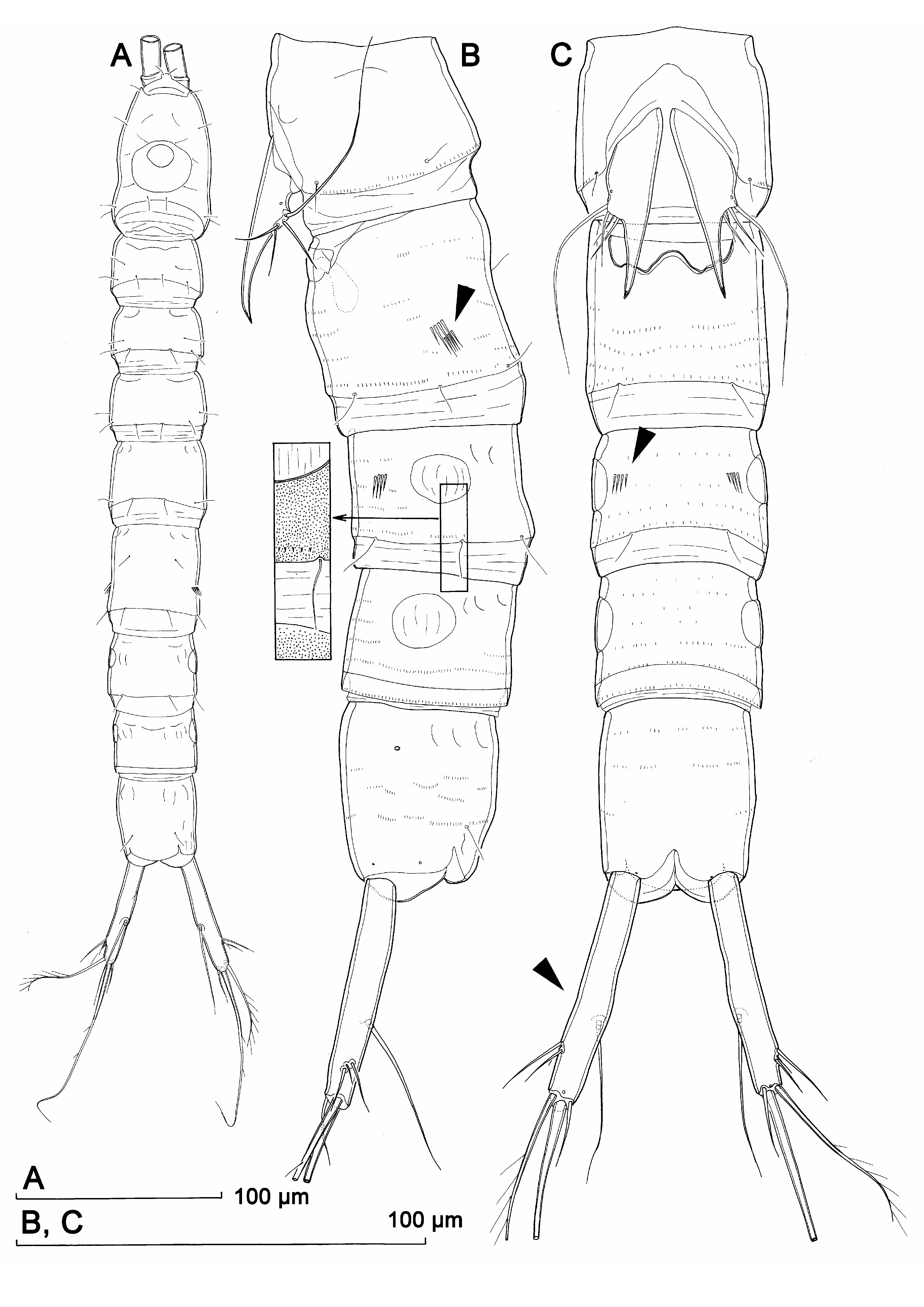
Female (based on allotype and one paratype from bore B14M). Body length 405 µm in allotype and 425 µm in paratype. Habitus ( Fig. 6A View FIGURE 6 ), ornamentation of prosomites, colour and nauplius eye similar to those in male, except genital and first abdominal somites fused into double-somite and middle part slightly less slender. Prosome/urosome ratio 0.78; greatest width from dorsal view hard to establish; body length/width ratio about 8.2; cephalothorax less than 1.1 times as wide as genital double-somite.
Genital double-somite ( Fig. 6A, B, C View FIGURE 6 ) only slightly longer than wide in dorsal view, without any trace of subdivision except for pair of ancestral dorso-lateral sensilla in anterior half; additionally ornamented with six posterior sensilla, many short rows of minute spinules, and two parallel short rows of large spinules (five and each spinules respectively); latter similar to that in K. esbe but not overlapping (arrowed in Fig. 6C View FIGURE 6 ). Genital complex ( Figs. 6B View FIGURE 6 ) as in K. lakewayi , sixth legs also with large spiniform processes.
Third urosomite ( Fig. 6B, C View FIGURE 6 ) similar to that in male, with four rows of four large spinules ventro-laterally, one row of three large spinules dorsolaterally (arrowed in Fig. 6C View FIGURE 6 ), and clearly visible lateral cuticular windows.
Fourth (preanal), and fifth (anal) urosomites also very similar to those in male ( Fig. 6B, C View FIGURE 6 ), without any large spinules, except those in anal sinus.
Caudal rami ( Figs 5C View FIGURE 5 , 6A, B, C View FIGURE 6 ) similar to those in male but slightly more divergent.
Antennula ( Fig. 6D View FIGURE 6 ) very similar to that in K. lakewayi , with only slightly shorter proximal aesthetascs (arrowed in Fig. 6D View FIGURE 6 ).
Antenna ( Fig. 6E View FIGURE 6 ), mouth appendages ( Fig. 6F View FIGURE 6 ), first swimming leg, second swimming leg ( Fig. 6G View FIGURE 6 ), and exopod of fourth swimming leg very similar to those in male and almost without any difference from those in K. lakewayi .
Endopod of second swimming leg ( Fig. 6G View FIGURE 6 ) 5.6 times as long as wide, its apical seta 0.9 times as long as segment.
Third swimming leg ( Fig. 6H, I View FIGURE 6 ) similar to K. lakewayi , with only slightly proportionately longer apical seta on second segment and more strongly scletotised cuticular plates on praecoxa and coxa.
Endopod of fourth swimming leg ( Fig. 6J View FIGURE 6 ) about 7.8 times as long as wide, half as long as first exopodal segment, armed with single robust bipinnate element apically, ornamented with four spinules along distal margin, at base of apical element. Exopod similar to that in male.
Fifth leg ( Fig. 6B, C View FIGURE 6 ) similar to that in male, but slightly more elongated, with narrower distal part and cuticular window on anterior surface better defined (although not easily visible with light microscope).
Sixth legs ( Fig. 6K View FIGURE 6 ) vestigial, fused into simple bilobate cuticular plate, covering gonopore, unornamented and unarmed; outer distal corners produced into sharp processes like in previous species, longer than inner lobes.
Etymology. The species name comes from its type locality (Barrambie), but should be treated as comprising an arbitrary combination of letters that can be treated as a Latin word and may be conceived as a noun in apposition to the generic name.
Variability. Only one male and two females were examined and no variability or asymmetry was observed, except in their body length. Note that the male is significantly smaller than females (328 µm vs. 405 µm and 425 µm), but that is probably a consequence of a small sample size rather than a specific character.
Remarks. This species differs from other eight Australian congeners in the shape of the caudal rami (which have much narrower base than the rest of the ramus and are inflated in lateral view; arrowed in Fig. 6C View FIGURE 6 ), armature of the genital double-somite in female (arrowed in Fig. 6C View FIGURE 6 ), as well as in the presence of some large spinules on the third urosomite in female dorso-laterally (arrowed in Fig. 6C View FIGURE 6 ). Similarities and differences between K. barrambie sp. nov. and K. lakewayi sp. nov. are discussed in the remarks section for the latter species (see above), as the two seem to share the greatest number of morphological characters. The only other Australian Kinnecaris Jakobi, 1972 with two rows of large spinules on the genital double-somite laterally is K. esbe sp. nov. (see below), but this species has much longer caudal rami, more ornamentation on most somites, as well as a very different shape of the third leg apophysis in male. Besides, the rows of large spinules are much closer to each other and clearly parallel in K. esbe (compare Fig. 6C View FIGURE 6 and Fig. 9B View FIGURE 9 ), which makes us to question their homology (or at least one of them). These structures have not been reported for any other member of the genus Kinnecaris , but it is fair to say that fine urosomal ornamentation did not always receive full attention in early descriptions.

| WAM |
Western Australian Museum |
| V |
Royal British Columbia Museum - Herbarium |
No known copyright restrictions apply. See Agosti, D., Egloff, W., 2009. Taxonomic information exchange and copyright: the Plazi approach. BMC Research Notes 2009, 2:53 for further explanation.