
Cycloporus variegatus Kato 1934
|
publication ID |
https://doi.org/ 10.11646/zootaxa.4652.2.5 |
|
publication LSID |
lsid:zoobank.org:pub:951C3124-F209-4E02-B489-695CBF71543D |
|
persistent identifier |
https://treatment.plazi.org/id/039187B7-D269-3B4D-FF35-FD256339FF52 |
|
treatment provided by |
Plazi |
|
scientific name |
Cycloporus variegatus Kato 1934 |
| status |
|
Cycloporus variegatus Kato 1934 View in CoL
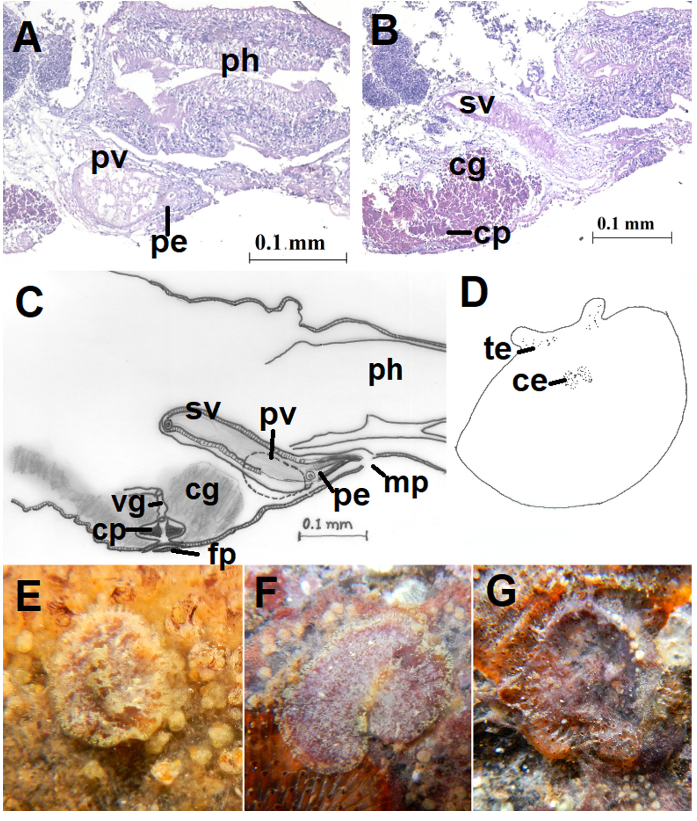
( Figure 2 View FIGURE 2 , 3 View FIGURE 3 )
Material examined: One specimen (BNHS-Pclad 118, 6.51 x 5.45 mm) dissected for sagittal sections of reproductive structure (26 slides), collected on 20-10-2013 at Alawa in Ratnagiri district, Maharashtra. One specimen (BNHS-Pclad 166, 6.48 x 4.16 mm) intact, collected 17-05-2014 at Undi in Ratnagiri district, Maharashtra .
Distribution: Originally described from Japan ( Kato 1934), re-described from West Australia, North Great Barrier Reef, Lizard Island and Vietnam (Newman and Canon 2002, 2003), Colombia from Caribbean ( Quiroga et al. 2004) and Cabo Frio, Brazil ( Bahia et al. 2014).
Diagnosis: Translucent body with intermittently arranged prominent yellowish spots. Thick grouping of these spots observed on the dorsal median line. Margin clear with yellow dash-like markings or spots. Pseudotentacles yellow.
Description: Colour: The colouration observed within intestinal branches across the dorsal surface appears brown to dark reddish ( Fig. 2A, B View FIGURE 2 ). Fine yellow spots are found throughout the surface which even clump together and become more prominent. Median line is slightly raised, commences at the base of the translucent cerebral area and ends prior to posterior margin. This raised portion shows brownish-red and yellow patches across its length. Main branch of the intestine with accumulated brownish-black food material visible as darker median colouration. Translucent margin possesses yellow stripes or spots of peripheral glands. Widely spaced pseudotentacles show prominent yellow pigmentation ( Fig. 2B View FIGURE 2 ).
Form: Slightly rounded or oval
Pseudotentacles: Small bumps, sometimes raised and pointed ( Fig. 2B, D View FIGURE 2 ), measuring 0.35 mm.
Eyespots: Cerebral eyespots arranged in two elongated clusters 50–55 in each and 0.8 mm away from anterior margin. Tentacular eyespots arranged dorsally and ventrally, ranging between 25–40 ( Fig. 2D View FIGURE 2 and 3D View FIGURE 3 ).
Digestive system: Pharynx tubular 0.72 mm long and mouth situated about 1.32 mm from the anterior margin. Main branch of the intestine 3.07 mm long with eight lateral branches ( Fig. 2C, D View FIGURE 2 ).
Gonopores: Male gonopore present immediately behind the pharynx and 1.46 mm distant from anterior margin, measuring 0.064 mm. Female gonopore 0.46 mm behind the male gonopore and 1.86 mm distant from anterior margin, measuring 0.088 mm ( Fig. 2E, F View FIGURE 2 ).
Male reproductive system: elongated-oval seminal vesicle (0.214 mm x 0.045 mm) is present diagonal to the ventral body wall ( Fig. 3B, C View FIGURE 3 ). Prostatic vesicle (0.076 mm x 0.101mm) ventrally placed enters penis via prostatic duct ( Fig. 3A, C View FIGURE 3 ). Stylet 0.0320 mm enters short male atrium measuring 0.0493 mm.
Female reproductive system: slender female atrium measures 0.045 mm. Cement pouch with lateral invagination (0.022 mm), densely disposed cement glands. Vagina 0.064 mm ( Fig. 3B, C View FIGURE 3 ). Uteri visible through dorsal surface ( Fig. 3C, F View FIGURE 3 ).
Taxonomic Remarks: round/oval body, tubular pharynx, small bumps of tentacles, prominent peripheral vesicles of intestinal branches, lateral branches of intestine range within 4-10 and multiple uterine vesicles place these specimens into the genus Cycloporus as defined in Newman and Canon (2002).
The present specimens exactly match with the description provided by Newman and Canon (2002; 2003) except for the white spot posterior to the cerebral eyespots mentioned by them. In fact, this region shows more prominent yellow patches in both the specimens studied.
Both the specimens collected were found on the unidentified compound ascidian and nearly of similar colouration ( Fig. 3E, F, G View FIGURE 3 ).

No known copyright restrictions apply. See Agosti, D., Egloff, W., 2009. Taxonomic information exchange and copyright: the Plazi approach. BMC Research Notes 2009, 2:53 for further explanation.
|
Kingdom |
|
|
Phylum |
|
|
Order |
|
|
Family |
|
|
Genus |