
Anthalona acuta, Van Damme, Sinev & Dumont, 2011
|
publication ID |
https://doi.org/ 10.11646/zootaxa.2875.1.1 |
|
persistent identifier |
https://treatment.plazi.org/id/0390471D-FFFD-1820-FF22-D0946B75C860 |
|
treatment provided by |
Felipe |
|
scientific name |
Anthalona acuta |
| status |
sp. nov. |
Anthalona acuta n. sp.
( Figs 1–6 View FIGURE 1 View FIGURE 2 View FIGURE 3 View FIGURE 4 View FIGURE 5 View FIGURE 6 )
Alona View in CoL n. sp. (verrucosa- group) in Van Damme & Dumont (2010: 759).
Etymology. The name “acuta” refers to the sharp, thickened setae on the exopod of the second antenna, unique for this species. Outside of the genus Anthalona , such an adaptation in the Aloninae is only found in the Austral(as)ian Armatalona Sinev, 2004 .
Material examined. Holotype. One adult parthenogenetic female mounted in glass slide labelled “ Anthalona acuta n. sp. holotype ”; from the Lençóis Maranhenses, NE Brazil, temporary dune pool near Atins (S3A in Van Damme & Dumont 2010), coordinates 2° 34’ 31” S and 42° 43’ 20” W, 16.VIII.2006, Leg. K. Van Damme GoogleMaps . Paratypes. two adult parthenogenetic females, two adult ephippial females, two adult males and one ephippium, slides labelled “ Anthalona acuta paratypes ”, same data as holotype. Type material from Brazil deposited at the Royal Belgian Institute for Natural Sciences, Brussels ( RBIN) under accession number IG 31782, INV 96691 GoogleMaps -93.
Additional specimens. Five adult parthenogenetic females, Lençóis Maranhenses (Sample series Lençóis Maranhenses from UG collection labeled 1996.001–1996.020 or SI–SIV in UG collection, Leg. K. Van Damme and D. Van Damme; see Van Damme & Dumont 2010). Paratypes (males) from dune pool at the Lençóis Maranhenses, collected 17.VIII.2006, 2° 36’ 45” S and 42° 43’ 30” W (S 5 in Van Damme & Dumont 2010; record not included) by K. Van Damme & D. Van Damme: slide with two adult parthenogenetic females, two ephippial females and one adult male. 11 adult parthenogenetic females in separate slides (dissected), three complete specimens, temporary pool near Orinoco River , Venezuela, Brown stream crossing Orinoco River, Venezuela, 04.V.2001, Leg. H.J. Dumont. Sample 2001.021 ( UG collection). All slides from Venezuela deposited under RBIN IG 31782 INV 96694 GoogleMaps - 704. Tube with seven complete specimens of A. acuta n. sp. from the latter locality were deposited together with the types, under RBIN IG 31782 INV 96705 .
Description. Adult parthenogenetic female. Habitus ( Figs 1A–C View FIGURE 1 , 3A–B View FIGURE 3 ). Medium sized animals, 0.35–0.45 mm, average length 0.38mm (n=12). Body 1.5 times as long as high. Colour yellow-brown to dark brown. Dorsum strongly convex, with high maximum dorsal point; posterior margin straight to moderately convex ( Figs 1A–C View FIGURE 1 ). Appearance in lateral view hunchbacked. Rostrum reaching ventral maximum of carapace margin. Ventral carapace margin moderately convex, deepest point just before midline ( Fig. 1A View FIGURE 1 ). Posteroventral corner round, without notch ( Fig. 1I View FIGURE 1 ). Head. Ocellus smaller than eye, 0.6 to half its diameter ( Fig. 1C View FIGURE 1 ). Well developed rostrum, broadly rounded, deep lateral embayment in head shield delineating rostrum ( Fig. 1C View FIGURE 1 ). Aesthetascs of antennules projecting laterally from this embayment ( Fig. 1B View FIGURE 1 ).
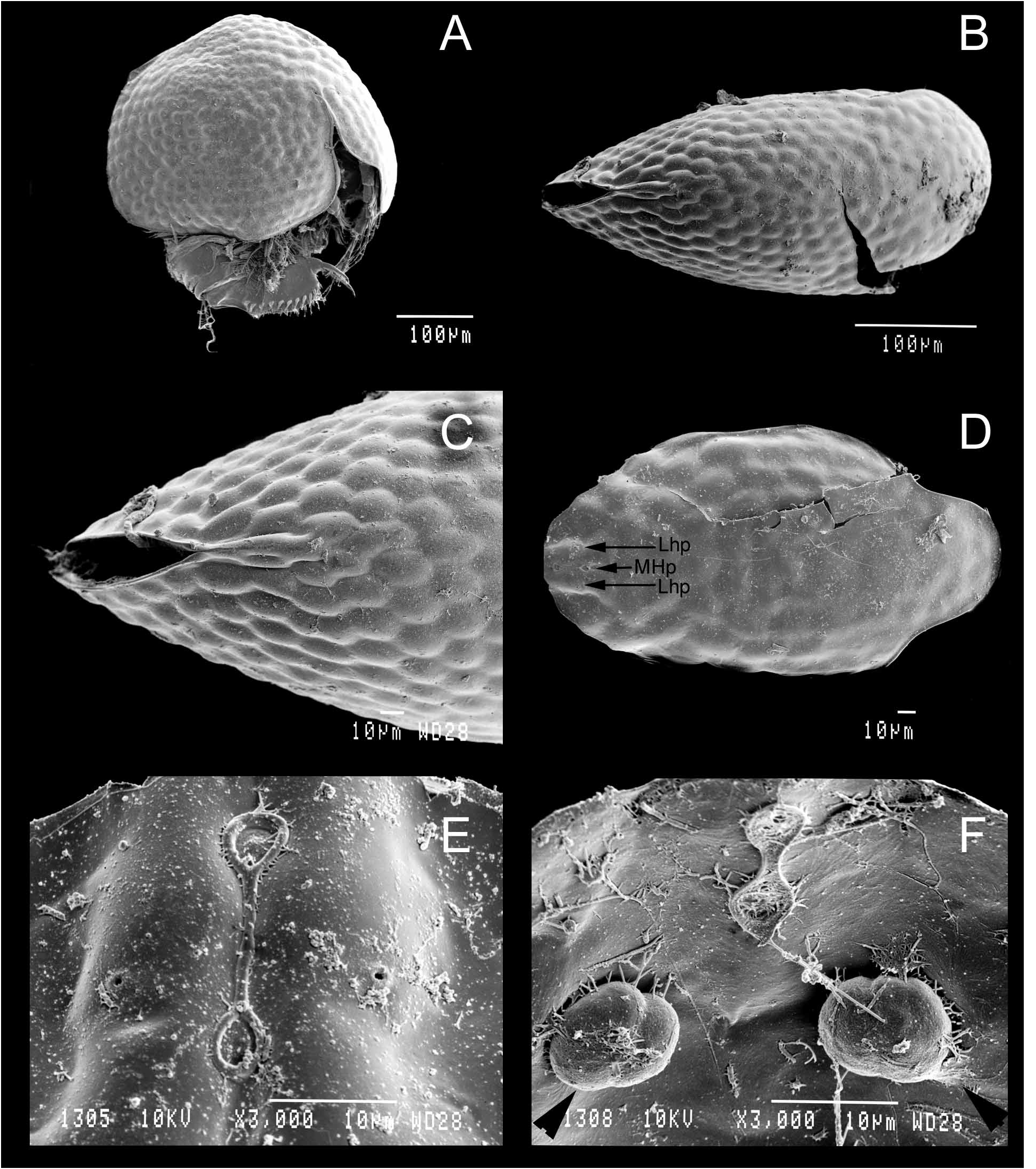
Headshield ( Figs 2F View FIGURE 2 & 3D View FIGURE 3 ) with broad, flat rostrum. Two main head pores ( Figs 2F–I View FIGURE 2 & 3E View FIGURE 3 ), interpore distance long. Distance between the two pores is two to three times the length of one main pore. PP distance short, one fifth of IP distance, lateral pores at about half of IP distance from midline and situated next to main pores, rarely posterior. Posterior margin situated at maximally half IP distance from posterior main pore. Typical arrangement in Fig. 2I View FIGURE 2 (= Fig. 1J View FIGURE 1 ), posterior margin of head shield strongly divided. Sacks under small pores large, their circular size about twice of a main head pore. Comparison with A. verrucosa in Figs 2A–E View FIGURE 2 and Fig. 3F View FIGURE 3 .
Carapace ( Figs 1A–C View FIGURE 1 , 3A–C View FIGURE 3 ). Ornamentation without striation but smooth or with verrucae. The latter may be faintly to strongly pronounced and are not arranged in rows ( Figs 3A–C View FIGURE 3 ). Marginal setae 35–45 ( Figs 1A–B View FIGURE 1 ), differentiated into two main groups (anterior and median groups of similar size). Setae in the middle of the carapace shortest. Posterior group of 10–20 setae in second half of the ventral margin makes up the longest group of setae. These are thickened, rigid and spiniform, bearing few strong setules on posterior side ( Fig. 1I View FIGURE 1 ). Setae decreasing in size towards the posteroventral corner, followed by fine setules not arranged in groups ( Fig. 1I View FIGURE 1 ). These setules of similar size, reaching beyond carapace margin in posteroventral corner and continuing in a posterior row of fine long setules towards posterodorsal corner of the valves ( Fig. 1I View FIGURE 1 ).
Labrum ( Fig. 1E View FIGURE 1 ). Labral keel in lateral view “axe-shaped” with straight to moderately convex margin, sometimes with ventral indentation. Single proximal denticle on labral keel.
First antennae or antennules ( Fig. 1F View FIGURE 1 ). About 2.5 times as long as wide, sensory seta implanted at one third of antennular corm. Three to four groups of short denticles on margin. Longest aesthetascs about half of antennular corm, shortest half as long.
Second antennae ( Fig. 1G View FIGURE 1 ). Basal spine large, conical. Spinal formula (exo/endo) 001/101, setal formula 113/ 003 ( Fig. 1G View FIGURE 1 ). A group of short spinules ( Fig. 1G View FIGURE 1 ) at base of first exopod segment. First exopod seta on antenna narrow ( Fig. 1G View FIGURE 1 ), not reaching beyond ultimate exopod segment, second exopod seta twice as long as previous; on external side of second exopod segment, two groups of three strong spiniform setules ( Fig. 1G View FIGURE 1 ). Spine on first endopod segment not longer than second segment; main terminal spines on endo- and exopod well developed, each as long as its apical segment or just shorter ( Fig. 1G View FIGURE 1 ; apical exopod spine relatively shorter than endopod spine). Terminal setae on antennal exopod strongly modified ( Figs 1A–B&G View FIGURE 1 ). These are chitinized and thickened, with acute apex and short setules in distal half ( Fig. 1H View FIGURE 1 ). Shortest seta most spiniform, about as long as three exopod together. Preanal, anal and postanal margins of similar length ( Fig. 1K View FIGURE 1 ). Anal margin straight to slightly concave, postanal margin stronger curved, convex. Distal margin not protruding, distal embayment dorsal to basal claw maximally as deep as claw width at base ( Fig. 1K View FIGURE 1 ). Overall dorsal margin S-shaped. Preanal corner triangular, not protruding far beyond maximal dorsal point of margin (neither postanal margin or preanal corner reach dorsally beyond each other). Marginal postanal teeth in six to seven groups ( Figs 1K–L View FIGURE 1 ). Each distal tooth with adjacent smaller elements on anterior side, partly merged towards distal end of postabdomen. Lateral setae in six fascicles in postanal portion, consisting of four to six elements in each group, parallel to each other. Distalmost lateral element spiniform, long and thick, protruding two thirds of its length beyond dorsal margin of postabdomen ( Fig. 1M View FIGURE 1 ). Most distal lateral elements in postanal portion reach half their size beyond the marginal teeth ( Fig. 1M View FIGURE 1 ). Smaller elements per fascicle at least half as long as distalmost spiniform element in each group ( Fig. 1M View FIGURE 1 ). Three to four clusters of smaller marginal teeth, close to each other, almost a continuously armed anal margin, and three to four fascicles in anal portion ( Fig. 1K View FIGURE 1 ).
Terminal claw ( Figs 1K–L & 1N View FIGURE 1 ). Longer than anal margin, moderately curved, implanted with setules along dorsal side. Proximal pecten ending in long spine about width of claw at this point and just before half of claw length. Basal spine short, just about claw width at base ( Fig. 1N View FIGURE 1 ) and about one sixth of claw length. Group of three to four thick, short basal spinules, about half of basal spine length ( Fig. 1N View FIGURE 1 ).
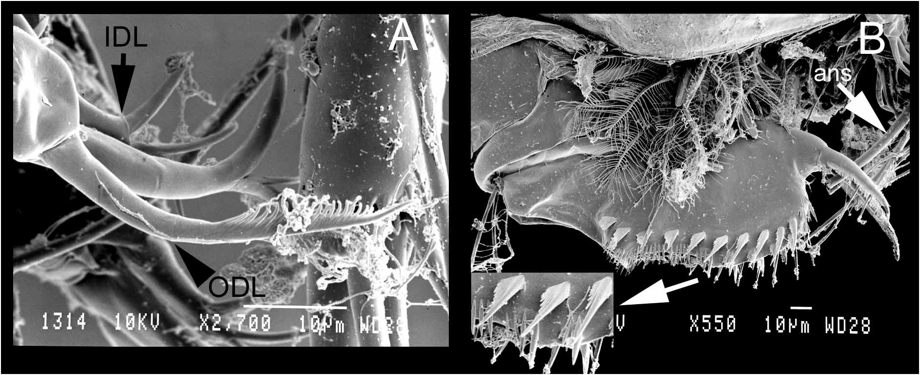
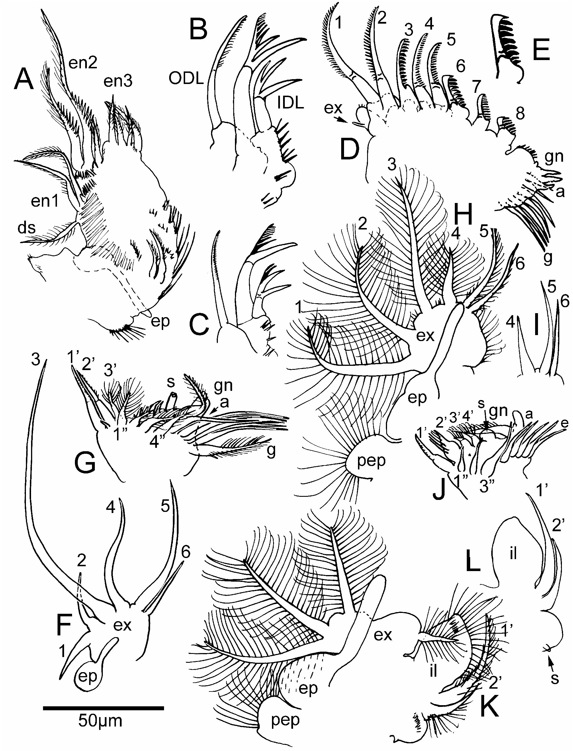
Five pairs of limbs. First limb ( Figs 4A View FIGURE 4 & 5A–C View FIGURE 5 ). Epipodite round with long projection, reaching beyond limb corm ( Fig. 5A View FIGURE 5 , ep). First endite with one dorsal and two marginal setae, second endite with three setae of which two longer (lengths differ strongly) and with thick teeth on anterior side ( Fig. 5A View FIGURE 5 ); third endite with four setae of similar length ( Fig. 5A View FIGURE 5 ); anterior elements on en1–2 present but minute, about as wide as long ( Fig. 5A View FIGURE 5 ). ODL with one slender seta, as long as largest IDL seta and with strong setules in distal half ( Fig. 4A View FIGURE 4 ); two setae in IDL, with modified and chitinized distal half ( Fig. 4A View FIGURE 4 ). On largest IDL seta, one large spine followed by spines decreasing in size distally and reduced distal part ( Figs 5B–C View FIGURE 5 ); spine in longest IDL seta mostly longer than distal part beyond it ( Figs 5B–C View FIGURE 5 ). On shortest IDL seta, two long spines of similar size, basal spine as long as distal part ( Fig. 5B View FIGURE 5 ). Accessory seta present (not shown). Four to five anterior setule groups with two to three setules in each group, decreasing in size ventrally ( Fig. 5A View FIGURE 5 ). Ejector hooks subequal and gnathobase triangular with setulated apex ( Fig. 5A View FIGURE 5 ).
Second limb ( Fig. 5D View FIGURE 5 ). Exopodite ( Fig. 5D View FIGURE 5 , ex) oval round, with short seta not reaching beyond exopodite apex; minute denticles on exopodite apex; endites with eight scrapers gradually decreasing in size towards gnathobase, eighth scraper shortest ( Fig. 5D View FIGURE 5 ). First two scrapers relatively slender and finely setulated, third stouter and shorter than two and four, with stronger denticles than scrapers one to five. Scrapers four and five similar, with fine denticles, scraper six modified with eight thick teeth ( Fig. 5E View FIGURE 5 ); final two scrapers decreasing in size towards gnathobase, scraper eight thickest and with longest denticles, this scraper half the size of sixth. Gnathobasic ‘brush’ short and round, implanted with short denticles ( Fig. 5D View FIGURE 5 ). Gnathobase with a sensillum and three elements, of which first a short seta, second a plump seta with small denticles in distal half and third a short seta; filter comb ( Fig. 5D View FIGURE 5 ) with seven setae of which only the first two shorter.
Third limb ( Figs 5F–G View FIGURE 5 ). Pre-epipodite round, epipodite round with long projection reaching half of exopodite; exopodite ( Fig. 5F View FIGURE 5 ) with quadrangular corm and six large setae in 2+4 arrangement; first exopodite seta as long as second; third exopodite seta about two times as long as fifth exopodite seta, fourth seta just shorter than fifth seta and a third longer than sixth seta; the latter half the size of fifth exopodite seta; all these setae plumose, except for fifth and sixth ( Fig. 5F View FIGURE 5 ) which are plumose-serrulate (fifth seta) or shortly plumose (sixth) in distal half. External endite ( Fig. 5G View FIGURE 5 ) with three setae (1’–3’) of which first two long, with short setules in distal half and minute element in between, third (3’) shorter by half and with long setules; four well developed plumose setae on inner side (1”– 4”) of similar length; one naked element and four small naked setae on internal endite ( Fig. 5G View FIGURE 5 ) preceding gnathobase; the latter with bottle-shaped sensillum and large bent plumose seta with two naked elements at its base ( Fig. 5G View FIGURE 5 ). Filter comb with seven setae of which the last one relatively thicker ( Fig. 5G View FIGURE 5 ).
Fourth limb ( Figs 5H–I View FIGURE 5 ). Pre-epipodite round, epipodite oval with long projection reaching beyond half of exopodite. Exopodite ( Fig. 5H View FIGURE 5 ) round, with six marginal plumose setae; first three exopodite setae long, third longest of the three, fourth half as long as preceding seta; fifth and sixth setae narrow and longer by a third of preceding seta ( Fig. 5I View FIGURE 5 ); fifth exopodite seta longer (by one fifth) than sixth ( Fig. 5I View FIGURE 5 ). Endite ( Fig. 5J View FIGURE 5 ) with marginal row over endite and two reduced naked elements; on inner side, three long plumose setae (1”–3”) gradually increasing in size towards gnathobase and a filter comb with five relatively short setae ( Fig. 5J View FIGURE 5 ). long as wide, with concave expanded margin between setae three and four; four exopodite setae, first (dorsal) two longest, oriented dorsally, longer by one third of exopodite width; third shorter than second exopodite seta, fourth exopodite seta narrower than other setae and one third shorter than third seta; inner portion of limb ( Fig. 5L View FIGURE 5 ) with broad triangular inner lobe and long apical setules; two thick endite setae (1’–2’) of which first longer, reaching over inner lobe; second endite seta between half and two thirds of latter. Gnathobase with one setulated round hillock and small naked projection, no filter comb ( Fig. 5L View FIGURE 5 ).
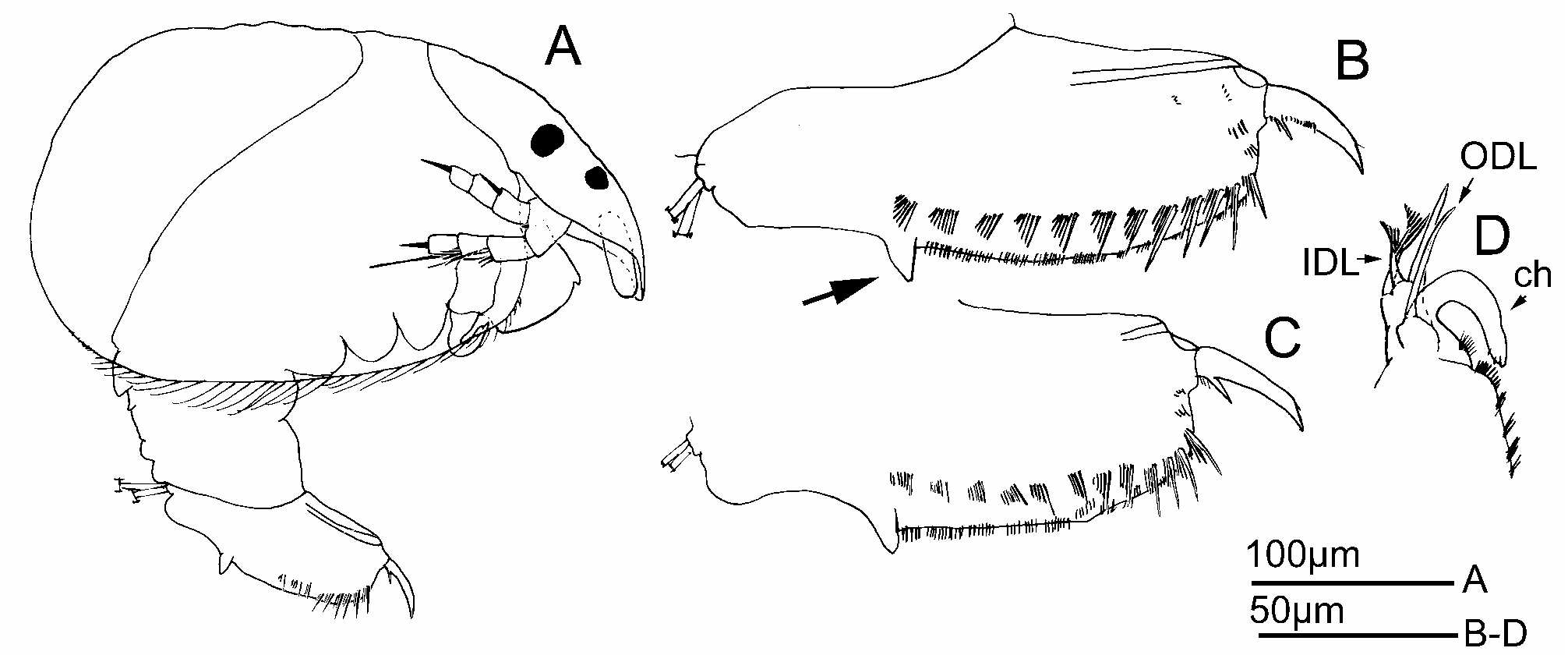
Adult male ( Fig. 6 View FIGURE 6 ). Size 0.3mm. (n=3), body 1.7 as long as high. Body widening posteriorly ( Fig. 6A View FIGURE 6 ). Second antennae with modified swimming setae, as in females. Postabdomen ( Figs 6B–C View FIGURE 6 ) about two times as long as wide, with strongly developed triangular preanal projection. Distalmost spine in each lateral fascicle on postabdomen long and reaching beyond dorsal margin of postabdomen. Terminal claw thick and short (shorter than anal margin) with basal spine about one fourth of terminal claw length. Gonopores opening ventrally, adjacent to the basal claw. First limb ( Fig. 6D View FIGURE 6 ) with IDL bearing three setae, of which two modified in distal portion, though less as in females. Third IDL seta naked and longer than two modified setae. Copulatory hook strongly curved and short, in inner side with broad base and U-shaped, distal part as long as proximal part.
Ephippial female and ephippium. Ephippial female larger than parthenogenetic female (up to 0.50mm), ephippium orange brown to (rarely) dark brown.
Differential diagnosis. The South American A. acuta n. sp. cannot be confused with other Anthalona species. Its major character are the thick spiniform apical setae on the antennal exopod, oriented forward after fixation ( Fig. 1A View FIGURE 1 ). This character is only shared with the Australasian Armatalona Sinev, 2004 . A. acuta n. sp. can be recognized also by a strongly “hunchbacked” body in lateral view (high anterior half) ( Fig. 3A View FIGURE 3 ). Rostrum relatively long, broad and curved inwards; main head pores long, lateral pores with relatively largest “sacks” for the genus ( Figs 2F–I View FIGURE 2 ); marginal setae on posterior portion of carapace thick and spiniform, relatively long (but not as strong as in A. brandorffi ). On postabdomen, distalmost spine in lateral fascicles thick, long and reaching far beyond the margin and basal spine on terminal claw not exceeding claw width ( Figs 1K–N View FIGURE 1 ). When sympatric, this species is larger (on average 0.38mm) than A. verrucosa (average 0.34mm). A comparison with head shields and pores of A. verrucosa can be seen in Figure 2 View FIGURE 2 . Males differ from A. verrucosa males by a deep preanal corner in the postabdomen ( Fig. 6B View FIGURE 6 , arrow). The ephippia are not black ( verrucosa ) but lighter brown. For comparison with other species, see Table 1 (after Systematic part).
Distribution and biology. NE-Brazil (Lençóis Maranhenses) and Venezuela (Orinoco Basin). These are now the only two certain records, but the species may have a wider distribution in the Neotropics, confused with Anthalona verrucosa ( Sars, 1901) . Found in the Lençóis Maranhenses in Brazil, in temporary pools on sandy substrate were not included in latter publication and were found later. Occurs together with true Neotropical Chydoridae Leydigiopsis curvirostris Sars, 1901 , Alona iheringula Kotov & Sinev, 2004 , Chydorus dentifer Daday, 1905 , Anthalona verrucosa verrucosa ( Sars, 1901) , Alona ossiani Sinev, 1998 (Lençóis) ; Alona dentifera ( Sars, 1901) , Anthalona verrucosa verrucosa ( Sars, 1901) and Chydorus dentifer Daday, 1905 (Orinoco) ( Van Damme & Dumont, 2010)


| UG |
Museo del Departamento de Estratigrafia y Paleontologia |
No known copyright restrictions apply. See Agosti, D., Egloff, W., 2009. Taxonomic information exchange and copyright: the Plazi approach. BMC Research Notes 2009, 2:53 for further explanation.
|
Kingdom |
|
|
Phylum |
|
|
Class |
|
|
Order |
|
|
Family |
|
|
Genus |
Anthalona acuta
| DAMME, KAY VAN, SINEV, ARTEM YU & DUMONT, HENRI J. 2011 |
Alona
| Van Damme, K. & Dumont, H. J. 2010: 759 |