
Morbakka virulenta ( Kishinouye, 1910 )
|
publication ID |
https://doi.org/ 10.1080/00222933.2012.717645 |
|
publication LSID |
lsid:zoobank.org:pub:6AFA1E11-810D-4829-8751-A65D41FAF3EC |
|
persistent identifier |
https://treatment.plazi.org/id/0390350A-FFAD-DF32-FE60-167AFE6196AA |
|
treatment provided by |
Felipe |
|
scientific name |
Morbakka virulenta ( Kishinouye, 1910 ) |
| status |
|
Morbakka virulenta ( Kishinouye, 1910) View in CoL
( Figures 4I. J View Figure 4 , 7 View Figure 7 , 8 View Figure 8 )
Tamoya virulenta Kishinouye (1910: 7 , Pl. I, fig. 10); Uchida (1947: 316); Williamson et al. (1996: 414).
Tamoya alata Uchida (1929: 172 , figs 8–88). non Tamoya alata Reynaud [refers to Copula sivickisi View in CoL ]: Uchida (1929: 178–80; figs 86, 87).
Tamoya bursaria Uchida (1947: 314–16 , figs. 2, 3); Uchida (1954: 209–19).
Tamoya haplonema Uchida (1970: 289 View in CoL , 293–4, figs 3, 4); Yamasu and Yoshida (1976: 325–6); Yamaguchi (1982: 249); Kubota (1998: 33); Iwama (2001: 109).
Tamoya bursaria (? gargantua View in CoL ) Williamson et al. (1996: 414).
Tamoya gargantua Williamson et al. (1996: 414) View in CoL .
Material examined
Neotype. USNM 1124253 View Materials , female, 150 mm BH, 60 mm IRW, Hiroshima Bay , Japan.
Other material. USNM 1124251, 150 mm BH, 68 mm IRW, Hiroshima Bay , Japan ; USNM 1124252, 140 mm BH, 50 mm IRW, Hiroshima Bay , Japan .
Type locality
Hiroshima Bay, Honshu, Japan.
Common name
Hikurage (“fire jellyfish” in Japanese).
Diagnosis
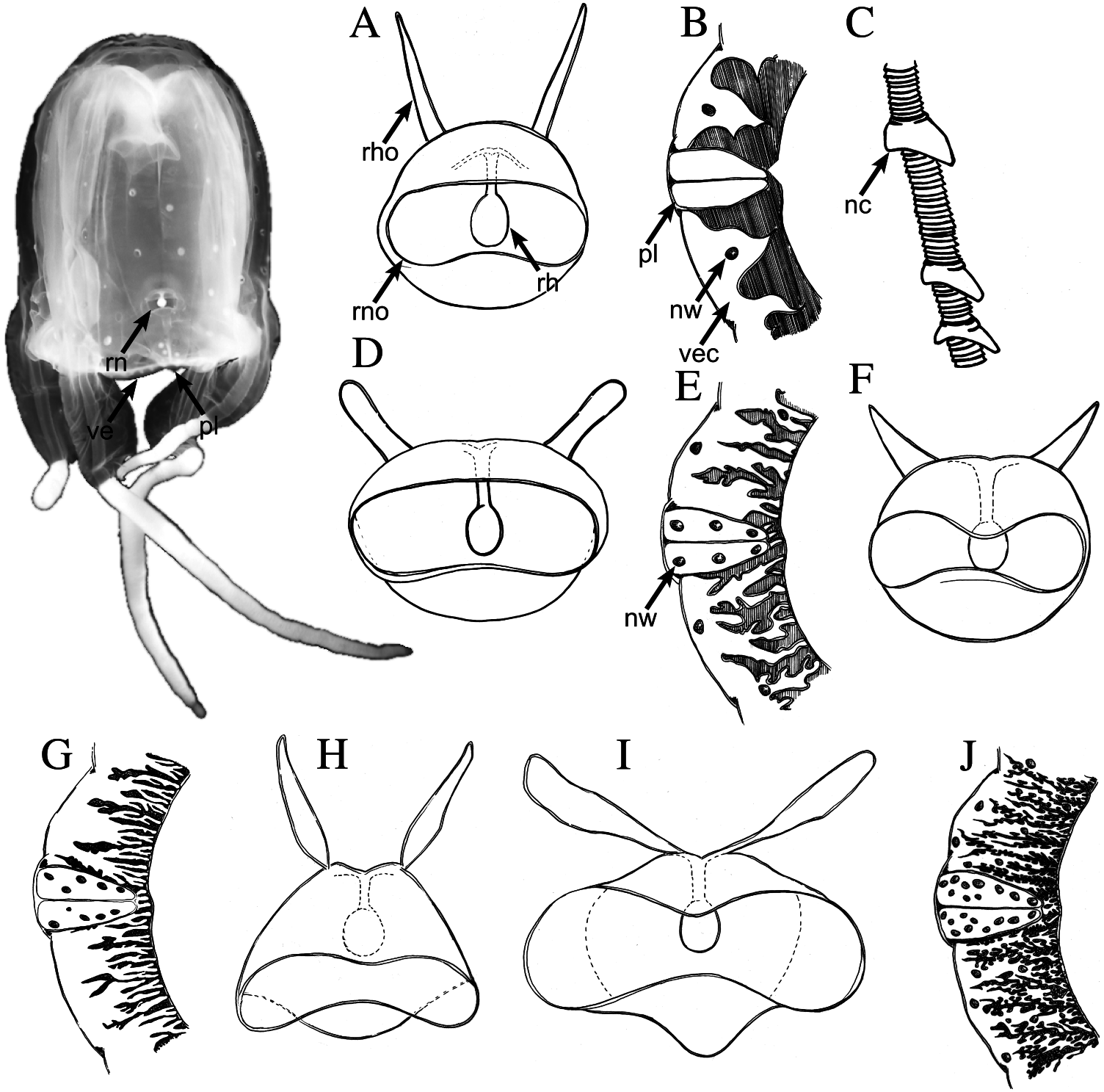
Large carybdeid medusa (up to 150 mm BH) lacking gastric phacellae; robust bell densely covered with nematocyst warts ( Figure 7A View Figure 7 ). Morbakka virulenta possesses “rabbit-ear”-shaped rhopaliar horns and differs from its congener, M. fenneri , in that M. virulenta ’s rhopaliar horns are swollen rather than pointed at the tips, and project from the top centre of the rhopaliar niche at a more oblique angle ( Figure 4H View Figure 4 versus 4I). Further, M. virulenta lacks the nematocyst wart on the rhopaliar stalk that is characteristic of M. fenneri .
Description
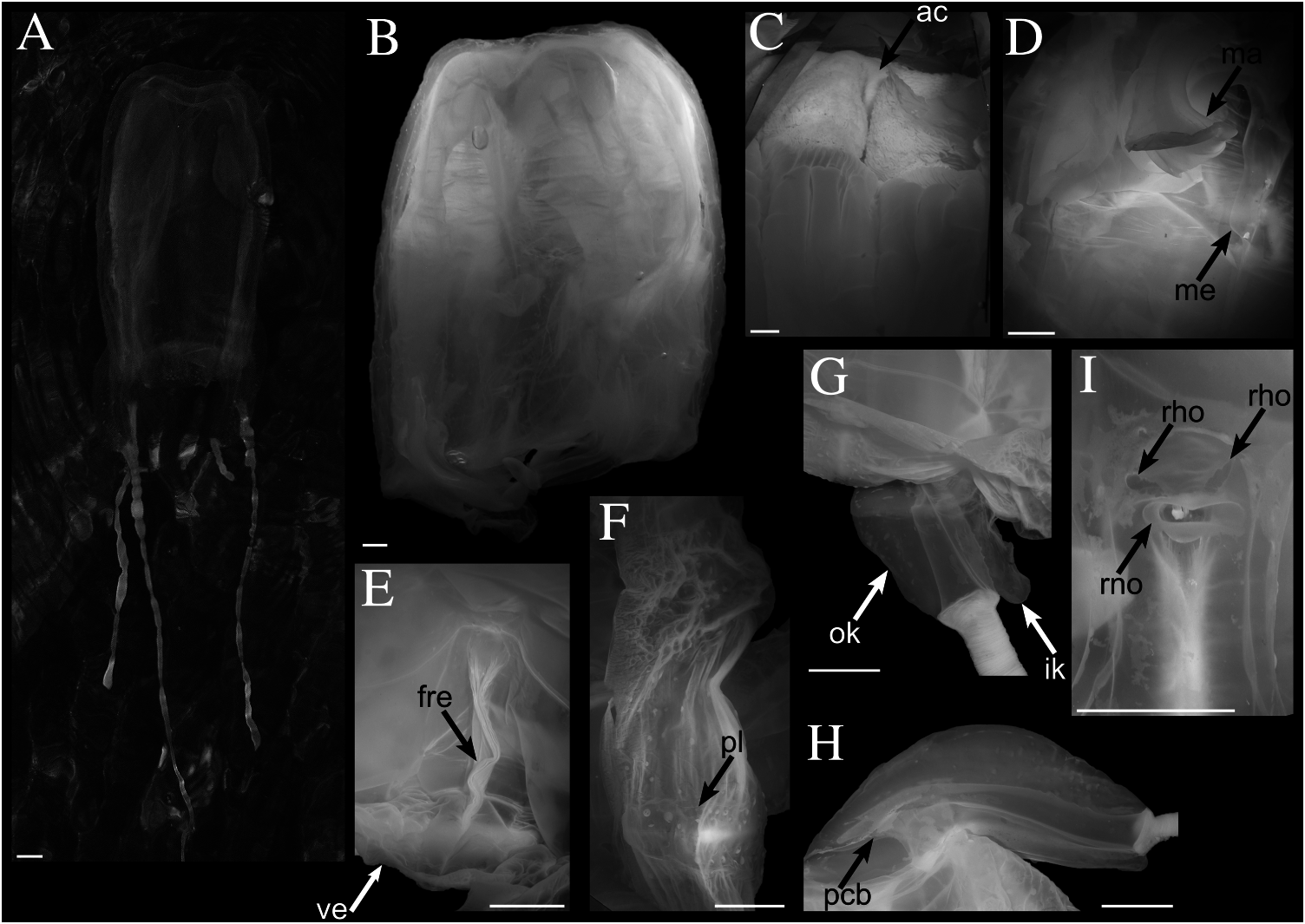
Carybdeid medusa, bell taller than wide, rectangular, with flat apex, and leaflike gonads in mature individuals ( Figure 7A, B View Figure 7 ); extended tentacles flat, cord-like in live specimens ( Figure 7B View Figure 7 ). Exumbrella densely covered with nematocyst warts ( Figure 7A View Figure 7 ). Maximum BH about 150 mm (observed range from 140 to 150 mm in mature individuals) and maximum IRW about 68 mm (observed range from 50 to 68 mm in mature individuals). Gastric phacellae absent; stomach with welldeveloped musculature (area corrugata; Figure 7C View Figure 7 ). Manubrium with rounded, smooth-edged lips ( Figure 7D View Figure 7 ) extending to about one-third of BH from subumbrellar ostium. Mesenteries well developed in upper half of the subumbrella, extending cord-like to rhopaliar window ( Figure 7D View Figure 7 ). Frenulum consisting of a single sheet that splits longitudinally near the rhopaliar niche, extending onto the lower half of the rhopaliar window ( Figure 7E View Figure 7 ); frenulum covering three-quarters of the distance between velarial turnover and velarial margin ( Figure 7E View Figure 7 ). Perradial lappets broad, triangular, not reaching subumbrellar edge of velarium ( Figure 7F View Figure 7 ); approximately 7–10 nematocyst warts on each side of the perradial lappet, mostly arranged in single rows but some scattered ( Figures 4J View Figure 4 and 7F View Figure 7 ). Velarium with six to eight complex dendritic canals per octant with lateral diverticula per octant ( Figures 4J View Figure 4 and 7F View Figure 7 ). Pedalium keeled on both sides of lateral median line; inner keel with overhang ( Figure 7G View Figure 7 ). Outer keel with warts ( Figure 7G View Figure 7 ), that appear to have mostly rubbed off after collection. Pedalial canal with thorn-like extension ( Figure 7H View Figure 7 ). Rhopaliar niche ostium frown-shaped with “rabbit-ear”-like rhopaliar horns ( Figures 4I View Figure 4 and 7I View Figure 7 ).
Cnidome
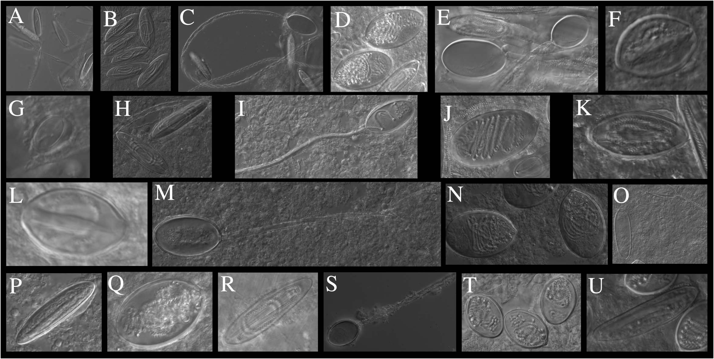
Tentacles were truncated in the preserved specimens, but nematocysts were sampled from both the distal end of the truncated tentacles as well as the proximal base of the tentacles near the pedalia. Distal tentacle tip: rod-shaped, microbasic p-mastigophores (L 58.2- 66.2 -71.2 µm, W 14.1 - 18.2 -16 µm, n = 6; Figure 8A, B View Figure 8 ); large, oval, holotrichous isorhizas (L 59.1- 60.1 -64.8 µm, W 31.2 - 38.6 -39.3 µm, n = 20; Figure 8C, D View Figure 8 ); oval, microbasic p-euryteles (L 18- 21.8 -23.6 µm, W 13.5 - 15.5 -17.6 µm, n = 5; Figure 8E, F View Figure 8 ); small, oval amastigophores (L 10.6 µm, W 6.4 µm, n = 1; Figure 8G View Figure 8 ). Proximal tentacle/tentacle base: rod-shaped, microbasic p-mastigophores (L 69.4- 71.7 -74.6 µm, W 14.9 - 16.7 -19.1 µm, n = 20; Figure 8H View Figure 8 ); large, oval, holotrichous isorhizas (L 61.8- 66.9 -77.6 µm, W 30.1 - 35.9 -43.3 µm, n = 20; Figure 8I, J View Figure 8 ); oval, microbasic p-euryteles (L 43.3- 43.3 -44.5 µm, W 18.6 - 18.6 -21.1 µm, n = 3; Figure 8K View Figure 8 ). Manubrium:?oval, microbasic p-euryteles (L 20.7 µm, W 12.8 µm, n = 1; Figure 8L View Figure 8 ). Pedalial warts: large, oval, holotrichous isorhizas (L 60.9- 64.5 -69.5 µm, W 37.8 - 40.6 -43.2 µm, n = 20; Figure 8M, N View Figure 8 ); rod-shaped, microbasic p-mastigophores (L 61.7- 67.2 -71 µm, W 15.7 - 18.9 -21.3 µm, n = 13; Figure 8O, P View Figure 8 ). Exumbrellar warts: large, oval, holotrichous isorhizas (L 42.9- 45.9 -50.2 µm, W 27.6 - 31 -33.6 µm, n = 20; Figure 8Q View Figure 8 ); rod-shaped, microbasic p-mastigophores (L 62.8- 70 -79.4 µm, W 14.7 - 17.2 -19.1 µm, n = 20; Figure 8R View Figure 8 ). Apex warts: oval, holotrichous isorhizas (L 33.6- 36.8 -40.4 µm, W 22.8 - 25.5 -27.6 µm, n = 20; Figure 8S, T View Figure 8 ); rod-shaped, microbasic p-mastigophores (L 67.9 and 68 µm, W 16.8 and 19.9 µm, n = 2; Figure 8U View Figure 8 );?large, oval, holotrichous isorhizas (only empty capsules observed, L 48.3 and 53 µm, W 27.6 and 31.6 µm, n = 2).
Remarks
A neotype is designated for M. virulenta to stabilize its taxonomic status and clarify its identity, with particular emphasis on differentiating it from its congener M. fenneri . The description and drawing of M. virulenta in the original description (as Tamoya haplonema ; Kishinouye 1910) agree well with the material examined by us, but lack the characters typical of Morbakka , in particular the rhopaliar horns. We believe Kishinouye (1910) overlooked these characters, as did his contemporaries, because the potential importance of these characters had not been recognized. The original description does not mention the presence of vertical gastric phacellae that are characteristic of Tamoya , which suggests that the specimen(s) investigated by Kishinouye (1910) did not possess gastric phacellae – a trait characteristic of Carukiidae . Additionally, the common name used by Kishinouye (1910) for T. virulenta is “Hikurage”, the same name applied to the specimens we studied. The original type locality was the Inland Sea off Kagoshima and Innoshima; Hiroshima Bay is about 50 km east of Innoshima.
| USNM |
Smithsonian Institution, National Museum of Natural History |
No known copyright restrictions apply. See Agosti, D., Egloff, W., 2009. Taxonomic information exchange and copyright: the Plazi approach. BMC Research Notes 2009, 2:53 for further explanation.
|
Kingdom |
|
|
Phylum |
|
|
Class |
|
|
Order |
|
|
Family |
|
|
Genus |
Morbakka virulenta ( Kishinouye, 1910 )
| Bentlage, Bastian & Lewis, Cheryl 2012 |
Tamoya bursaria
| Williamson JA & Fenner PJ & Burnett JW & Rifkin J 1996: 414 |
Tamoya gargantua
| Williamson JA & Fenner PJ & Burnett JW & Rifkin J 1996: ) |
Tamoya haplonema
| Iwama Y 2001: 109 |
| Kubota S 1998: 33 |
| Yamaguchi M 1982: 249 |
| Yamasu T & Yoshida M 1976: 325 |
| Uchida T 1970: 289 |
Tamoya bursaria
| Uchida T 1954: 209 |
| Uchida T 1947: 16 |
Tamoya alata
| Uchida T 1929: 172 |
| Uchida T 1929: 178 |
Tamoya virulenta
| Williamson JA & Fenner PJ & Burnett JW & Rifkin J 1996: 414 |
| Uchida T 1947: 316 |
| Kishinouye K 1910: 7 |