
Truttaedacnitis sphaerocephala (Rudolphi, 1809) Petter, 1974
|
publication ID |
https://doi.org/10.11646/zootaxa.4185.1.1 |
|
publication LSID |
lsid:zoobank.org:pub:0D054EDD-9CDC-4D16-A8B2-F1EBBDAD6E09 |
|
DOI |
https://doi.org/10.5281/zenodo.5626964 |
|
persistent identifier |
https://treatment.plazi.org/id/038FB248-FF2B-FF2C-89B9-C5C723C9991A |
|
treatment provided by |
Plazi (2016-11-08 07:41:12, last updated by Guilherme 2025-02-13 19:53:50) |
|
scientific name |
Truttaedacnitis sphaerocephala (Rudolphi, 1809) Petter, 1974 |
| status |
|
Truttaedacnitis sphaerocephala (Rudolphi, 1809) Petter, 1974
Synonym: Bulbodacnitis sphaerocephala (Rudolphi, 1809) Maggenti, 1971
Description (after Choudhury & Dick 1996b, and Moravec 2013). With characteristics of the genus. Moderately large worms with thick cuticle. Anterior end often dorsally flexed and mouth opening markedly oblique. Mouth opening bordered by cuticular collarette bearing weakly developed inconspicuous longitudinal riblike structures (“teeth”). Ventral cephalic ridge traverses ventral side at level of pseudobuccal capsule; excretory pore occurs between the two hemispheres of this ridge. Two very narrow alae extend along anterior part of body, starting below level of posterior end of pseudobuccal capsule. Two broad closely apposed cephalic plates are on either side of the mouth opening ( Fig. 86 View FIGURE 86 B). The pseudobuccal capsule has a thickened cuticular lining surrounded by musculature that becomes progressively thinner posteriorly. Hemizonid and asymmetrical deirids behind nerve ring. Tail conical. [Measurements in square parentheses are taken from Moravec’s (2013) description of European material.]
Males: 11.9–16.2 long. [11.24–22.71 long; maximum width 0.31–0.48. Nerve ring 0.468–0.640 from anterior end. Oesophagus 1.64–2.12 long. Tail conical, 0.265–0.374 long, pointed tip.] Testis single. Posterior end of body bent ventrally, with well developed pre-cloacal sucker. Spicules equal, bent [0.270–0.440 long] ( Fig. 89 View FIGURE 89 ). [Gubernaculum 0.075–0.096 long.] Arrangement of caudal papillae similar to that of T. clitellarius above ( Fig. 87 View FIGURE 87 B).
Females: 15.9–18.8 long. [17.44–28.56 long; maximum width 0.30–0.48. Nerve ring 0.468–0.640 from anterior end. Oesophagus 1.89–2.28 long. Deirids asymmetrical, 0.075–0.160 from anterior end. Tail 0.374–0.562 long, conical, tip rounded with mucron.] Vulva in posterior half of body, anterior lip large, overlapping posterior lip. Eggs oval, thin-walled, [0.070–0.092 x 0.045–0.064].
Site: spiral valve
Host: Acipenser oxyrinchus
Distribution: Atlantic, New Brunswick
Records: Linkletter et al. 1977 (AT); Appy & Dadswell 1978 (NB)
Appy, R. G. & Dadswell, M. J. (1978) Parasites of Acipenser brevirostrum LeSueur and Acipenser oxyrhynchus Mitchill (Osteichthyes: Acipenseridae) in the Saint John River estuary, N. B., with a description of Caballeronema pseudoargumentosus sp. n. (Nematoda: Spirurida). Canadian Journal of Zoology, 56, 1382 - 1391. http: // dx. doi. org / 10.1139 / z 78 - 191
Choudhury, A. & Dick, T. A. (1996 b) Observations on the morphology, systematics, and biogeography of the genus Truttaedacnitis (Nematoda: Cucullanidae). Journal of Parasitology, 82, 977 - 987. http: // dx. doi. org / 10.2307 / 3284209
Linkletter, L. E., Lord, E. L. & Dadswell, M. J. (1977) A checklist and catalogue of the marine fauna and flora of the lower Bay of Fundy shore of New Brunswick. Huntsman Marine Laboratory, St. Andrews, New Brunswick, vii + 68 pp.
Maggenti, A. R. (1971) A review of the family Cucullanidae Cobbold, 1864 and the genus Bulbodacnitis Lane, 1916 with a description of Bulbodacnitis sp. n. (Nematoda: Cucullanidae) from Salmo gairdneri Richardson. Proceedings of the Helminthological Society of Washington, 38, 80 - 85.
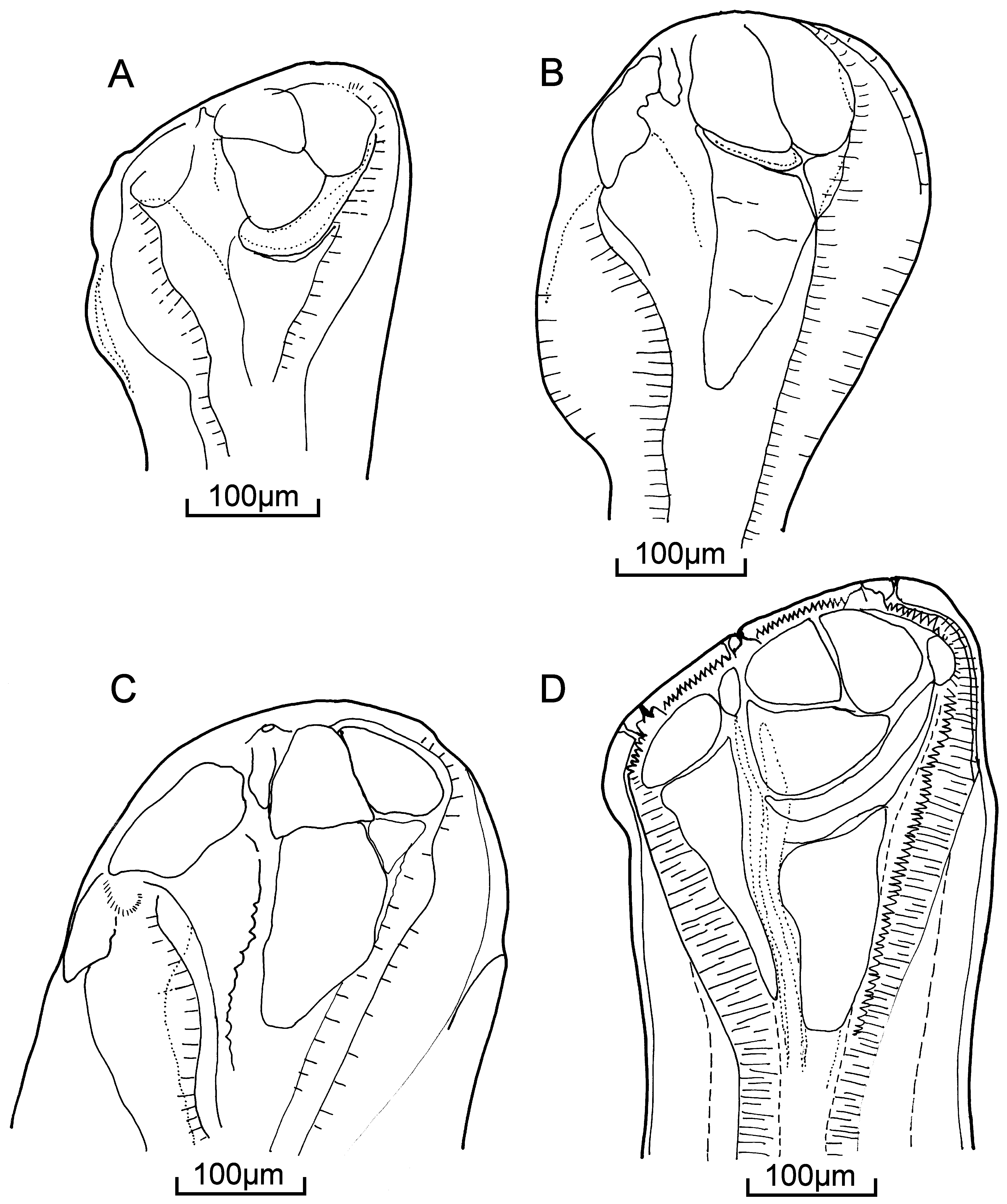
FIGURE 86. Truttaedacnitis spp.: heads showing cephalic plates. A. T. truttae; B. T. sphaerocephala; C. T. clitellarius; D. T. pybusae. (A., B. and C. redrawn from Choudhury & Dick 1996 b; D. redrawn and rotated from Pybus et al. 1978 a)
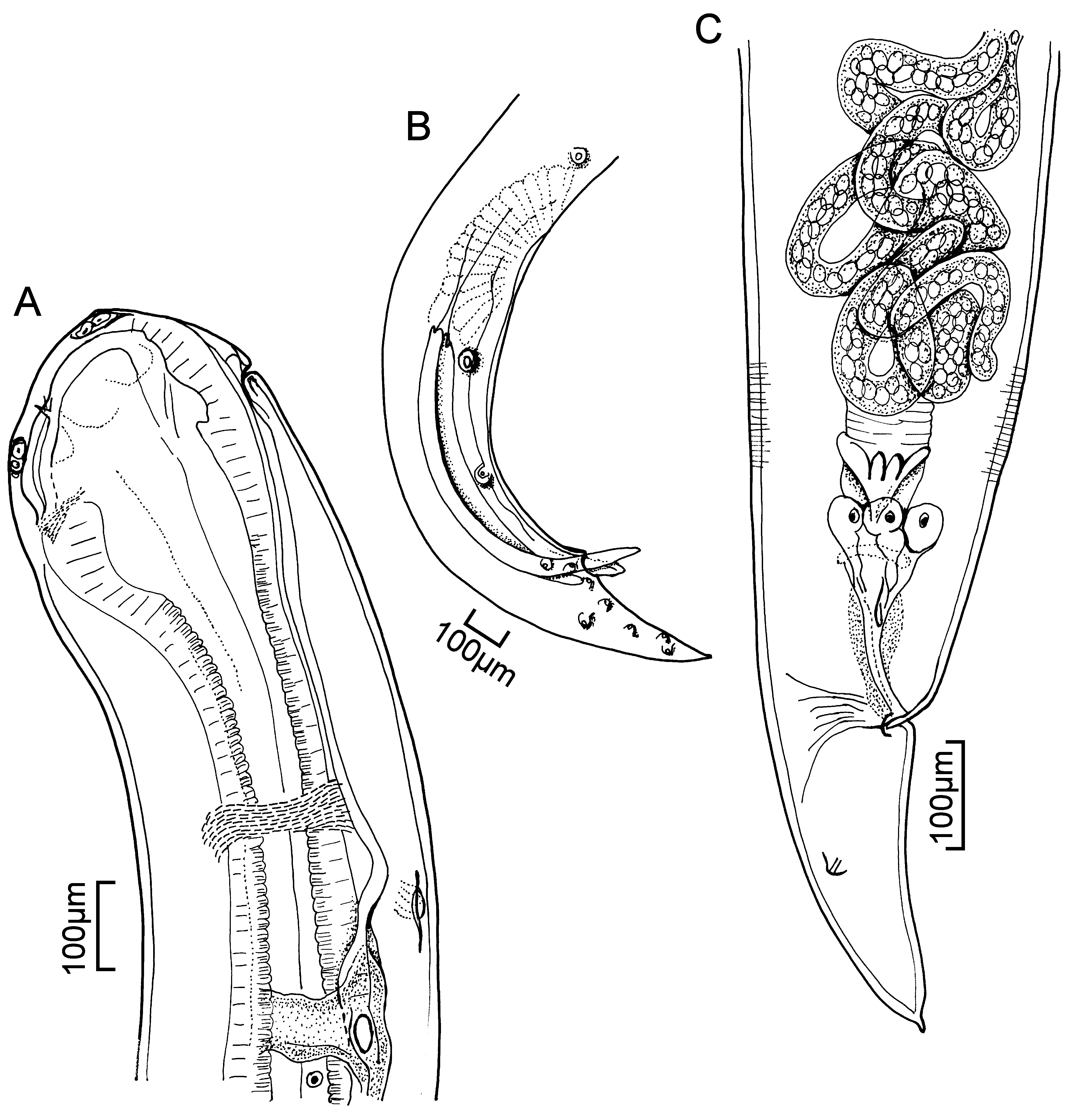
FIGURE 87. Truttaedacnitis clitellarius (Ward & Magath, 1917) Petter, 1974. A. male, anterior end showing excretory pore and duct, lateral view; B. male, posterior region showing pre-cloacal sucker, spicules and papillae, lateral view; C. female, posterior end showing posterior coiled ovary, rectal glands, phasmid and mucron, lateral view. (Redrawn from Choudhury & Dick 1996 a)

No known copyright restrictions apply. See Agosti, D., Egloff, W., 2009. Taxonomic information exchange and copyright: the Plazi approach. BMC Research Notes 2009, 2:53 for further explanation.
|
Kingdom |
|
|
Phylum |
|
|
Class |
|
|
Order |
|
|
InfraOrder |
Oxyuridomorpha |
|
SuperFamily |
Spiruroidea |
|
Family |
|
|
Genus |
1 (by plazi, 2016-11-08 07:41:12)
2 (by ImsDioSync, 2016-11-08 22:36:35)
3 (by ImsDioSync, 2016-11-08 23:22:06)
4 (by ImsDioSync, 2016-11-09 00:24:56)
5 (by ImsDioSync, 2016-11-09 01:39:36)
6 (by ImsDioSync, 2016-11-09 02:10:29)
7 (by ImsDioSync, 2017-01-30 16:56:53)
8 (by ImsDioSync, 2017-01-30 17:04:12)
9 (by ImsDioSync, 2019-03-29 21:03:45)
10 (by ExternalLinkService, 2019-09-26 07:54:39)
11 (by ExternalLinkService, 2021-10-29 03:00:38)
12 (by ExternalLinkService, 2021-10-29 07:52:09)
13 (by ExternalLinkService, 2021-10-30 14:51:33)
14 (by ExternalLinkService, 2021-10-30 14:51:33)
15 (by ExternalLinkService, 2021-10-30 14:51:33)
16 (by ExternalLinkService, 2021-10-30 14:51:33)
17 (by plazi, 2023-10-27 07:43:52)
18 (by ExternalLinkService, 2023-10-27 18:26:09)