
Fridericia raxiensis, Dózsa-Farkas & Felföldi, 2018
|
publication ID |
https://doi.org/ 10.17109/AZH.64.1.1.2018 |
|
persistent identifier |
https://treatment.plazi.org/id/038D3B07-EC7D-FF9B-FE3D-F60DAC99FE3B |
|
treatment provided by |
Felipe |
|
scientific name |
Fridericia raxiensis |
| status |
sp. nov. |
Fridericia raxiensis sp. n.
( Figs 4 – 5 View Fig View Fig )
Type material – Holotype. F.27. slide No. 2125, Rax Mountain , close to the Rax ca- ble car terminal, under Pinus mu go 47°43.036N, 15°46.024E, 1620 m a.s.l., leg. Farkas, J., 15.05.2012. GoogleMaps
Paratypes. In total 12 specimens P . 111.1.1.–111.1.4. slide No. 2229–2233 four speci- mens Rax Mountain, close to the Rax cable car terminal, under Pinus mugo , 47°43.23.3 N , 15°45.164E 1612 m a.s.l., leg. Farkas, J., 15.05.2012; P . 111.2.1–111.2.3 slide No. 2126–2128 three specimens Rax Mountain, close to the Rax cable car terminal, subalpine meadow, 47°43.172N, 15°45.218E, 1613 m a.s.l., leg. Farkas, J., 15.05.2012; P GoogleMaps . 111.3.1–111.3.2 slide No. 2227–2228 two specimens Rax Mountain, close to the Rax cable car terminal, under Pinus mugo 47°43.007N, 15°45.401E, 1613 m a.s.l., leg. Farkas, J., 15.05.2015; P GoogleMaps . 111.4.1–111.4.3 slide No. 2239–2241 three specimens Rax Mt. 47°71.666N, 15°77.305E, 1613 m a.s.l., under Pinus mugo leg. Bauer, R., 10.06.2008 .
Etymology – Named after the Rax Mountain where this species was found.
Diagnosis – The new species can be recognized by the following combination of characters: (1) medium size (14–18 mm in vivo), segments 51–59; (2) maximum 5 chaetae per bundle; (3) clitellum girdle-shaped: hyalocytes and granulocytes arranged in transverse rows but weakly developed; (4) five preclitellar pairs of nephridia; (5) coelomo-mucocytes numerous, c/b-type (according to Möller 1971), scarce, 30–44 μm in vivo, lenticytes 8–10 Μm long; (6) chylus cells in XIII–XVI (3–4 segments long); (7) bursal slit T-shaped, the transverse component is short; (8) seminal vesicle large; (9) a small subneu- ral gland in XIII; (10) sperm funnel pear-shaped, approximately half as long as body diameter, collar narrower as funnel body, sperm 340–370 Μm long, heads 75–85 Μm in vivo; (11) spermatheca with long ectal duct, large ectal gland, ampulla entally separate, with about 8 sessile, sphaerical diverticula varying in size.
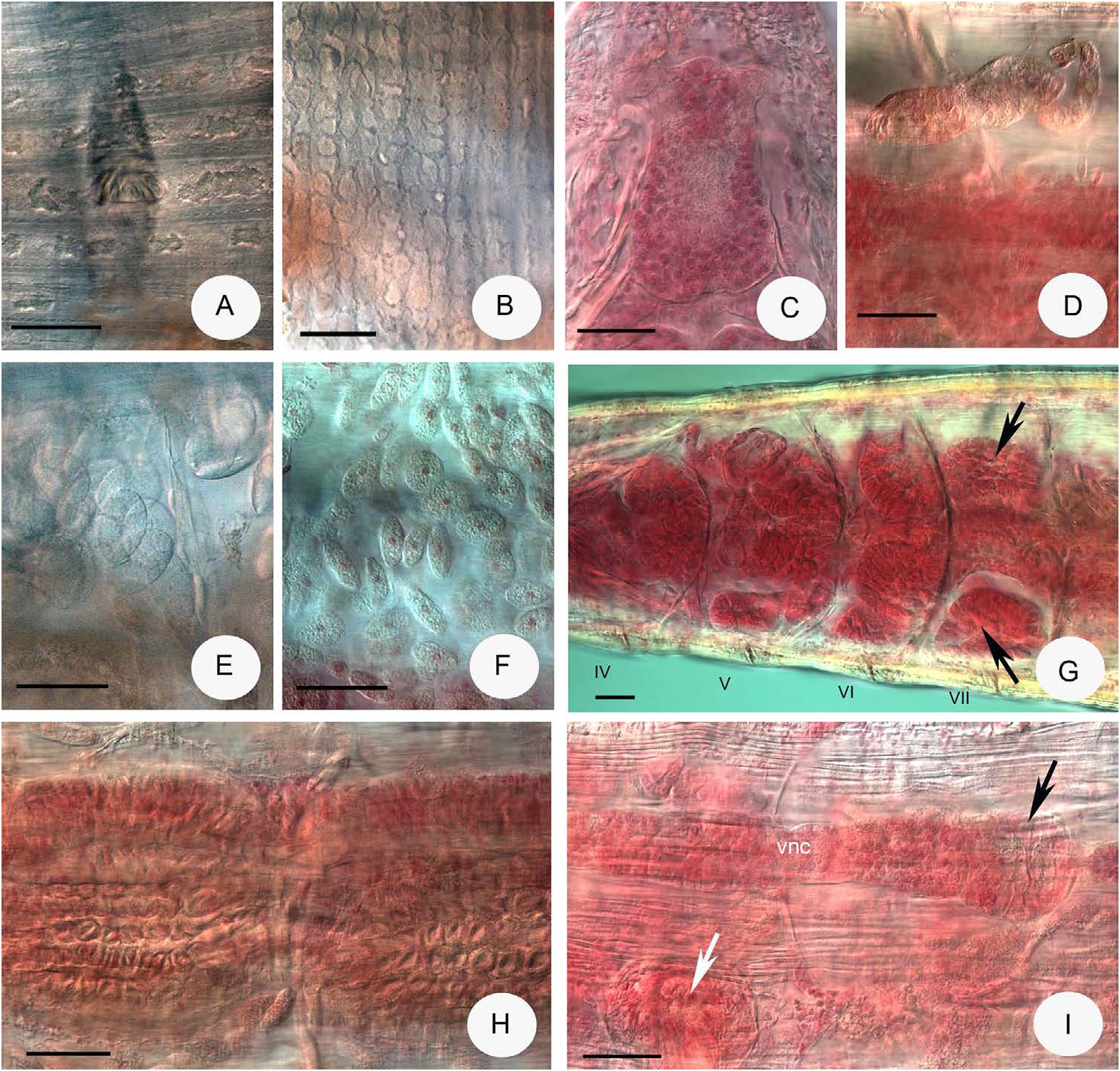
Description – Holotype 11.7 mm long, 380 μm wide at VIII and 380 μm at the clitellum (fixed), 51 segments. Body length of the paratypes 14–18 mm, width 350–410 Μm at VIII and 370–480 μm at the clitellum (in vivo). Length of fixed specimens 8–13 mm, width 350–440 μm at VIII and 380–470 μm at the clitellum. Segments 51–59. Chaetal formula: 2,3,4 – 4,3,2,(1): 3,4,5 – 4,3,2. Chaetae in bundles arranged in pairs with the outer pair being longer and thicker than the inner pair: 45–60 by 5 Μm against 35–40 by 2.5–3 Μm. Chaetal lengths about the same also in postclitellar segments. From about XXX only two chaetae per bundle, but in one case (slide No. 2229) already from XXV, these about 60 μm long and 5 μm wide in terminal segments. Head pore at 0/I. Dorsal pores from VII. Epidermal gland cells in 5–9 transverse rows per segment ( Fig. 4A View Fig ). Body wall thick, about 50 Μm, the cuticle 3–5 Μm, so that internal organs are often difficult to investigate in vivo. Clitellum in XII–1/2XIII, girdle-shaped, glands arranged in transverse rows, weakly developed ( Fig. 4B View Fig ).
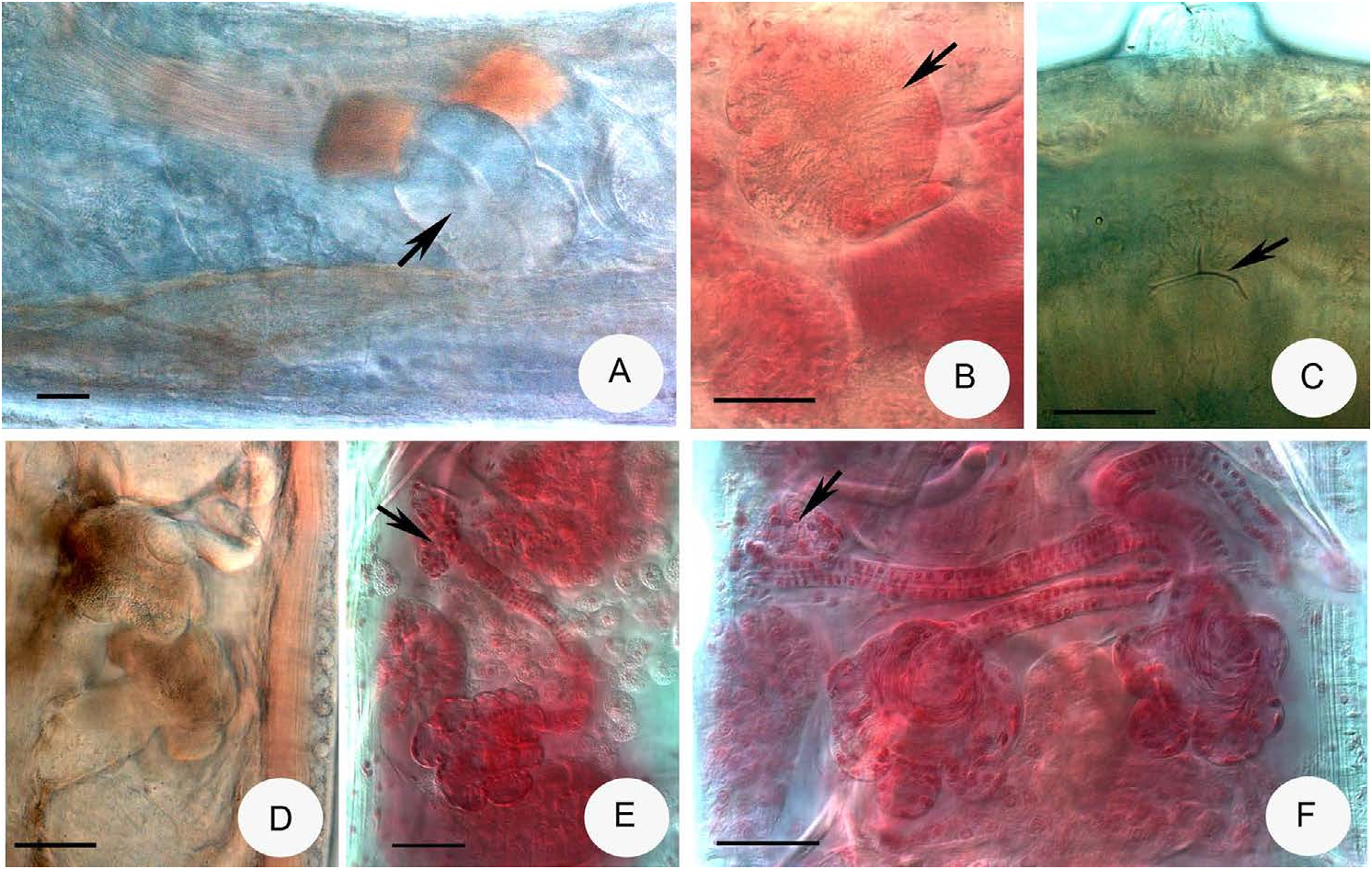
Brain egg-shaped, about 140–160 μm long, 2 times longer than wide in vivo and 120– 160 Μm and 1.5 times longer than wide in the fixed specimens ( Fig.4C View Fig ). Oesophageal ap- pendages extending into V, without branches. Pharyngeal glands are very characteristic. All pairs connected dorsally (sometimes the third is free), ventral lobes absent in IV. Large additional ventral lobes in segment VII ( Fig. 4G View Fig ). Chloragocytes from V, brown in vivo. Dorsal vessel from XVII–XX, blood colourless. Midgut pars tumida not visible. Five pairs of preclitellar nephridia from 6/7 to 10/11 ( Fig. 4D View Fig ), large anteseptale, the length ratio anteseptale: postseptale 1:1.2–1.6, posteroventral origin of efferent duct. Coelomo-mucocytes oval, numerous, c/b-type, matrix fine granulous with some refractile grains, length 30–44 Μm in vivo ( Fig. 4E View Fig ), but in the fixed specimens the matrix of the mucocytes (24–36 Μm long) considerably granulous with well stained nucleus ( Fig. 4F View Fig ). Lenticytes scarce, 8–10 Μm long. Chylus cells ( Fig. 4H View Fig ) between XIII–XVI, occupying 3–4 segments. Seminal vesicle large, in X–XI. Sperm funnels nearly pear-shaped, about 170–250 μm long and 1.5–2 times as long as wide (in vivo) ( Fig. 5A View Fig ). Funnel length in fixed specimens 120–190 Μm and about 1.4 times longer than wide ( Fig. 5B View Fig ). Collar narrower than the funnel. Spermatozoa about 340–370 Μm long, heads 75–85 Μm in vivo, in fixed specimens 250–300 Μm and 45–60 Μm, respectively. Diameter of sperm ducts 6–8 Μm (fixed). Male copulatory organs ( Figs 4I View Fig , 5C View Fig ) small, 140–160 Μm long, 70–90 Μm wide and 60 Μm high (in vivo), (100–120, 60–90 and 40–60 μm in fixed specimens, respectively). Bursal slits T-shaped, but the transverse component is short and the longitudinal component at the two ends with two short transverse components too ( Fig. 5C View Fig ). One small subneural gland in XIII ( Fig. 4I View Fig ). Spermathecae ( Figs 5D–F View Fig ): one large, 40–60 Μm long (in vivo and fixed equally) ectal gland at the orifice ( Figs 5E–F View Fig ). Ectal ducts about 320–390 Μm long and 18–20 Μm wide (250–330 Μm long and 16–18 Μm wide, fixed), projecting into ampullae, ental bulbs about 40 Μm wide in vivo, canals not widened. About 6–9 sessile diverticula (mostly 8) of varying size: diameter (16)–30–50 μm (fixed). Sperm in a circle in lumen of ampullar distal part. Diameter of ampulla and diver- ticula together 90–120 Μm. The epithelium of diverticula in vivo thick and warty ( Fig. 5D View Fig ), mostly no sperm in the diverticula. Separate openings into oesophagus dorso-laterally. One or two mature eggs at a time.
Distribution and habitat – Only known from the type locality.
Differential diagnosis – The number of valid Fridericia species with more than two diverticula per spermatheca is 18: F. agilis Smith, 1895 ; F. agricola Moore, 1895 ; F. bernini Dózsa-Farkas, 1988 ; F. douglasensis Welch, 1914; F. dura Eisen, 1879 ; F. firma Smith et Welch, 1913 ; F. glandifera Friend, 1911 ; F. galba (Hoffmeister, 1843) ; F. gigantea Dequal, 1912 ; F. hegemon (Vejdovksy, 1878) ; F. minor Friend, 1913 ; F. oconeensis Welch, 1914 ; F. paraunisetosa Xie, Liang et Wang, 2000 ; F. pyrenaica Gianni, 1979 ; F. regularis Nielsen et Christensen, 1959 ; F. terrarossae Sesma et Dózsa-Farkas, 1993 ; F. vixdiverticulata Sesma et Dózsa- Farkas, 1993; F. callosa (Eisen, 1878) (type with diverticula).
The new species differs from all these species, leaving other characters out of consideration, by the presence of the additional large ventral lobes of phar- yngeal glands in VII, which is a very rare character among the Fridericia species.
Molecular results
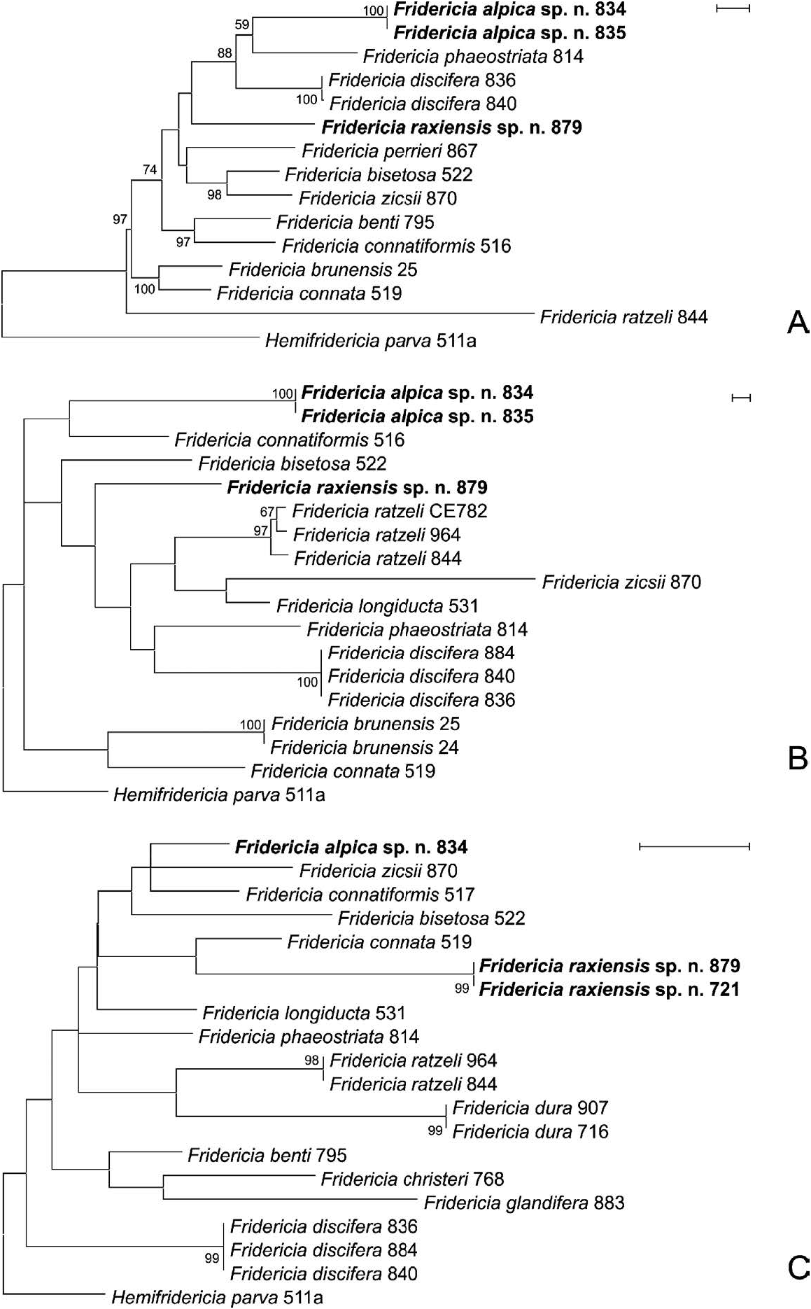
In total, 8, 10 and 12 sequences were obtained from various Fridericia spec- imens in the case of ITS, CO1 and H3, respectively, and some additional se- quences obtained previously were also used for comparison ( Table 3). Results of this molecular analysis confirmed that F. discifera and F. alpica sp. n. are distinct species, since they separated on the phylogenetic trees constructed based on the three studied regions ( Fig. 6 View Fig ). Additionally, the other novel spe- cies, Fridericia raxiensis sp. n. also had a position on the trees distinct from any other similar species.
| F |
Field Museum of Natural History, Botany Department |
| J |
University of the Witwatersrand |
| P |
Museum National d' Histoire Naturelle, Paris (MNHN) - Vascular Plants |
| N |
Nanjing University |
| R |
Departamento de Geologia, Universidad de Chile |
No known copyright restrictions apply. See Agosti, D., Egloff, W., 2009. Taxonomic information exchange and copyright: the Plazi approach. BMC Research Notes 2009, 2:53 for further explanation.
|
Kingdom |
|
|
Phylum |
|
|
Class |
|
|
Order |
|
|
Family |
|
|
Genus |