
Acanthocope annulatus Menzies, 1962
|
publication ID |
https://doi.org/ 10.5281/zenodo.157199 |
|
publication LSID |
lsid:zoobank.org:pub:CB3CB22A-685B-4FBE-963D-413F86933923 |
|
DOI |
https://doi.org/10.5281/zenodo.6272066 |
|
persistent identifier |
https://treatment.plazi.org/id/038CC302-FFF0-7D00-FED3-3D66686FFA3E |
|
treatment provided by |
Plazi |
|
scientific name |
Acanthocope annulatus Menzies, 1962 |
| status |
|
Acanthocope annulatus Menzies, 1962 View in CoL ( Figs 6–10 View FIGURE 6 View FIGURE 7 View FIGURE 8 View FIGURE 9 View FIGURE 10 )
Acanthocope annulatus Menzies, 1962: 155 View in CoL , fig. 44 B, C.
Material examined: Acanthocope annulatus Menzies, 1962 , holotype ɗ (3.2 mm), Stn. Ve m a– 14–31, southeast Atlantic, southwest of Cape Town, 36°34’S, 14°08’E, 4885 m, 4 April 1958 ( AMNH N 1–156); 1 ɗ, 6 Ψ (2.1–5.1 mm), ANDEEP stn. 42–S, 59°40.32’S, 57°35.64’W, 3689 m, ANDEEP stn. 42–E, 1 ɗ (3.2 mm), same locality ( ZMH K–40719); 1 Ψ (5.0 mm), ANDEEP stn. 43–E, 0 3 February 2002, 60°27.19’S, 56°04.81’W, 3962 m, RV Polarstern ( ZMH K–40720).
Additional material examined: Acanthocope argentinae Menzies, 1962 , holotype Ψ (3.3 mm), Vema stn. 12–1, Argentine Basin, 38°58.5’S, 41°45’W, 5024 m, 6 April 1957 ( AMNH N 1–155).
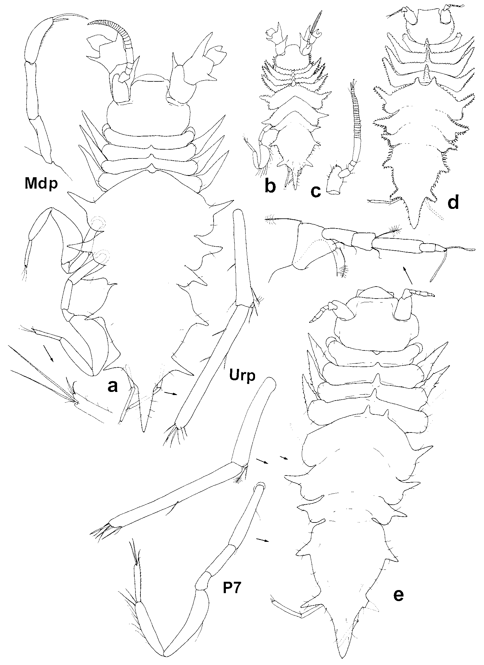
Diagnosis: Dorsal spines on pereonites 1–4 not longer than pereonite, straight, directed upwards; coxae of pereopods 2–4 triangular, about half as long as pereonite width; natasome with a pair of dorsal projections on anterior margin of pereonite 5, and 4 low transverse elevations anteriorly of pleotelson; anterolateral spines of pereonites 5–7 as long as pereonite laterally, almost half as long and wide as coxal spines of pereopods 2–4; lateral spines of pleotelson subequal in size, both pairs directed like lateral spines of pereonite 7, lateral margin in between rounded; terminal spine a third as long as pleotelson; preanal ventral process short, rounded; uropod subequal in length to terminal spine of pleotelson, tiny exopod present.
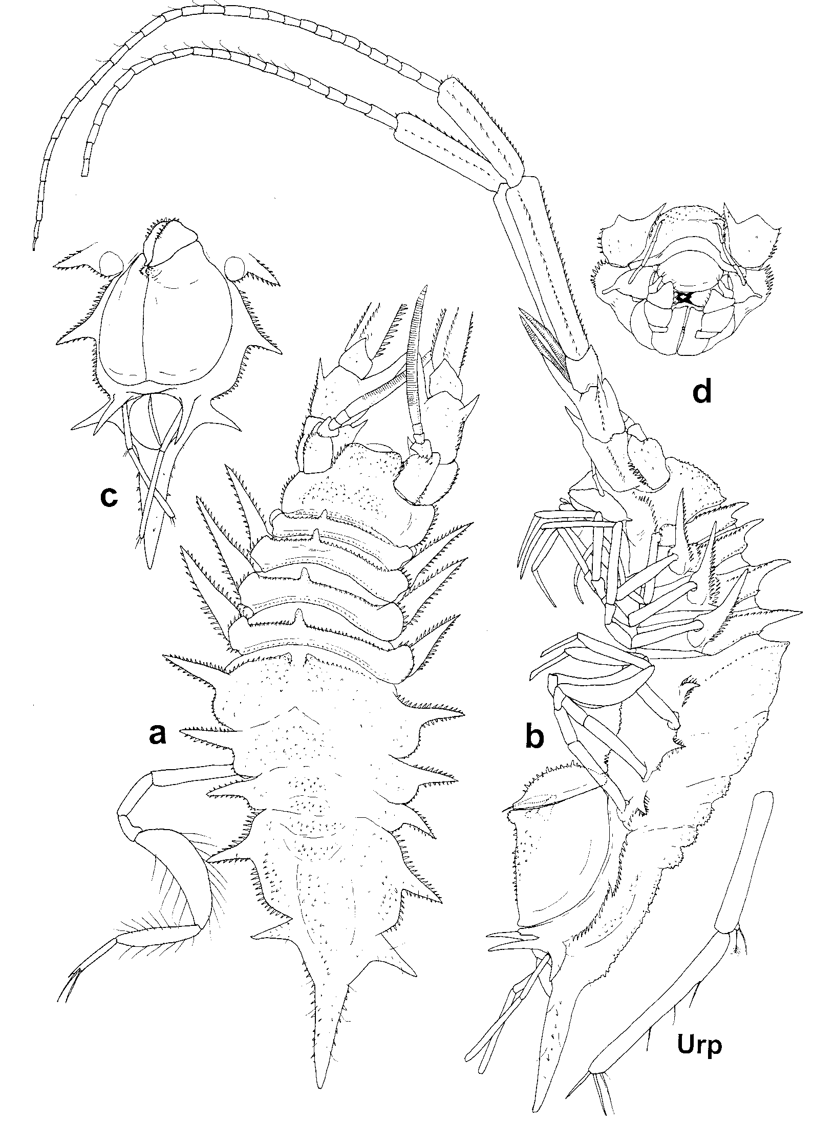
Description: Body ( Figs 6–8 View FIGURE 6 View FIGURE 7 View FIGURE 8 ) about 2.2 times as long as wide (greatest width of pereonite 5 with lateral spines); all lateral margins of body and spines with transparent scale spinules, dorsal surface granulated; pleotelson, especially terminal spine with setae; head about 0.5 times as long as wide, dorsum domeshaped, with acute tubercles, lateral margins in dorsal view slightly convex, with spinule row; frontal margin sloping, smooth; interantennular distance 0.4 times as wide as head and twice as wide as antenna 1 basally, with serrated lateral margins.
Pereonite 1 slightly wider than head; pereonites 1–4 in males ( Figs 6 View FIGURE 6 and 7 View FIGURE 7 ) slightly, in females ( Fig. 8 View FIGURE 8 ) more broadening and lengthening from 1 to 4, in female pereonite 4 as wide as pereonite 5 without lateral spines, in males the ratio is 0.8–0.9; pereonite 1 shortest, pereonites 2–4 subequal in length in males, pereonite 3 longest in females; pereonites 1–4 with a dorsomedial spine anteriorly, shorter than pereonites in females, in males spine on pereonite 1 slightly shorter, on pereonites 2–4 slightly longer than corresponding pereonite; lateral margins of pereonites 1–4 rounded, anterior margin with spinules; coxal spines of pereopods 2–4 subequal in length and in triangular shape, directed somewhat anteriorly, half as long as pereonite width in males, and about third in females.
Natasome 2.6 times as long as anterior body part; pereonites 5–7 decreasing in width and length from 5 to 7; anterolateral spines of pereonite 5 directed slightly anteriorly, those of pereonite 6 perpendicular to body axis, and of pereonite 7 – posteriorly; only pereonite 5 with a pair of short stout dorsal projections anteriorly, 4 low transversal dorsal elevation anteriorly of pleotelson; pleotelson about 1.3 times as long as wide, with 2 subequal, straight lateral spines, directed slightly posteriorly, lateral margin convex; terminal spine from dorsal pore to tip about 0.4 times as long as pleotelson; dorsal surface without spines; preanal ventral process short, rounded; 2 ventrally directed spines anteriorly of uropod insertion, invisible in dorsal view, as long as uropod protopod.
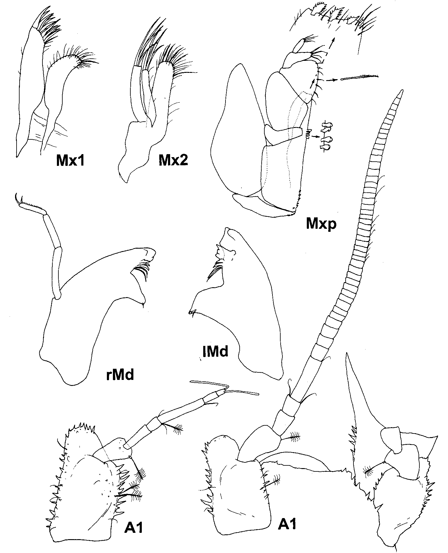
Antenna 1 ( Fig. 9 View FIGURE 9 ) of males 0.3 times as long as body; article 1 1.8 times as long as wide, with many lateral spinules, distolateral lobe relatively narrow, rounded; article 2 almost twice as long as distal lobe of article 1, widening distally, with distomedial broom seta; articles 3–5 0.5, 0.3, and 1.0 times as long as article 2, flagellum with 45 articles: proximal articles as long as article 4, distal articles shorter than article 4.
Antenna 1 ( Fig. 9 View FIGURE 9 ) of females 0.1 times as long as body, with 7 articles, article 1 1.9 times as long as wide, with many lateral spinules and dorsal acute tubercles, two broom setae distomedially; article 2 subequal in length to distolateral lobe of article 1, slightly widening distally, with 1 or 2 distomedial broom setae; articles 3–7 0.9, 0.4, 1.5, 0.5, 0.3 times as long as article 2, terminal article with 2 aesthetascs.
Antenna 2 in females broken off, in male ( Fig. 7 View FIGURE 7 ) 1.4 times as long as body; article 2 with distomedial spine shorter than article; article 3 slightly larger than article 2, with distomedial spine as long as article and twice as long as lateral one; article 4 0.7 times as long and wide as article 3, article 5 1.4 times as long as articles 1–4 together, article 6 slightly narrower and 0.7 times as long as article 5, both articles with 3 rows of spinules along, flagellum 2.3 times as long as peduncle, with approximately 30 elongate articles.
Mandibles ( Figs 9 View FIGURE 9 ) incisors with 3 cusps, lacinia mobilis of left mandible 0.7 as long as incisor, with a few narrow distal teeth, spine row with 4 and 5 spines on left and in right mandibles respectively, molar process distally denticulated; palp slender, 0.7 times as long as body, article 1 longest, article 3 shortest, with 1 long and one short distal setae, in males having a ventral row of short setules.
Maxilla 1 ( Fig. 9 View FIGURE 9 ) mesial endite 0.75 times as wide as lateral endite, with 11 clawlike setae, some of them serrated and 1 small seta distally.
Maxilla 2 ( Fig. 9 View FIGURE 9 ) mesial endite longest, as wide as lateral and middle together; with dense row of distal setae, including 5 serrated setae and 1 long setulated seta distomedially; lateral and middle endites with 4 spinelike distal setae.
Maxilliped ( Fig. 8 View FIGURE 8 ) endite with 4 coupling hooks, distal margin slightly concave, with 3 fansetae, and numerous simple setae; palp article 2 as wide as basis and 1.4 times as long as wide, lateral margin 2.3 times as long as medial one, article 3 0.9 times as wide as article 2, 1.8 times as long as article 2 medially and 0.1 laterally, medial margin slightly convex, with 7 setulated setae; article 4 3.2 times as long as article 3 laterally, medial lobe as long and about twice as wide as article 5, the lobe and article 5 with 4 simple distal setae each; epipod 2.3 times as long as wide, slightly longer than and 1.3 times as wide as basis, tip angular, lateral margin convex in basal half.
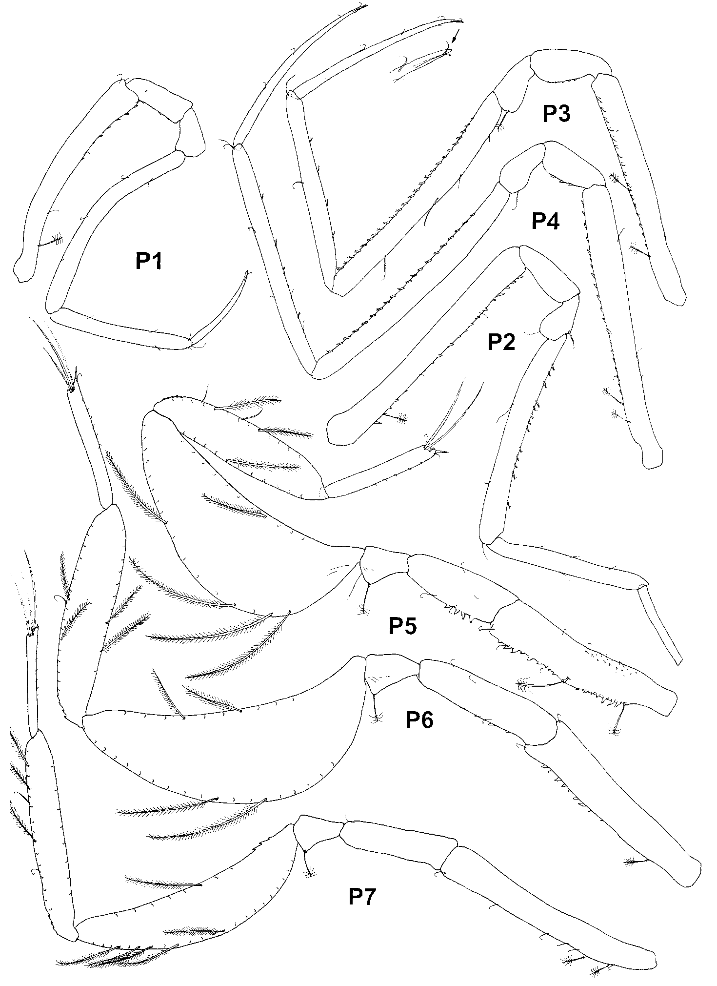
Pereopods ( Fig. 10 View FIGURE 10 ) increasing in size from 1 to longest 4, pereopods 5 and 7 shorter and pereopod 6 longer than pereopod 3; basis of pereopod 4 longest, of pereopod 5 shortest and broadest, others pereopod bases subequal in length, with dorsal denticles and spinules, few broom setae on proximodorsal part and small sparse setae along; dactyli of pereopods 1–4 slender, tapering, of pereopods 5–7 elongate, of the same width through the length, projected distoventral corner with short robust seta and a fine seta, 3 long setae at base of the projection.
Pereopod 1 ( Fig. 10 View FIGURE 10 ) ratios of lengths of ischium–dactylus to basis: 0.3, 0.2, 1, 0.7, 0.4; carpus and propodus with few small marginal simple setae, propodus with tuft of setulated distal setae.
Pereopod 2 ( Fig. 10 View FIGURE 10 ) ratios of lengths of ischium–propodus to basis (dactylus broken off): 0.3, 0.2, 0.8, 0.6; carpus with 13 ventral short robust setae and propodus with 3 ventral and 1 dorsal simple setae.
Pereopod 3 ( Fig. 10 View FIGURE 10 ) basis only slightly shorter than carpus, ratios of lengths of ischium–dactylus to basis: 0.3, 0.2, 1, 0.9, 0.8; carpus with 25 stout ventral setae and 4 dorsal setae, propodus with 4 ventral short spinelike setae, 1 simple dorsal seta, distodorsal whip seta.
Pereopod 4 ( Fig. 10 View FIGURE 10 ) basis longest, ratios of lengths of ischium–dactylus to basis: 0.3, 0.2, 0.9, 0.8, 0.7; carpus with 20 short ventral setae, propodus with 4 ventral spinelike setae and 2 simple dorsal setae.
Pereopods 5–7 carpi dorsal plumose setae almost twice as long as ventral, propodi plumose setae on both margins of the same length, dorsal margin with a few short whip setae.
Pereopod 5 ( Fig. 10 View FIGURE 10 ) ratios of lengths of ischium–dactylus to basis: 0.6, 0.3, 1.3, 1, 0.6; carpus 2.6 times, propodus 4.4 times as long as wide.
Pereopod 6 ( Fig. 10 View FIGURE 10 ) ratios of lengths of ischium–dactylus to basis: 0.7, 0.2, 1.3, 1, 0.6; carpi 3 times, propodus 5 times as long as wide.
Pereopod 7 ( Fig. 10 View FIGURE 10 ) ratios of lengths of ischium–dactylus to basis: 0.5, 0.2, 1, 0.9, 0.5; carpi 4.5 times, propodus 7 times as long as wide.
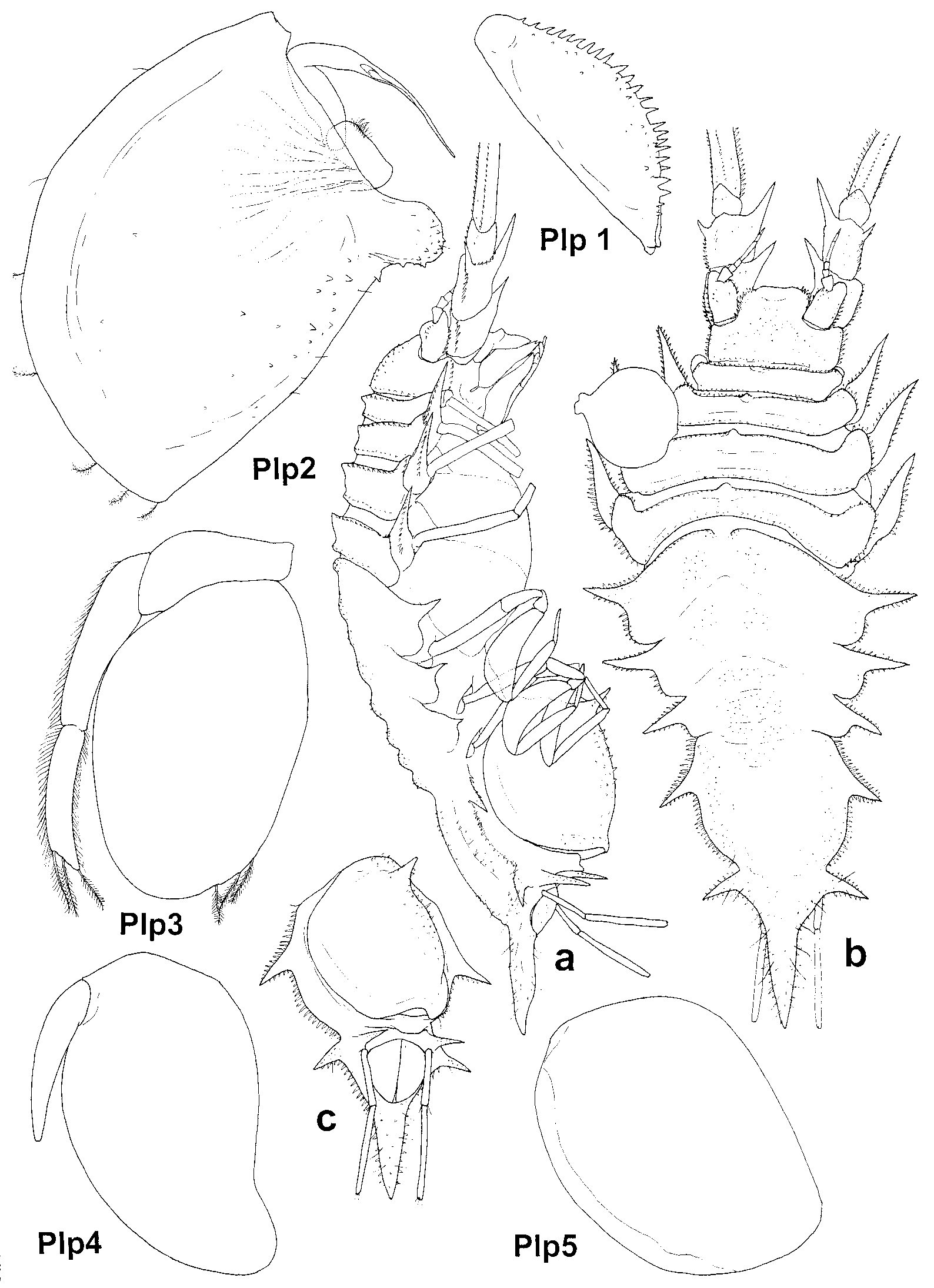
Pleopod 1 of male ( Figs 7 View FIGURE 7 , 8 View FIGURE 8 ) 3.0 times as long as high, ventral keel with spinules.
Pleopod 2 of female ( Fig. 8 View FIGURE 8 a, c) 1.2 times as long as wide, margins with plumose setae, ventral spine short, longitudinal keel low, with spinules.
Pleopod 2 of male ( Figs 7 View FIGURE 7 b, c, 8) protopod about 1.5 as long as wide; ventromedial projection rounded, serrated, dividing medial margin on two parts: distal part 1.4 times as long as proximal one; endopod inserted in midlength of the proximal part, stylet 0.8 times as long as this proximal margin, exopod 0.7 times as long as proximal article of endopod, with tuft of thin setae on medial margin.
Pleopod 3 ( Fig. 7 View FIGURE 7 ) endopod 1.6 times as long as wide, with 3 distomedial short plumose setae, exopod 0.2 times as wide and almost as long as endopod, with row of thin simple lateral setae, apical article 0.8 times as long as proximal one, with 6 distal plumose setae.
Pleopod 4 ( Fig. 7 View FIGURE 7 ) endopod 1.7 times as long as wide, exopod a half as long and 0.2 times as wide as endopod, without seta.
Pleopod 5 ( Fig. 7 View FIGURE 7 ) 1.4 times as long as wide.
Uropod ( Figs 6 View FIGURE 6 , 7 View FIGURE 7 ) 0.4 times as long as pleotelson, almost reaching tip of terminal spine of pleotelson in adults, but not in juveniles; protopod 7.5 times as long as wide, almost not broadening distally; endopod 1.3 times as long and almost as wide as protopod, with 3 lateral setae, exopod tiny, with 1 broom and 1 unequal bifid distal setae.
Remarks: Menzies (1962) noted just two differences between A. annulatus and A. argentinae : the structure of antenna 1 (the antenna 1 of A. annulatus consists of numerous articles, that of A. argentinae of only 7) and the different shape of the head frontal margin (concave in A. argentinae and convex in A. annulatus ). Malyutina (1999) suggested that A. annulatus may be a male of A. argentinae because the species are similar and the difference in antennae 1 might be sexually dimorphic in Acanthocope . We examined the holotypes of both species but they are in poor condition and it was not possible to study lateral views or any ventral structures. The specimens have lost almost all the transparent spinules on the lateral margins of the body that were illustrated by Menzies ( Fig. 6 View FIGURE 6 b, d). Although we could not study the pleopods, we have no doubt that the holotype of A. annulatus is a male, not a female, as stated by Menzies, because it has a long multiarticulated antenna 1 and a ventral row of setules on the last article of the mandibular palp. We have found that the holotype of A. argentinae actually differs from the holotype of A. annulatus in having a wider terminal spine of pleotelson, longer dorsal spines of pereonite 5 and stouter articles 2–7 of antennae 1. The frontal margins of the head of both holotypes have the same shape, the uropods are almost identical and have tiny exopods, not mentioned by Menzies. The anterolateral projections of pereonites 2–4, drawn by Menzies, actually are coxal plates, separated from rounded lateral margins of pereonites. We compared the holotypes with the specimens from the new ANDEEP material, and the specimens described by Malyutina from the collection of RV Akademik Kurchatov in the Argentine Basin ( Malyutina, 1999: 321, figs 1, 2 a–g) and identified our specimens as A. annulatus . Although the dorsal spines of pereonites 1–4 of males and females from the same sample are different lengths, they all have only short anterodorsal projections on pereonite 5, not spines as in A. argentinae , the difference in thickness of the terminal spine is not significant. The females from the ANDEEP material also have slender articles 2–7 of antenna 1, as A. annulatus . We hesitate to synonymise these two species until we have more material at hands.
Distribution: Abyssal southeast Atlantic (southwest off Cape Town) and Southern Ocean (off Elephant Island).
No known copyright restrictions apply. See Agosti, D., Egloff, W., 2009. Taxonomic information exchange and copyright: the Plazi approach. BMC Research Notes 2009, 2:53 for further explanation.
|
Kingdom |
|
|
Phylum |
|
|
Class |
|
|
Order |
|
|
Family |
|
|
Genus |
Acanthocope annulatus Menzies, 1962
| Malyutina, Marina & Brandt, Angelika 2004 |
Acanthocope annulatus
| Menzies 1962: 155 |