
Imeri lomanhungae, Pinto-Da-Rocha, Ricardo & Tourinho, Ana Lúcia, 2012
|
publication ID |
https://doi.org/ 10.5281/zenodo.281385 |
|
DOI |
https://doi.org/10.5281/zenodo.6177635 |
|
persistent identifier |
https://treatment.plazi.org/id/038C87C8-290C-E44A-AAF6-F889E81CFBC9 |
|
treatment provided by |
Plazi |
|
scientific name |
Imeri lomanhungae |
| status |
sp. nov. |
Imeri lomanhungae View in CoL sp. nov.
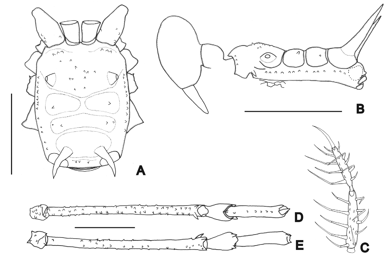
( Figs 1 View FIGURE 1 A–E, 12A–C)
Diagnosis: see the genus diagnosis.
Type locality: Brazil, Amazonas, São Gabriel da Cachoeira (Pico da Neblina , 860 m. a.s.l.),
Type material: All material collected in Brazil, Amazonas, São Gabriel da Cachoeira (Pico da Neblina mountain). Holotype, 860 m. a.s.l., 28.IX.2007, N. Lo Man Hung leg., male (INPA-OP 916). Paratype: idem, 860 m. a.s.l. male (MZSP-36523); 860 m. a.s.l., 30.IX.2007, N. Lo Man Hung leg., female (MZSP-36525); idem, 860 m. a.s.l., 28.IX.2007, female (MZSP-36535); idem, 860 m. a.s.l., D. Candiani leg., female (MZSP-36534); idem, 860 m. a.s.l., female (INPA-OP 917); idem, 860 m. a.s.l., 30.IX.2007, female (INPA-OP 928); idem, 860 m. a.s.l., 29.IX.2007, N. Lo Man Hung leg., female (INPA-OP 929); idem, 860 m. a.s.l., 29.IX.2007, A. Nogueira leg., female (INPA-OP 930); idem, 860 m. a.s.l., 29.IX.2007, 2 males (INPA-OP 331); idem, 860 m. a.s.l., 30.IX.2007, female (INPA-OP 332); idem, 400 m. a.s.l., 26.IX.2007, female (INPA-OP 333).
Etymology: In honor of the Brazilian arachnologist Nancy Lo Man Hung who has sampled and enriched Brazilian arachnid collections with Amazonian specimens from many new localities.
Male description (holotype): Measurements: DSL 2.5; PL 1.2; DSW 2.1; PW 2.0; ID 1.4; chelicera II 2.1; III 0.9; pedipalpus 7.9; leg I 8.6; II 18.8; III 11.6; IV 13. 4.
Dorsum ( Fig. 1 View FIGURE 1 A–B): Anterior margin with three tubercles each side. Prosoma with 10 tubercles between eye mounds. Lateral margin with one row of 11–15 tubercles from coxa II to posterior margin. Area I with one tubercle each side; II with two each side; III with two very long and divergent spines backwards. Posterior margin with four minute tubercles. Free tergite I–II with eight minute tubercles; III with 12. Anal operculum with one anterior row of minute tubercles and some on posterior margin.
Venter: Coxa I with six large tubercles in median row, one small posterior; II with one median row of large tubercles, one anterior row of 10 small tubercles, one posterior row of seven small tubercles; III with one median row of seven small tubercles; IV irregularly tuberculate. Posterior margin with one row of small tubercles, some scattered. Anal operculum with small scattered tubercles.
Chelicera: Bulla of segment I smooth; II–III with one large basal tubercle, three small distal. With interchelar space.
Pedipalpus: Coxa with four ventral tubercles (two larger), four dorsal tubercles. Trochanter with two ventral tubercles. Femur with one small ventral basal tubercle, slightly swollen at distal seventh. Patella swollen at distal fifth. Tibia and tarsus slender. Tibia ( Fig. 1 View FIGURE 1 C): ectal IIiIi, mesal IIIIi, with four small ventral tubercles. Tarsus ( Fig. 1 View FIGURE 1 C): ectal iIiIIi, mesal IiIII, with nine ventral tubercles.
Legs: Coxa I with two tubercles; II with one anterior, one posterior; III with one anterior fused with one of coxa IV; IV smooth dorsal and ventrally. Trochanter I smooth dorsally; II with one ventral tubercle, one dorsal; III with two ventral, one retrolateral apical, one dorsal; IV with three dorsal tubercles, five ventral. Femora I–III with small tubercles; II–IV with two dorsoapical tubercles (retrolateral much larger); IV ( Fig. 1 View FIGURE 1 D–E) with one retrolateral, one prolateral and one dorsal row of small tubercles, two ventral rows of tubercles increasing in size at distal third. Patella IV triangular-shaped, with six ventral tubercles, one ventroapical larger. Claws with six teeth. Tarsal segmentation: 7, 13, 7, 7.
Penis ( Fig. 12 View FIGURE 12 A–C): Truncus with five pairs of lateral setae, plus two pairs on the base of the stylus. Ventral plate slender, slightly elliptical, distal margin with U-cleft; base elongated; with three pairs of setae and with papillae on dorsal side and, one pair of setae on ventral side; six pairs of basal setae. Dorsal process absent. Basal part of glans long and membranous.
Color: Dark-brown on scutal areas, reticulate on prosoma, dorsal femur apex, patella and tibia IV. Pedipalpus yellowish with some small black patches. Anterior and posterior margins with one black stripe each. Posterior margin with two large white patches on corners. Coxa-tibia IV orange-brown.
Female description (MZSP-36525): Measurements: DSL 2.9; PL 1.2; DSW 2.3; PW 2.0; ID 1.6; chelicera II 1.2; III 0.8; pedipalpus 8.0; leg I 8.6; II 17.8; III 11.8; IV 15. 7.
Anterior margin of dorsal scutum with two–three tubercles each side. Prosoma with 14 tubercles. Area I with one tubercle each side; II with one–two tubercles each side. Lateral margin with 11–12 tubercles. Patella IV with two dorsoapical tubercles. Tibia with minute tubercles. Pedipalpus: tibia ectal IIiIi, mesal IIIIi, six ventral tubercles; tarsus ectal iIiIi, mesal IiIiIIi, five ventral tubercles. Femur IV with two rows of small tubercles. Patella IV with two dorsal apical tubercles half size of male ones. Tibia IV with small tubercles. Claws with seven teeth. Tarsal segmentation: 7, 17, 6, 7.
Distribution: Recorded only from the type locality, from 400 to 860 m.a.s.l.

No known copyright restrictions apply. See Agosti, D., Egloff, W., 2009. Taxonomic information exchange and copyright: the Plazi approach. BMC Research Notes 2009, 2:53 for further explanation.