
Jime chifrudo, Pinto-Da-Rocha, Ricardo & Tourinho, Ana Lúcia, 2012
|
publication ID |
https://doi.org/ 10.5281/zenodo.281385 |
|
DOI |
https://doi.org/10.5281/zenodo.6177651 |
|
persistent identifier |
https://treatment.plazi.org/id/038C87C8-2900-E446-AAF6-F991E82EFC8A |
|
treatment provided by |
Plazi |
|
scientific name |
Jime chifrudo |
| status |
sp. nov. |
Jime chifrudo View in CoL sp. nov.
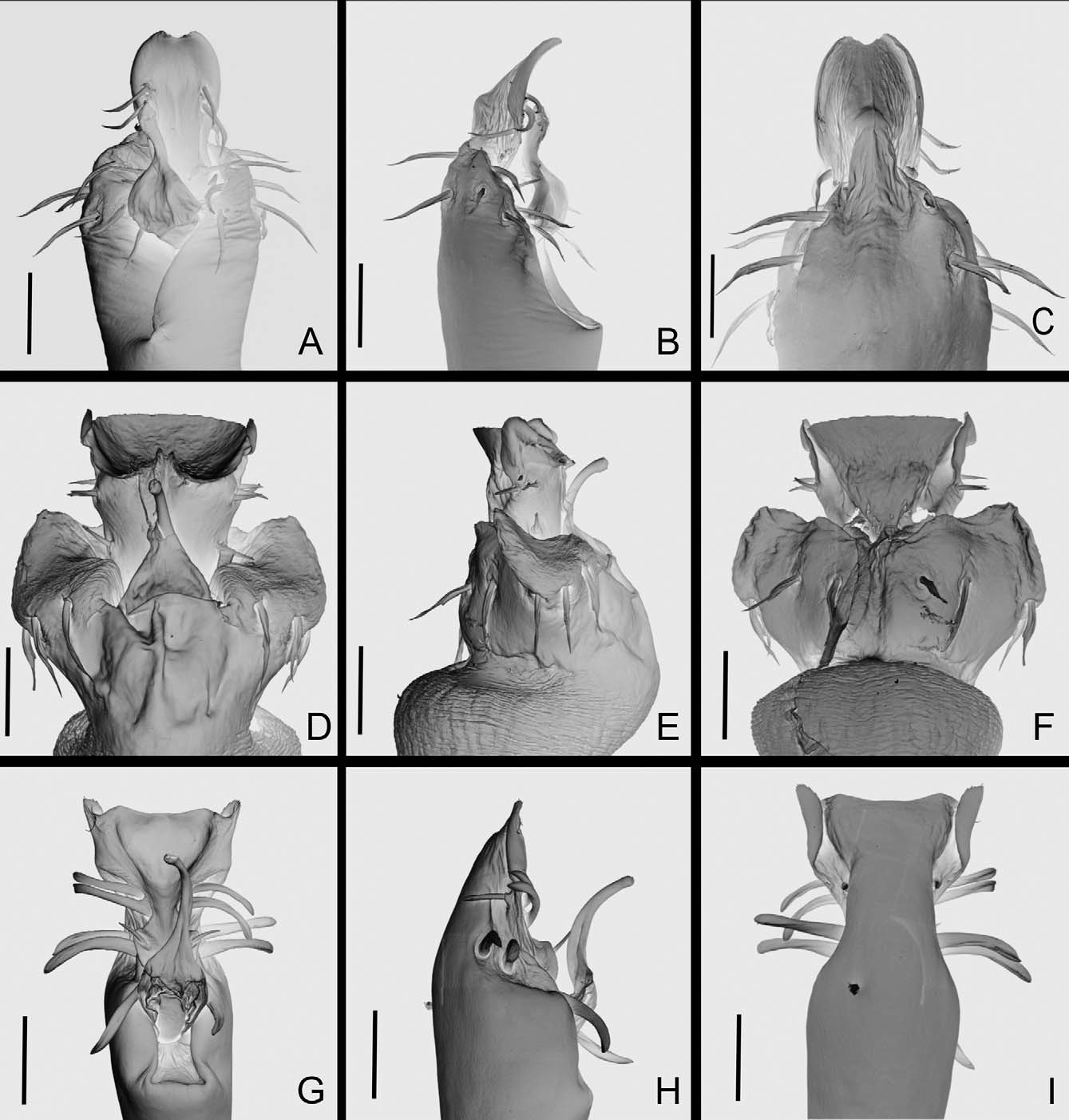
( Figs. 7 View FIGURE 7 A–E, 13G–I)
Diagnosis: see the genus diagnosis.
Type locality: Brazil, Amazonas, São Gabriel da Cachoeira (Pico da Neblina, 2400 m .a.s.l.).
Type material: All material collected in Brazil, Amazonas, São Gabriel da Cachoeira (Pico da Neblina ). Holotype, male, 400 m. a.s.l., 27.IX.2007, N. Lo Man Hung leg. (INPA-OP 1021). Paratypes: 400 m. a.s.l., 26.IX.2007, A. Nogueira leg., female (INPA-OP 1022); 2,000 m.a.s.l., 26.IX.2007, A. Nogueira leg., female (INPA-OP 1023); 400 m. a.s.l., 13.X.2007, D. Candiani leg, male (MZSP-36521); 2,400 m.a.s.l., X.2007, N. L.M. Hung, A. Nogueira & D. Candiani leg, female (MZSP-36531); 1,550 m.a.s.l., 2.X.2007, N. L.M. Hung, male (INPA-OP 1024).
Etymology: Noun in apposition, from the Portuguese “ chifrudo ” or “long horn”, in reference to the huge length of prosomal spine.
Male description (holotype): Measurements: DSL 2.0; PL 0.8; DSW 1.5; PW 1.6; ID 1.2; chelicera II 2.1; III 1.2; pedipalpus 6.2; leg I 7.2; II 10.4; III 8.4; IV 10. 6.
Dorsum ( Fig. 7 View FIGURE 7 A–B): Anterior margin with two–three small tubercles each side. Prosoma with one long spine slightly directed frontally, with tuberculate base. Lateral margin with one row of small tubercles from groove I to IV. Eye mounds smooth, with three–four small tubercles close each other. Area I with seven tubercles each side; II with 21 tubercles; III with two divergent spines backwards, with smooth base, 15 tubercles scattered. Posterior margin with one row of 16 tubercles. Free tergite I with 12 tubercles; II with 10 tubercles; III with eight tubercles. Anal operculum with two central tubercles, one row of tubercles on posterior margin.
Venter: Coxa I with three anterior tubercles, one median row of four, one posterior row of five, three apical; II with one median row of seven tubercles, seven posterior, three apical; III with three anterior tubercles, one median row of five, seven posterior; IV irregularly tuberculate. Posterior margin with one row of small tubercles. Anal operculum with small scattered tubercles.
Chelicera: Segment I with two tubercles on bulla; II with one median large, two distal small tubercles; III with three small tubercles on distal half. With large interchelar space.
Pedipalpus: Coxa with one ventral tubercle, six dorsal tubercles. Trochanter with two ventral tubercles; one dorsal. Femur smooth, slightly swollen at distal fifth. Patella swollen at distal third. Tibia ( Fig. 7 View FIGURE 7 C): ectal IIiIi, mesal IIiIi, with one ventral tubercle. Tarsus ( Fig. 7 View FIGURE 7 C): ectal iiIiii, mesal IiIii, one ventral tubercle.
Legs: Coxa I with two tubercles; II with one anterior, one posterior fused with one of coxa III; IV with some sparse tubercles on lateral. Trochanter I with three ventral tubercles, one retrolateral, one dorsal; II with three ventral, one retrolateral, three dorsal; III with four ventral, four retrolateral, three dorsal; IV with three dorsal, four ventral and four retrolateral tubercles. Femora I–III minute tuberculate; IV ( Fig. 7 View FIGURE 7 D–E) with one dorsal and two ventral rows of small tubercles. Patella IV ( Fig. 7 View FIGURE 7 D–E) irregularly tuberculate, with one dorsodistal large tubercle, one ventral row of tubercles ending in a large and tuberculate projection. Tibia IV ( Fig. 7 View FIGURE 7 D–E) smooth. Tarsal segmentation: 6, 12, 6, 7. Without tarsal process.
Penis ( Fig. 13 View FIGURE 13 G–I): Ventral plate thick, with divergent lateral sides, distal margin slightly straight; with three pairs of large steae on middle of ventral plate, being two pairs on dorsal side, and one pair on ventral side, one pair of small intermediary setae and three pairs of large (than distal margin) setae on basal region. Stylus long and thin, basal third membranous.
Color: Reddish-brown; margins of dorsal scutum and border of areas I–II darker, area III almost entirely blackish. Chelicerae not reticulate. Legs with black minute patches more concentrated distad. Pedipalpus yellowish with some small black patches.
Female description (INPA-OP 1023): Measurements: DSL 2.5; PL 1.1; DSW 1.9; PW 1.7; ID 1.3; chelicera II 1.4; III 0.6; pedipalpus 5.7; leg I 7.0; II 11.4; III 9.2; IV 10. 1.
Anterior margin with two tubercles each side. Prosomal spine smaller than that of male. Area I with two tubercles each side; II with two small tubercle each side; III with one tubercle each side and one behind each spine. Legs I–IV minute tuberculate. Patella IV normal. Tarsal segmentation: 6, 15, 6, 7.
Distribution: Recorded only from the type locality, from 400 to 2400 m.a.s.l.

No known copyright restrictions apply. See Agosti, D., Egloff, W., 2009. Taxonomic information exchange and copyright: the Plazi approach. BMC Research Notes 2009, 2:53 for further explanation.