
Sympodium omasum, Koido & Imahara & Fukami, 2024
|
publication ID |
https://doi.org/10.11646/zootaxa.5443.2.3 |
|
publication LSID |
lsid:zoobank.org:pub:E15C3C76-EF6A-4CED-839F-418BD7981670 |
|
DOI |
https://doi.org/10.5281/zenodo.11059908 |
|
persistent identifier |
https://treatment.plazi.org/id/038A87C1-FF85-FFF8-A190-FC6ABCFE9918 |
|
treatment provided by |
Plazi |
|
scientific name |
Sympodium omasum |
| status |
sp. nov. |
Sympodium omasum sp. nov.
( Figs. 2–9 View FIGURE 2 View FIGURE 3 View FIGURE 4 View FIGURE 5 View FIGURE 6 View FIGURE 7 View FIGURE 8 View FIGURE 9 )
New Japanese name: Senmai-chizimi-umiazami
Material
Holotype: KBF-OA-00081 (MUFS-COTUG 2 in Koido et al. 2019), Tsukishima Isl., Kushima City, Miyazaki Prefecture , depth < 15 m, December 3, 2014, coll. T. Koido. Paratypes: KBF-OA-00082 (MUFS-COTUK 16 in Koido et al. 2019), Tsukishima Isl., Kushima City, Miyazaki Prefecture, depth < 15 m, December 3, 2014, coll. T. Koido; KBF-OA-00083 (MUFS-COMO 63 in Koido et al. 2019), Oshima Isl., Nichinan City, Miyazaki Prefecture, depth < 10 m, December 25, 2012, coll. T. Koido; KBF-OA-00084, Tsukishima Isl., Kushima City, Miyazaki Prefecture, depth < 10 m, December 3, 2014, coll. T. Koido.
Sympodium sp. ; Koido et al. 2019: 5–13, Figs 2G View FIGURE 2 , 3G View FIGURE 3 , 4G View FIGURE 4 , 5–6 View FIGURE 5 View FIGURE 6 .
Descriptions
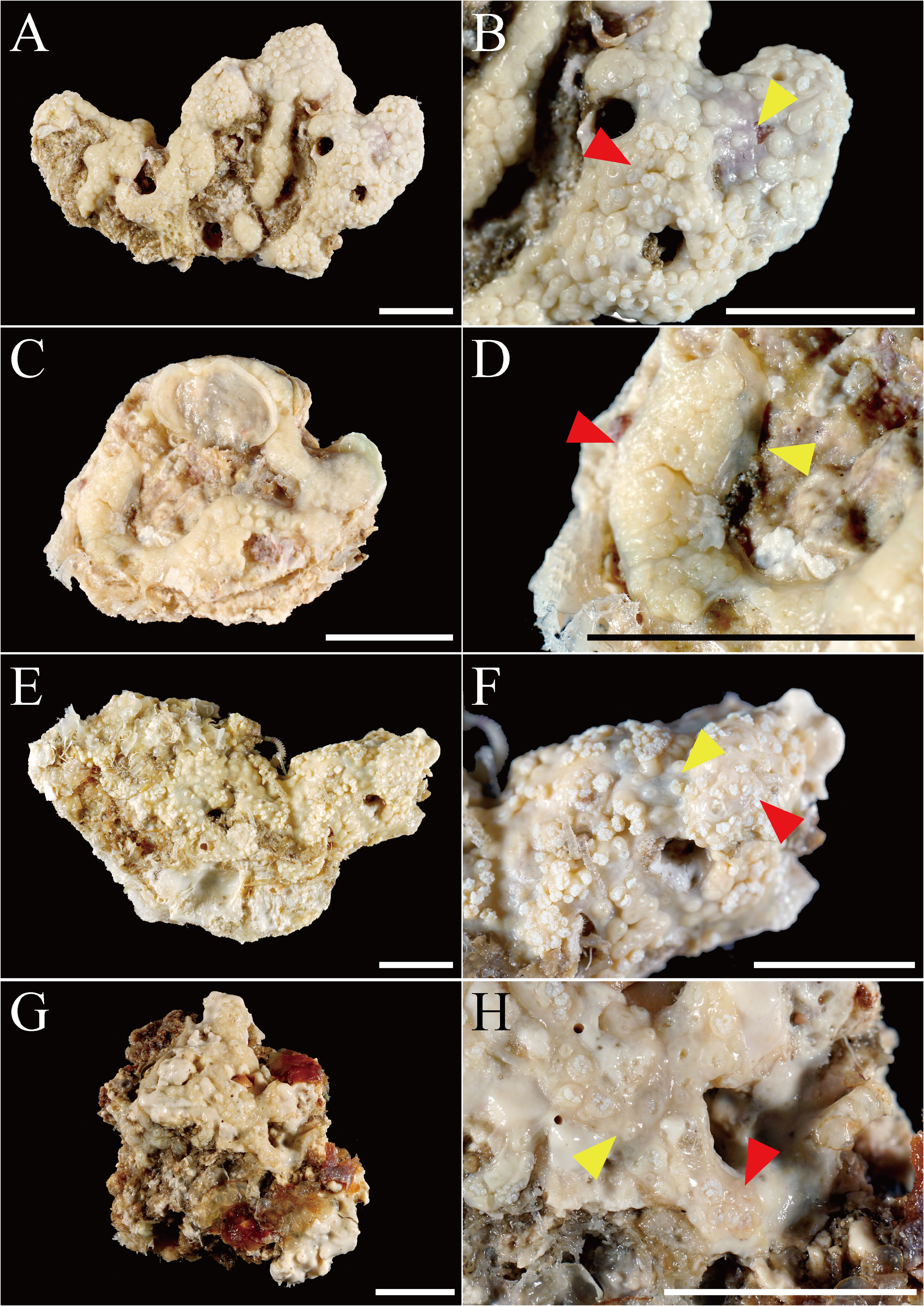
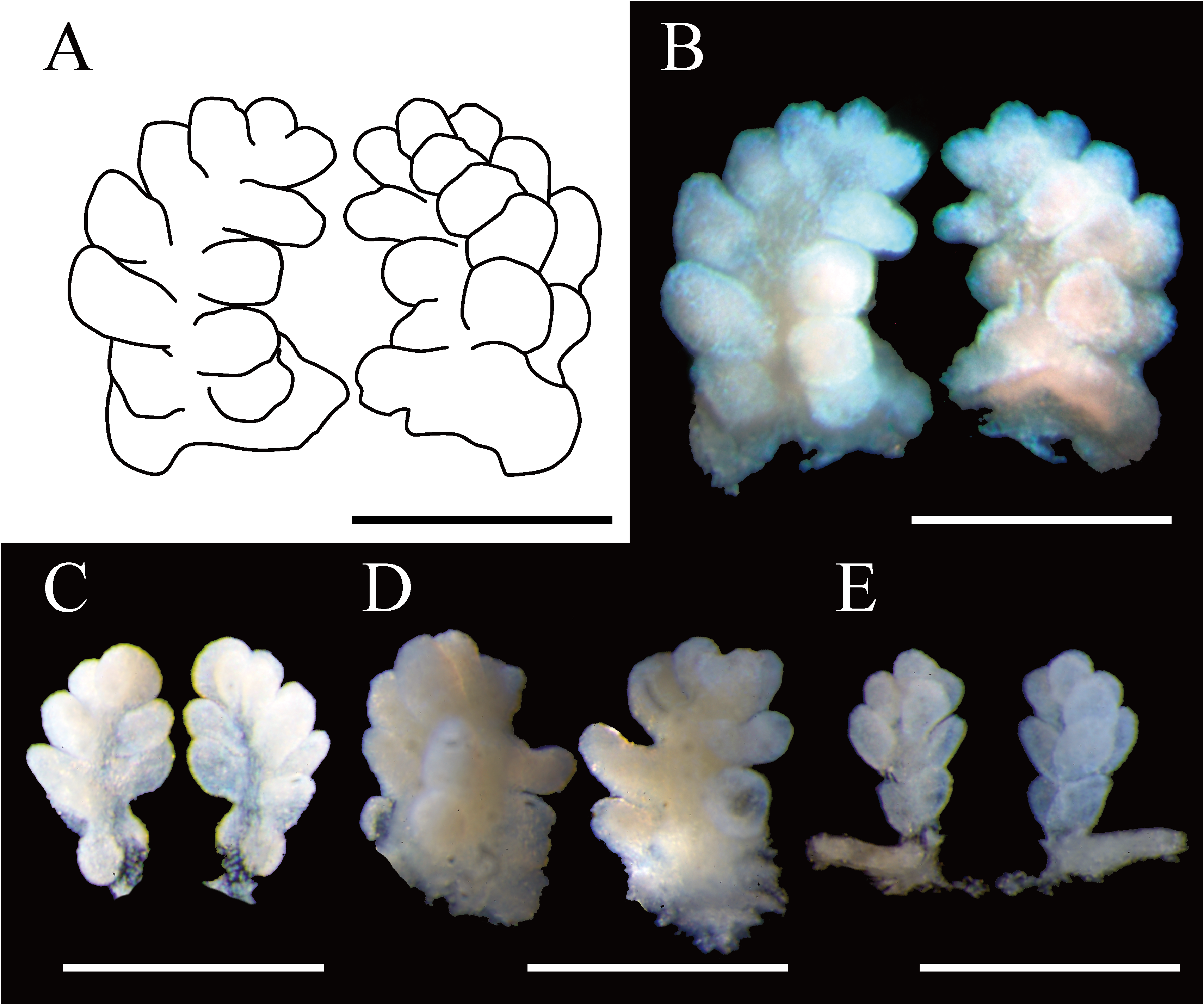
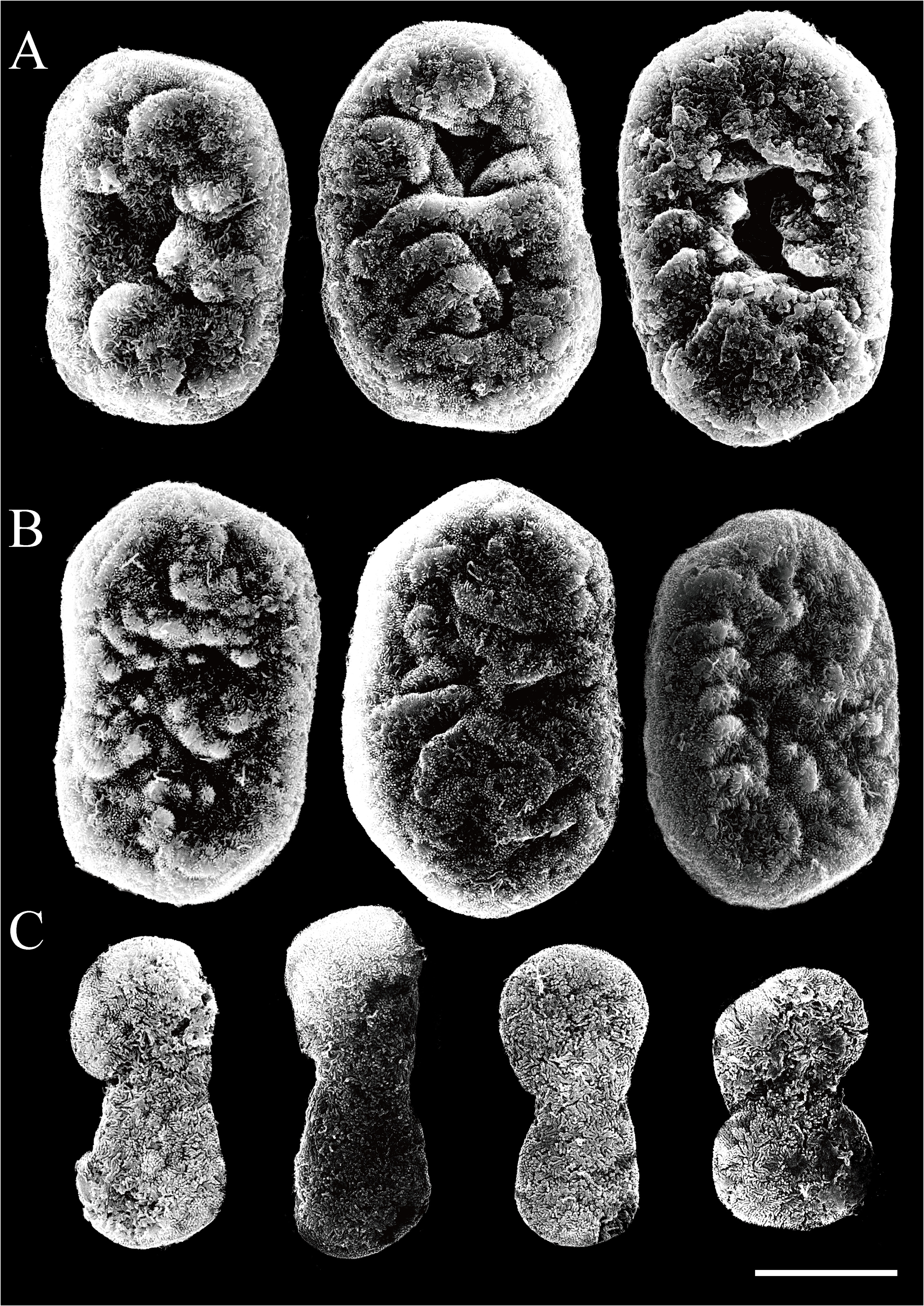
The holotype is an irregularly spreading ribbon-like membrane (approximately 1 mm thick) attached to a dead coral rock, measuring approximately 50 × 35 mm ( Fig. 2A View FIGURE 2 ). The width of the holotype colony is approximately 10 mm at the widest portion, and in some places, the colony extends in a reticulate pattern. These areas, where the polyps are densely packed (no gaps between polyps) form knob-like mounds (up to 5 mm high). Polyp density is lower at the edges of mounds and between mounds (gaps between polyps are up to 0.97 mm), where thin coenenchyme can be observed ( Fig. 2B View FIGURE 2 ). Most polyps are fully retracted while others are expanded a little. The most expanded polyps are up to 0.5 mm long and the tentacles are up to 0.67 mm long and up to 0.33 mm wide. One row of pinnules is arranged along each lateral side of the tentacles (outer row), and an additional single row is arranged in the median space of the oral side of the tentacle (central row) ( Fig. 3 View FIGURE 3 ). The outer row usually includes 4–6 pinnules, and the central row includes 4–5 pinnules. The pinnules up to 0.30 mm long and 0.25 mm wide at the proximal portion. No gap between adjacent pinnules exists.
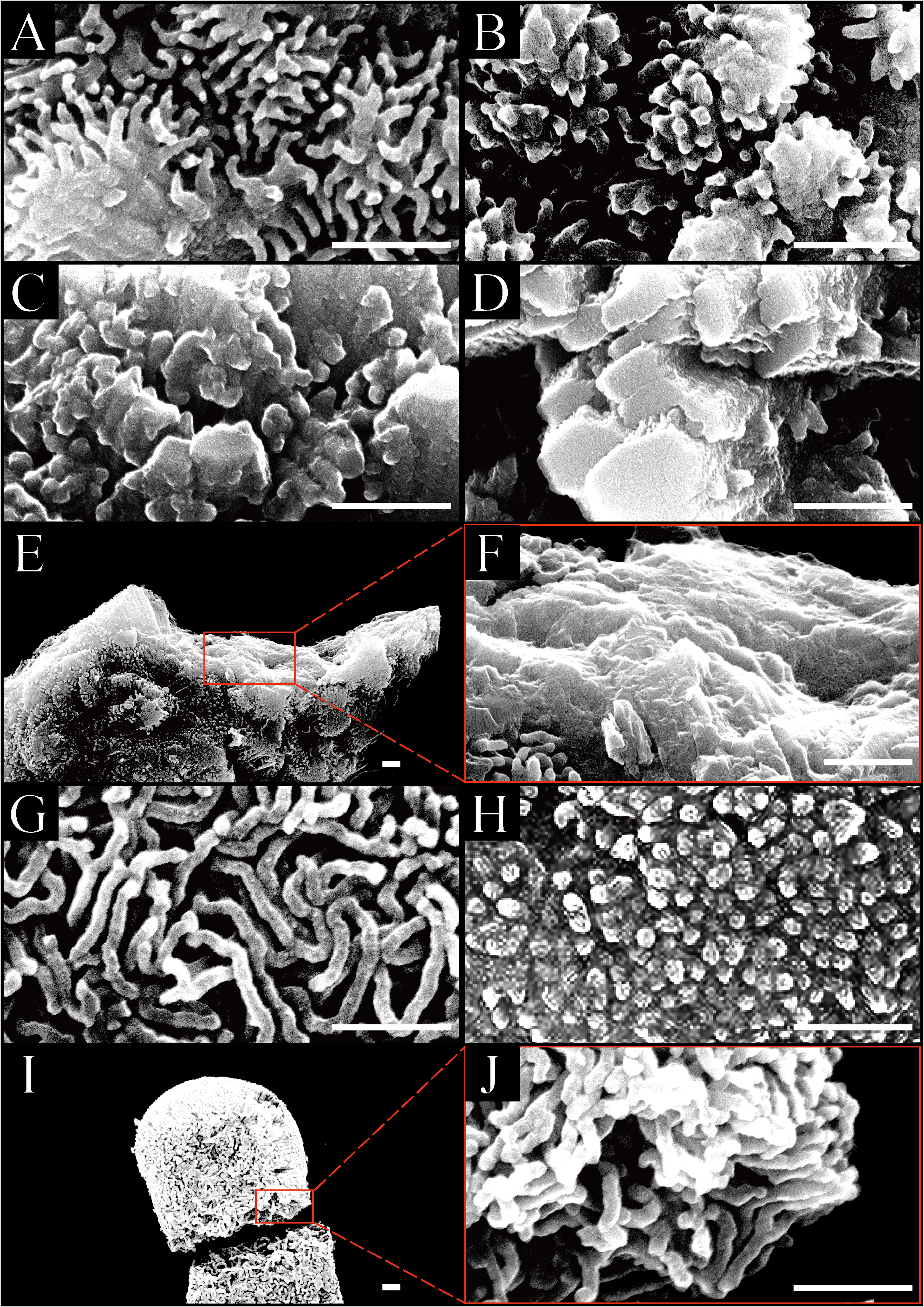
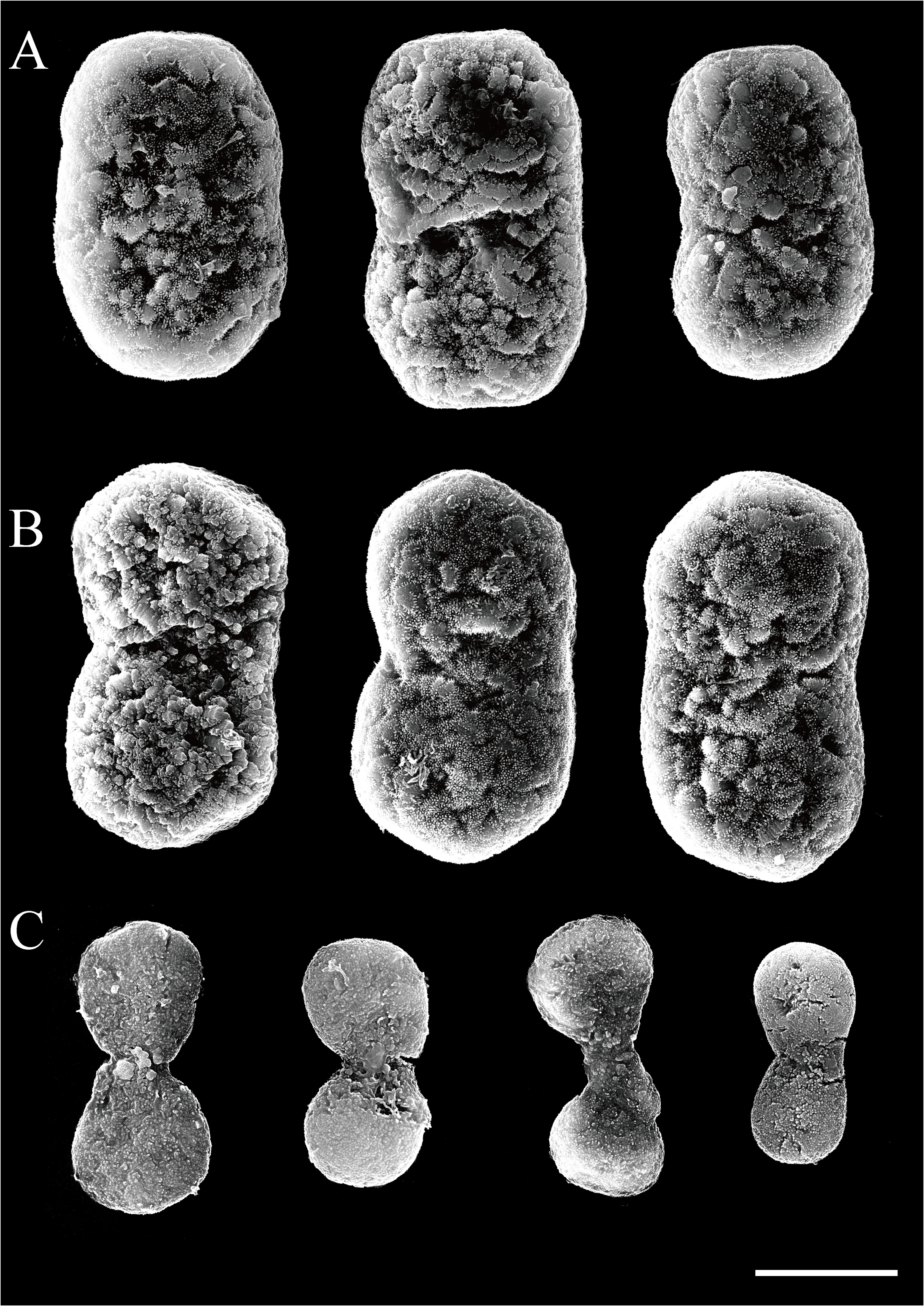
Sclerites are abundant throughout the colony. In addition, they are more densely distributed on the surface of the coenenchyme than those in the interior. Sclerites can be observed in three forms, ellipsoid platelets, hexagonal platelets, and double-heads. Under light microscopy, distinguishing between ellipsoid platelets and hexagonal platelets is difficult; however, they are clearly distinguishable under SEM. Ellipsoid platelets and hexagonal platelets are abundant in the polyp body, coenenchyme surface, and coenenchyme interior, but not in the tentacles. In contrast, double-heads are observed only in tentacles ( Fig. 4 View FIGURE 4 ). Under SEM, ellipsoid platelets are 0.026 –0.029 mm long and 0.014 –0.016 mm wide, and narrower in the middle by 0.001 –0.002 mm ( Fig. 5A View FIGURE 5 ). Hexagonal platelets are 0.025 –0.029 mm long and 0.015 –0.019 mm wide, occasionally with a central waist ( Fig. 5B View FIGURE 5 ). Double-heads ( Fig. 5C View FIGURE 5 ) are 0.018 –0.022 mm long and 0.007 –0.009 mm wide, and the central narrower part is 0.003 –0.007 mm wide. Ellipsoid platelets and hexagonal platelets are covered by sinuous rodlets, which are clearly fused and form many nodules on the sclerites surface ( Fig. 6A View FIGURE 6 ). The length of these rodlets vary from long to short, depending on the platelets ( Figs. 6A–C View FIGURE 6 ). The rodlets are up to 0.43 μm long ( Fig. 6A View FIGURE 6 ), short rodlets are 0.09–0.18 μm long ( Fig. 6C View FIGURE 6 ). The interior of the broken platelet is fused, and no calcite rods are observed ( Figs. 6E, F View FIGURE 6 ). Double-heads are composed of dendritic rods. The tips of the dendritic rods are parallel to the sclerites surface and are sinuous ( Figs. 6G View FIGURE 6 ). The interior of the broken double-heads consists of radially arranged rods ( Figs. 6I, J View FIGURE 6 ).
The whole colony is creamy white in ethanol. Living tentacles were purplish blue with light-brown anthosteles and coenenchyme ( Koido et al. 2019).
Variation. Two paratypes (KBF-OA-00083 and KBF-OA-00084) are attached to a dead coral rock, similar to the holotype (KBF-OA-00081) ( Figs. 2E, G View FIGURE 2 ). In contrast, the paratype KBF-OA-00082 is attached to a dead bivalve shell ( Fig. 2C View FIGURE 2 ). The paratype KBF-OA-00083 ( Fig. 2E View FIGURE 2 ) is sparsely distributed, with less dense polyps on the colony than the other paratypes. For the sclerites, the double-heads in the paratype KBF-OA-00082 ( Fig. 2B View FIGURE 2 ) differ from those in the other three paratypes, as the dendritic rods are fused and composed of densely packed rodlets whose tips provide a granular appearance ( Fig. 6H View FIGURE 6 ). The tentacles vary in length and width depending on the specimens; KBF-OA-00083 ( paratype) has the tentacle size same as the KBF-OA-00081 ( holotype), whereas KBF-OA-00082 ( paratype) and KBF-OA-00084 ( paratype) have smaller tentacles, 0.36–0.39 mm long and 0.09–0.10 mm wide ( Fig. 3 View FIGURE 3 ). In addition, two paratypes, KBF-OA-00083 and KBF-OA-00084 have fewer pinnules; the outer row includes 3 pinnules, and the center row includes 2–3 pinnules ( Fig. 3 View FIGURE 3 ). In the coenenchyme surface, coenenchyme interior, and polyp body of all specimens, double-heads are absent, and only ellipsoid platelets and hexagonal platelets are observed; ellipsoid platelets are approximately 74–82% more prevalent. The tentacles have only double heads and no other sclerites are present ( Table 2 View TABLE 2 ). All paratypes have the same platelet surface architectures as the holotype. Platelets with few or no rodlets were rarely observed, and their surfaces are composed of fused polygonal pillars of different heights. The side of the pillars are rough, and the tips are relatively smooth ( Fig. 6 View FIGURE 6 ). Platelets with such a surface architecture were observed in all specimens, although the proportion varies by part of the colony ( Table 3 View TABLE 3 ).
Locality. Common around Oshima and Tsukishima Islands, Miyazaki, Japan, from 10–20 m in depth. They attached themselves to the surface of the dead coral rock.
Etymology. The omasum is the third compartment of the ruminant stomach. The species is named after the shape of the microstructure of the platelet sclerites, which is particularly similar to that of the third stomach of cows, and because Miyazaki Prefecture, where the specimens were collected, is the producing area of Miyazaki beef. The present study also proposes the new Japanese name “Senmai-chizimi-umiazami” for S. omasum sp. nov., “senmai” meaning the third stomach of the cow in Japanese.
Remarks. Sympodium omasum sp. nov. (= Sympodium sp. 1 by Koido et al. 2019) has three forms of sclerites: ellipsoid platelets, hexagonal platelets, and double-heads, whereas the nine congeneric species have only ellipsoid platelets or hexagonal platelets ( Cohn 1908; Reinicke 1997; Benayahu et al. 2021). The double-heads of S. omasum sp. nov. are composed of dendritic rods aligned parallel to the surface of the sclerites. This is similar to the microstructure of the ellipsoid platelets or hexagonal platelets of nine congeneric species. In contrast, ellipsoid platelets and hexagonal platelets of S. omasum sp. nov. are covered by sinuous rodlets that are fused and form many nodules on the sclerites surface. These microstructural features of ellipsoid platelets and hexagonal platelets have not been reported in other species of the genus.


No known copyright restrictions apply. See Agosti, D., Egloff, W., 2009. Taxonomic information exchange and copyright: the Plazi approach. BMC Research Notes 2009, 2:53 for further explanation.