
Myiocephalus Marshall, 1898
|
publication ID |
https://doi.org/10.11646/zootaxa.4700.1.6 |
|
publication LSID |
lsid:zoobank.org:pub:BD3E014F-6A3C-478E-9E37-62C9287E65E2 |
|
persistent identifier |
https://treatment.plazi.org/id/038987BB-9803-7738-FF45-80C72016B9E9 |
|
treatment provided by |
Plazi |
|
scientific name |
Myiocephalus Marshall, 1898 |
| status |
|
Myiocephalus Marshall, 1898 View in CoL View at ENA
Figs 1–57 View FIGURES 1–2 View FIGURES 3–13 View FIGURES 14–16 View FIGURES 17–20 View FIGURES 21–30 View FIGURES 31–35 View FIGURES 36–38 View FIGURES 39–50 View FIGURES 51–54 View FIGURES 55–57
Loxocephalus Foerster, 1863: 252 (not Loxocephalus Eberhard, 1862 ). Type species (by monotypy): Loxocephalus longipes Foerster, 1863 [= Myiocephalus boops ( Wesmael, 1835) View in CoL ]. Unavailable name.
Myiocephalus Marshall View in CoL [in André], 1898: 218; Chen & van Achterberg 1997: 74; Belokobylskij 2000: 372. Type species (by monotypy): Microctonus boops Wesmael, 1835 .
Spilomma Morley, 1909: 211 . Type species (by monotypy): Spilomma falconivibrans Morley, 1909 [= Myiocephalus boops ( Wesmael, 1835) View in CoL ]. Synonymised with Myiocephalus Marshall View in CoL by Muesebeck (1936).
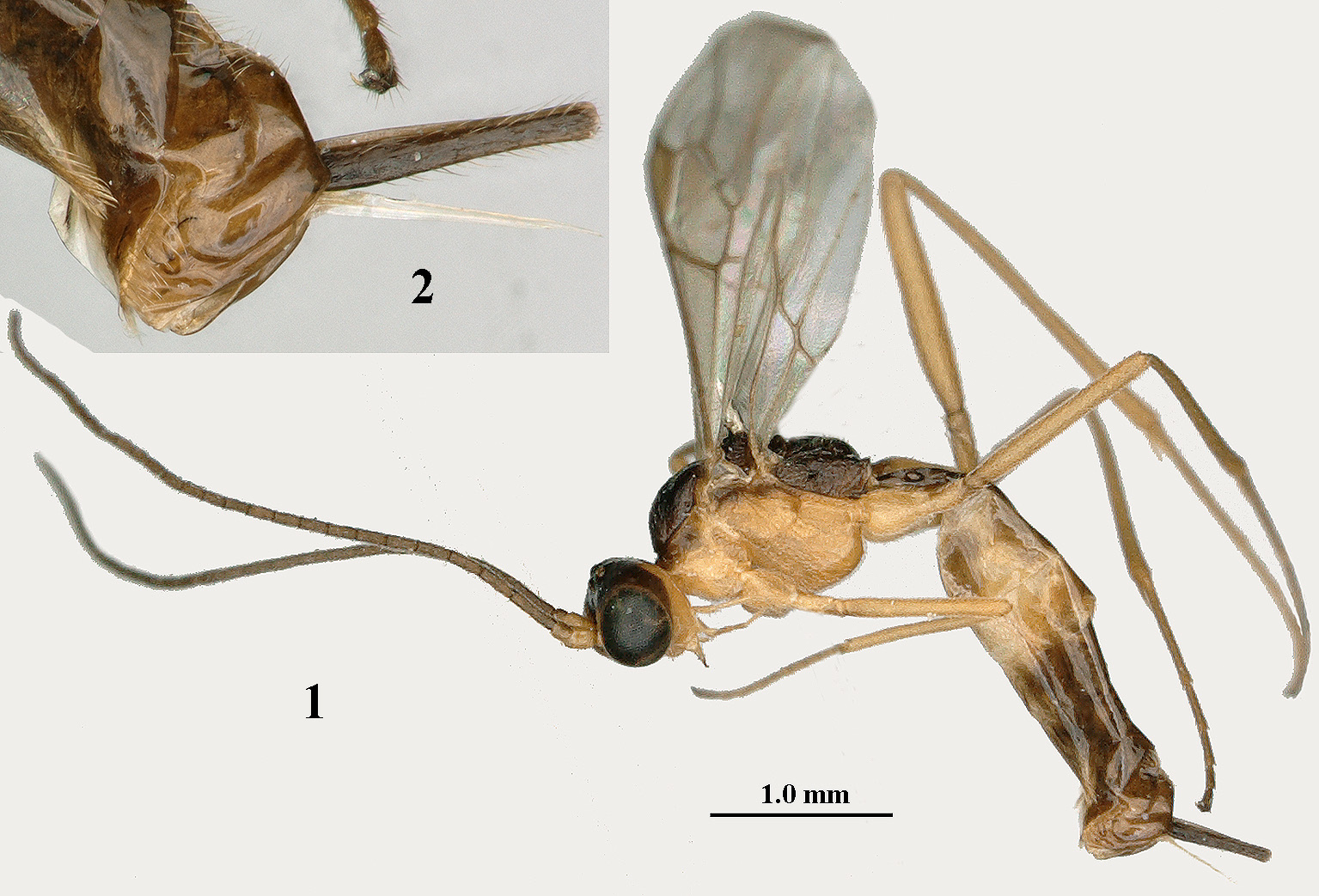
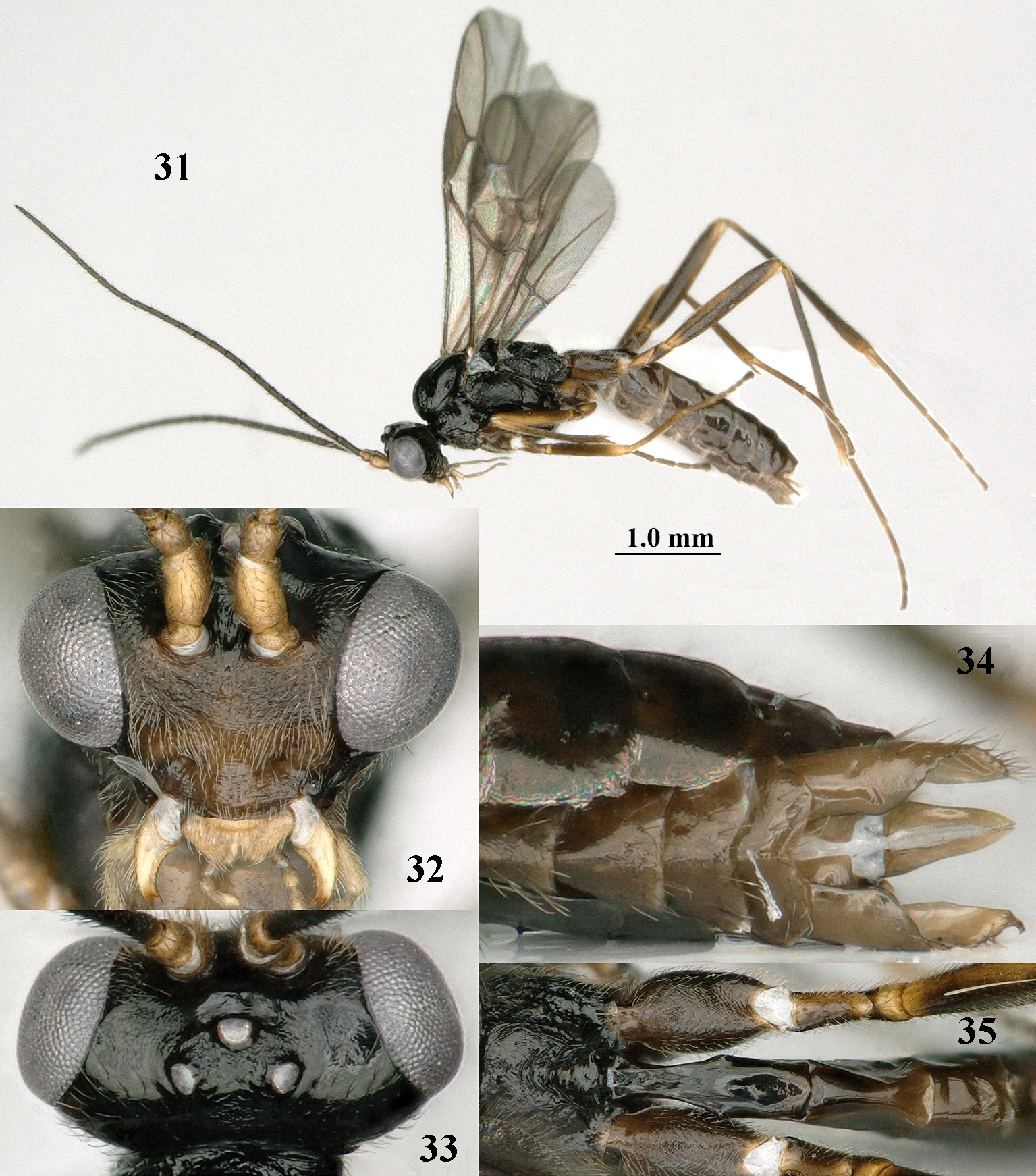
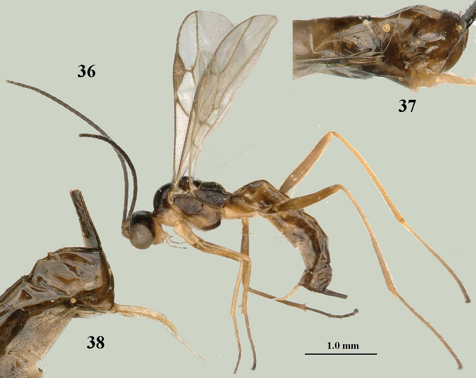
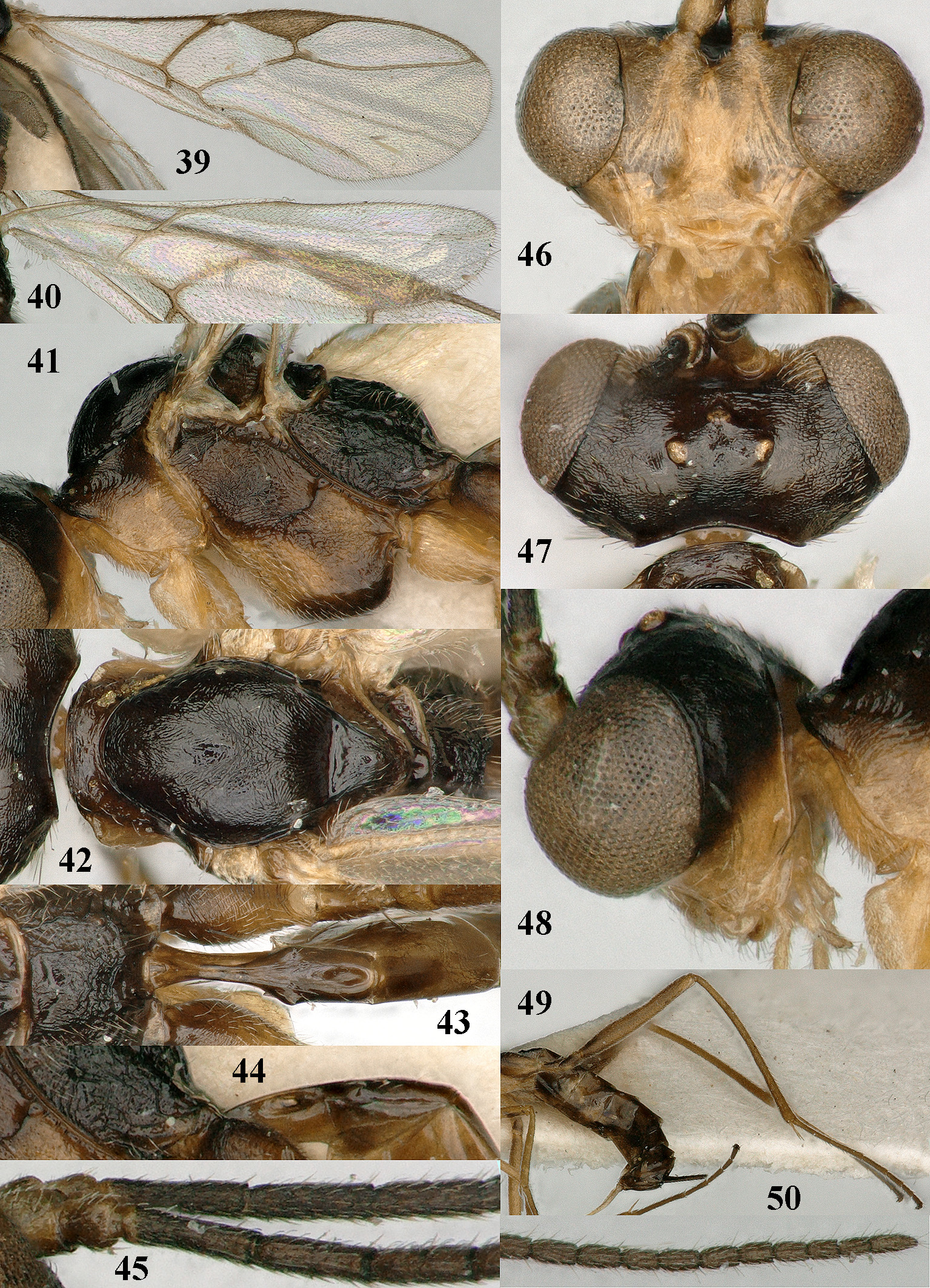
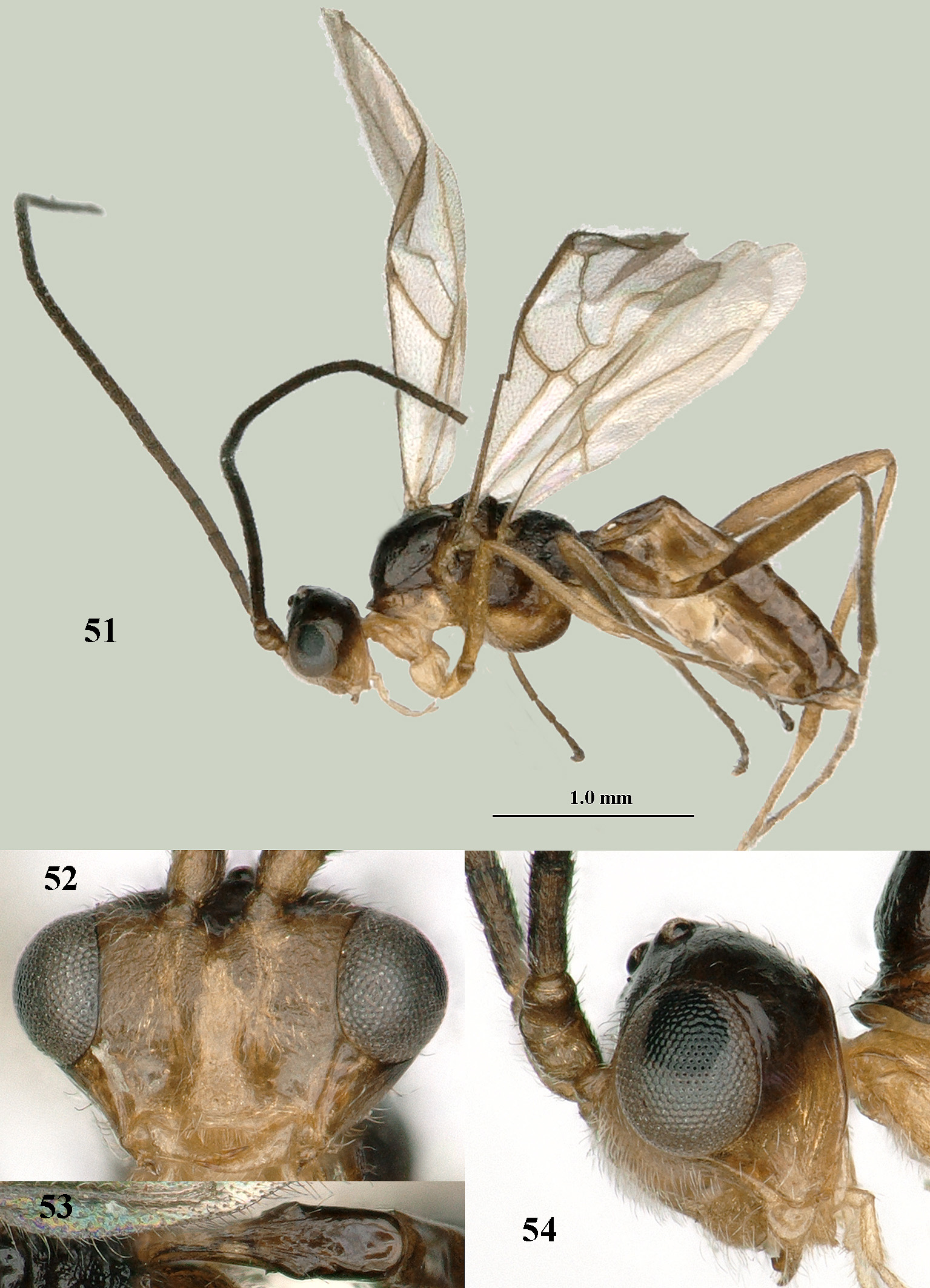
Short diagnosis. Laterope large, deep and submedially situated in slender first tergite ( Figs 7 View FIGURES 3–13 , 44 View FIGURES 39–50 ); head in dorsal view strongly transverse and usually slightly concave anteriorly ( Figs 10 View FIGURES 3–13 , 27 View FIGURES 21–30 , 47 View FIGURES 39–50 ); eyes enlarged and protruding ( Figs 26 View FIGURES 21–30 , 46 View FIGURES 39–50 ); clypeus rather narrow ( Fig. 26 View FIGURES 21–30 ); scapus normal, slightly or not enlarged and subequal to or shorter than third antennal segment ( Fig. 13 View FIGURES 3–13 ); maxillary and labial palpi with 5 and 3 segments, respectively; vein 1-SR+M of fore wing absent ( Fig. 3 View FIGURES 3–13 ); vein 1-R1 longer than pterostigma; vein M+CU1 largely unsclerotised; middle and hind legs elongated ( Figs 1 View FIGURES 1–2 , 17 View FIGURES 17–20 , 36 View FIGURES 36–38 ); metasoma of ♀ strongly compressed ( Figs 17, 19, 20 View FIGURES 17–20 ) with fifth sternite of ♀ fingerlike protruding posteriorly ( Figs 1 View FIGURES 1–2 , 8 View FIGURES 3–13 , 20 View FIGURES 17–20 , 37 View FIGURES 36–38 ); hypopygium of ♀ with long setae apically ( Figs 8 View FIGURES 3–13 , 37 View FIGURES 36–38 ) or hypopygium medially membranous ( Fig. 25 View FIGURES 21–30 ).
Biology. Unknown.
Distribution. Holarctic (four species).


No known copyright restrictions apply. See Agosti, D., Egloff, W., 2009. Taxonomic information exchange and copyright: the Plazi approach. BMC Research Notes 2009, 2:53 for further explanation.
|
Kingdom |
|
|
Phylum |
|
|
Class |
|
|
Order |
|
|
Family |
Myiocephalus Marshall, 1898
| Tan, Jiangli, Achterberg, Cornelis Van, Tian, Xiaoxia & Zhang, Ruonan 2019 |
Myiocephalus
| Belokobylskij, S. A. 2000: 372 |
| Chen, X. X. & van Achterberg, C. 1997: 74 |
Spilomma
| Morley, C. 1909: 211 |
Loxocephalus
| Foerster, A. 1863: 252 |