
Rhamphostomella echinata, Grischenko & Gordon & Taylor & Kuklinski & Denisenko & Spencer-Jones & Ostrovsky, 2022
|
publication ID |
https://doi.org/10.11646/zootaxa.5131.1.1 |
|
publication LSID |
lsid:zoobank.org:pub:CF550031-D6A9-48A3-A953-A1BD40C72F5E |
|
DOI |
https://doi.org/10.5281/zenodo.7628999 |
|
persistent identifier |
https://treatment.plazi.org/id/03892374-0B7A-336D-FF73-A84C1DE7FAC5 |
|
treatment provided by |
Plazi |
|
scientific name |
Rhamphostomella echinata |
| status |
sp. nov. |
Rhamphostomella echinata n. sp.
( Fig. 24 View FIGURE 24 )
Rhamphostomella sp. : Grischenko 1997, p. 176; 2002, p. 115.
Diagnosis. Colony encrusting, multiserial. Zooids small, hexagonal. Frontal shield moderately convex, uniformly tessellated, with a few marginal areolae. No interareolar ridges. Umbonuloid component small. Suboral mucro absent. Primary orifice irregularly oval to quadrangular; proximal margin bisinuate, with median bifurcated to multiply branched lyrula and small, lateral, triangular processes. Condyles absent. Secondary orifice irregularly oval, cormidial, just slightly above primary orifice. 2‒3 pairs of very long tubular, articulated oral spines with massive bases on lateral margins of secondary orifice, conferring spinose appearance to colony. Suboral avicularian cystid very small, with finely dimpled surface. Rostrum short, oval, slightly elevated. Mandible spatulate, with rounded distal end. Crossbar incomplete. No adventitious avicularia. Ovicell hyperstomial. Ectooecium with small scattered pseudopores, no secondary calcification. 2–3 pore chambers in distolateral wall, and 1–2 multiporous septula in transverse walls. Basal surface of zooids fully calcified, with tubular protuberances.
Material examined. Holotype: ZIRAS 1 /50126, one colony fragment, IMB Collection , Stn 158/393, 18 September 1973, Kitolovnaya Bank , Medny Island , coastal waters of Medny Island , Commander Islands, Bering Sea, 55°02.2ʹ N, 167°10.9ʹ E, depth 60 m, rock dredge, collector S.D. Vavilin. GoogleMaps Paratype: NHMUK 2013.10 About NHMUK .21.3, one colony encrusting oyster shell , RV Norseman, Stn LT –2, 3 July 2011, Longshot , East of Square Bay , coastal waters of Amchitka Island , Rat Islands , western Aleutian Islands, Bering Sea, 51°26.6ʹ N, 179°12.2ʹ E, depth 10 m GoogleMaps , SCUBA, collector P. Kuklinski.
NHMUK 2013.10 About NHMUK .21.4, one colony , RV Norseman, Stn LT –1, 4 July 2011, Longshot , South of Crown Reefer Point , coastal waters of Amchitka Island , Rat Islands , western Aleutian Islands, Bering Sea, 51°27.3ʹ N, 179°11.4ʹ E, depth 13 m GoogleMaps , SCUBA, collector P. Kuklinski.
Additional material. 14 specimens. IMB Collection (1973) Stns 108/284, 150/385, 151/386, 160/395, 173/408; KIENM Collection (1992) Stns 5, 39, 140 (see Appendix 1 for details).
Etymology. The specific name echinata alludes to the spinose appearance of the colony, with numerous oral spines.
Type locality. Kitolovnaya Bank , coastal waters of Medny Island , Commander Islands, Bering Sea, 55°02.2ʹ N, 167°10.9ʹ E, depth 60 m. GoogleMaps
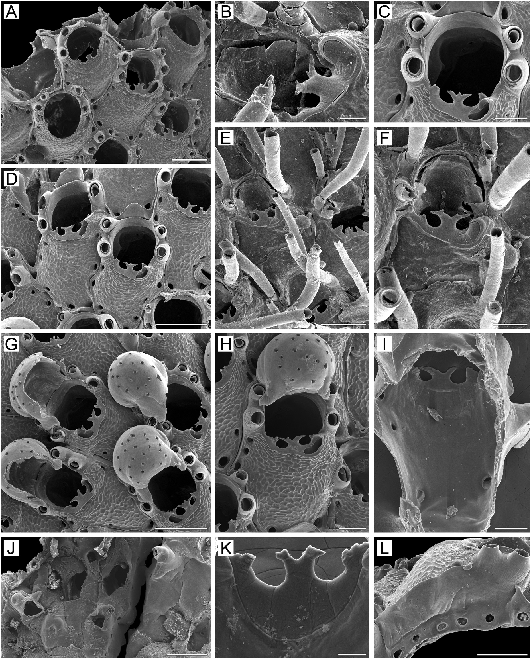
Measurements. ZIRAS 1/50126, Medny Island, Commander Islands, Bering Sea ( Fig. 24A–L View FIGURE 24 ). ZL, 0.52–0.78 (0.64 ± 0.06). ZW, 0.32–0.45 (0.39 ± 0.04). ZD, 0.28–0.35 (n = 2). OrL, 0.18–0.25 (0.23 ± 0.02). OrW, 0.20–0.25 (0.22 ± 0.02). OeL, 0.27–0.33 (0.30 ± 0.02) (n = 20). OeW, 0.27–0.33 (0.31 ± 0.02) (n = 20). Av(s)L, 0.06–0.11 (0.08 ± 0.01) (n = 20). Sp(or)L, 0.39–1.05 (0.77 ± 0.14). P(m)N, 5–10 (7) (n = 12). P(oe)N, 18–22 (20) (n = 5).
NHMUK 2013.10.21.3, Amchitka Island, Aleutian Islands, Bering Sea. ZL, 0.49–0.80 (0.61 ± 0.07). ZW, 0.25–0.48 (0.35 ± 0.05). ZD, 0.27–0.32 (n = 2). OrL, 0.18–0.27 (0.23 ± 0.02). OrW, 0.18–0.27 (0.22 ± 0.02). OeL, 0.21–0.31 (0.26 ± 0.02). OeW, 0.21–0.35 (0.31 ± 0.03). Av(s)L, 0.05–0.10 (0.08 ± 0.01). Sp(or)L, 0.16–0.69 (0.37 ± 0.11). P(m)N, 3–8 (6) (n = 20). P(oe)N, 6–21 (15).
Description. Colonies encrusting, multiserial, unilaminar ( Fig. 24A View FIGURE 24 ), irregular in form, small, attaining 5 mm in maximal dimension, bright brown when alive, light brown or pink when dry. Zooids small, oval, rectangular or hexagonal, arranged in checkered pattern, demarcated by fine undulating sutures between lateral and transverse walls; sutures recognisable in all parts of colony.
Frontal shield moderately convex in distal half, flattened proximally, uniformly tessellated, with a few circular to oval areolae along zooidal margins. Interareolar ridges absent. Interior of frontal shield ( Fig. 24I View FIGURE 24 ) with thin discrete ring scar ( Fig. 24K View FIGURE 24 ). Umbonuloid component small, occupying 15‒26% of length of frontal shield.
Primary orifice ( Fig. 24A, C, D View FIGURE 24 ) irregularly oval to quadrangular with broadly rounded angles; distal and lateral margins formed by upper part of distal transverse wall. Distal wall of orifice shallowly rounded or nearly straight, proximal margin bisinuate, with prominent, median, bifurcate, trifid or multiply branched lyrula and small, lateral, triangular processes, often looking like acute spinules, 1–4 in number on each side ( Fig. 24A, B View FIGURE 24 ). Condyles absent.
Secondary orifice ( Fig. 24D–H View FIGURE 24 ) irregularly oval to quadrangular, cormidial; distolateral curvature just slightly above primary orifice, formed by upper terminal part of distal transverse wall ( Fig. 24C, D View FIGURE 24 ); proximally restricted by asymmetrically placed cystid of suboral avicularium. Usually, two or three pairs of tubular, articulated oral spines along lateral margins of secondary orifice, two (less frequently three) pairs in ovicellate zooids ( Fig. 24A–H View FIGURE 24 ); spines very long, sometimes exceeding length of zooid, with thick bases, weakly bent ( Fig. 24E, F View FIGURE 24 ), giving distinctive spinose appearance to colony.
Cystid of suboral avicularium ( Fig. 24B–D, F–H View FIGURE 24 ) very small, flattened, surface finely dimpled, with single minute communication pore situated proximally, on left or right side relative to orifice. Frontal surface (rostral/ postmandibular areas) of avicularium curved to one side, out of zooidal midline, facing obliquely proximolaterally. Rostrum short, oval, slightly elevated, directed distolaterally and frontally. Mandible spatulate, with rounded distal end ( Fig. 24B, F View FIGURE 24 ), palate of similar shape, forming blunt angle with postmandibular area; opesia semioval. Crossbar incomplete, visible as two indictinst denticles.
No adventitious avicularia.
Ovicells hyperstomial ( Fig. 24G, H View FIGURE 24 ), ooecium not overgrown by secondary calcification. Ooecium formed by distal autozooid, ooecial fold developing at colony periphery, concurrently with frontal shield of distal zooid ( Fig. 24D View FIGURE 24 ). Ooecium flattened laterally, proximal margin weakly concave with minor wrinkles. Ectooecium with numerous small, circular uniformly scattered pseudopores. Bases of distalmost oral spines appressed to sides of ooecium.
Zooids interconnected by 2–3 mural pore chambers ( Fig. 24L View FIGURE 24 ) in each distolateral wall and 1‒2 multiporous septula (sometimes with individual pores in between) or line of individual pores in basal half of transverse walls.
Basal surface of zooids ( Fig.24J View FIGURE 24 ) fully calcified, flattened to moderately convex, with long, tubular protuberances (up to 0.24 mm long, 0.11–0.27 mm in diameter) ( Fig. 24J View FIGURE 24 ). Boundaries between zooids indicated basally by irregular undulation and sutures.
Ancestrula and early astogeny not observed.
Remarks. Rhamphostomella echinata n. sp. clearly differs from congeners in the following combination of characters: 1) small zooid size; 2) two or three pairs of very long, basally articulated oral spines, frequently exceeding zooid length and conferring a distinctive spiny appearance to the colony; 3) wide, massive spine bases; 4) a bisinuate proximal orificial margin, with a bifurcate or multi-pronged median process and irregular lateral processes; and 5) a very small, frontally curved suboral avicularium.
Rhamphostomella spinigera has a generally similar proximal margin of the primary orifice and distolateral oral spines with large bases, but in R. spinigera , the spines are straight and situated along the distal curvature of the orifice, whereas in R. echinata n. sp. they are slightly curved, and their bases are arranged laterally along the orifice. Among other differences, the frontal shield is tuberculate in R. spinigera but tesselated in R. echinata n. sp., and the suboral avicularium differ in morphology between the two species.
Ecology. This species has been collected from from depths of 10–100 m on hard bottoms, including rock, boulders and pebbles. Colonies encrust serpulid tubes, oyster shells, hydroid stolons, articulated red algae, other bryozoans ( Tricellaria beringia and lichenoporid cyclostomes) and crab carapaces.
Distribution. Based on known records, which include several localities from the shelf zone of the Commander Islands ( Grischenko 1997, 2002; and our data) and Beringian coastal waters of Amchitka Island, Rat Islands, and the western Aleutian Islands, R. echinata n. sp. is a Pacific high-boreal, sublittoral species.
| RV |
Collection of Leptospira Strains |
No known copyright restrictions apply. See Agosti, D., Egloff, W., 2009. Taxonomic information exchange and copyright: the Plazi approach. BMC Research Notes 2009, 2:53 for further explanation.
|
Kingdom |
|
|
Phylum |
|
|
Class |
|
|
Order |
|
|
SubOrder |
Flustrina |
|
SuperFamily |
Lepralielloidea |
|
Family |
|
|
Genus |
Rhamphostomella echinata
| Grischenko, Andrei V., Gordon, Dennis P., Taylor, Paul D., Kuklinski, Piotr, Denisenko, Nina V., Spencer-Jones, Mary E. & Ostrovsky, Andrew N. 2022 |
Rhamphostomella sp.
| Grischenko, A. V. 1997: 176 |