
Mixtoscutella ussowi (Kluge, 1908)
|
publication ID |
https://doi.org/ 10.11646/zootaxa.5131.1.1 |
|
publication LSID |
lsid:zoobank.org:pub:CF550031-D6A9-48A3-A953-A1BD40C72F5E |
|
DOI |
https://doi.org/10.5281/zenodo.6520709 |
|
persistent identifier |
https://treatment.plazi.org/id/03892374-0B62-3365-FF73-AA3F1CE3FD5D |
|
treatment provided by |
Plazi |
|
scientific name |
Mixtoscutella ussowi (Kluge, 1908) |
| status |
|
Mixtoscutella ussowi (Kluge, 1908)
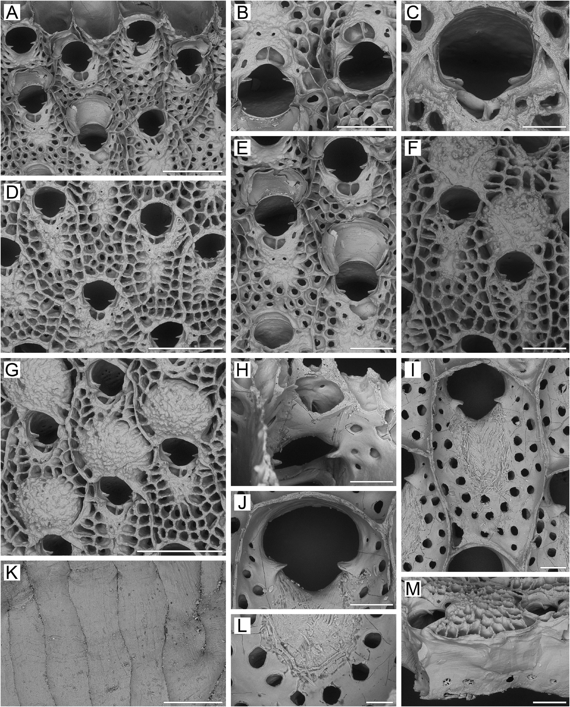
( Figs 28 View FIGURE 28 , 33H View FIGURE 33 )
Schizoporella ussowi Kluge, 1908a, p. 527 , figs 2a–c.
Hippodiplosia ussowi: Kluge 1953, p. 175 ; 1961, p. 135; 1962, p. 501, fig. 348; 1975, p. 609, fig. 348; Androsova 1958, p. 139, fig. 62; Kluge et al. 1959, p. 212.
Hippodiplosia smitti: Osburn 1933, p. 330 , pl. 9, fig. 6.
Hippoporina ussowi: Gontar & Denisenko 1989, p. 358 ; Denisenko 1990, p. 37; 2013, p. 184; Grischenko 1997, p. 189; Shunatova & Ostrovsky 2001, p. 115.
Rhamphostomella ussowi: Winston & Hayward 2012, p. 124 View in CoL , fig. 80.
Material examined. Lectotype: ZIRAS 4 /2331, single colony fragment, Stn 8, 27 June 1876, estuary of Mezen River , White Sea, dredge, collector K.S. Merezhkovsky.
ZIRAS 13 /13526, colony fragment, summer 1897, Zajatskie Islands of the Solovetskiy Archipelago, Onego Bay , White Sea , dredge, collector G.A. Kluge. ZIRAS 14 /50567, colony fragment , KIENM Collection , RV Nazarovsk, Stn 394, 18 September 1988, Korfa Gulf , eastern Kamchatka Peninsula, Bering Sea , 60°07.0ʹ N, 165°57.0ʹ E, depth 48 m, rock dredge, collector A GoogleMaps . V. Rzhavsky .
Additional material. Seven specimens. IMB Collection, Commander Intertidal Expedition (1972) Stn 194/2; KIENM Collection (1992) Stns 54, 88 (see Appendix 1 for details).
Measurements. ZIRAS 14/50567, Korfa Gulf, eastern Kamchatka, Bering Sea ( Fig. 28A–M View FIGURE 28 ). ZL, 0.67–1.13 (0.91 ± 0.11). ZW, 0.37–0.70 (0.54 ± 0.07). ZD, 0.60–0.78 (n = 2). OrL, 0.20–0.25 (0.23 ± 0.01). OrW, 0.21–0.25 (0.23 ± 0.01). OeL, 0.35–0.43 (0.39 ± 0.02) (n = 15). OeW, 0.36–0.44 (0.39 ± 0.02) (n = 15). Av(s)L, 0.09–0.14 (0.11 ± 0.01). P(f)N, 32–49 (42) (n = 10).
Description. Colonies encrusting, multiserial, unilaminar ( Fig. 28A View FIGURE 28 ), roughly circular, up to 11 × 8 mm in maximal dimension, bright-yellow to beige when alive, light yellow when dry. Zooids of medium size, oblonghexagonal, oval to irregularly polygonal, arranged in quincunx, delineated by undulating fused lateral walls.
Frontal shield mixed, comprising umbonuloid and lepralioid components ( Fig. 28A, D, F View FIGURE 28 ), flattened to moderately convex and coarsely granular medially. Proximal to orifice is narrow imperforate area, extending along zooidal midline and corresponding to umbonuloid part of shield. Extensive peripheral area, corresponding to lepralioid part of shield, bearing numerous pseudopores lying in large, deep, polygonal (square, trapezoid, rectangular) infundibular depressions. With age, frontal shield appearing increasingly reticulate ( Fig. 28F, G View FIGURE 28 ). Interior of frontal shield ( Fig. 28I View FIGURE 28 ) showing mixed structure. Ring scar indistinct ( Fig. 28L View FIGURE 28 ), forming irregular boundary between umbonuloid exterior wall and extra-umbonuloid interior wall microstructure. Narrow umbonuloid component occupying more than 50% length of frontal shield (55% in one measured zooid), corresponding to external imperforate area.Adjacent pseudoporous area surrounding imperforate region ( Fig. 28I, J, L View FIGURE 28 ), corresponding to lepralioid part of shield. One or two small areolae (connecting hypostegal and visceral coelomic cavities) visible lateral to primary orifice ( Fig. 28B, C View FIGURE 28 ), as smallest pores in shield interior ( Fig. 28I, J View FIGURE 28 ).
Primary orifice ( Fig. 28A, B, C, J View FIGURE 28 ) roughly circular to pyriform; distal and lateral margins formed by upper terminal part of distal transverse wall. Сondyles usually narrow triangular, with acute or rounded tips. Distal margin of orifice rounded, proximal margin with deep, wide U-shaped sinus. No oral spines or lyrula.
Secondary orifice ( Fig. 28D, E, G View FIGURE 28 ) rounded to transversely oval, generally repeating form of primary orifice, cormidial; restricted distally by vertical thickening of proximal wall of daughter zooid and, occasionally, by adjacent zooids ( Fig. 28C–E View FIGURE 28 ); proximal curvature formed by distal side of avicularian cystid connecting to weakly elevated, very small lateral lappets from frontal shield; in ovicellate zooids, these low lappets connecting with proximolateral corners of ooecium ( Fig. 28E, F, G View FIGURE 28 ).
Cystid of suboral avicularium small, only slightly broadened and elevated ( Fig. 28A–G View FIGURE 28 ), situated medially proximal to orifice, surface finely to coarsely granular, with 3–4 minute communication pores. Avicularian frontal surface (rostral/postmandibular areas) angular, crossing zooidal midline, facing obliquely distally. Rostrum low ( Fig. 28B, C View FIGURE 28 ), short, oval, transversely oriented, directed distolaterally to laterally, forming blunt angle with postmandibular area. Palate short, lingulate, foramen repeating shape of palate; opesia semicircular. Crossbar complete.
Ovicells initially hyperstomial, becoming subimmersed and even appearing endozooidal ( Fig. 28E–G View FIGURE 28 ) as consequence of overgrowth by secondary calcification from frontal shields of daughter and adjacent zooids; surface coarsely granular, with 1–2 divergent sutures subdividing secondary calcification from contiguous zooids ( Fig. 28F– G View FIGURE 28 ). Ooecium formed by distal autozooid, ooecial fold developing at colony margin concurrently with formation of frontal shield of distal zooid ( Fig. 28A View FIGURE 28 ). Fully completed ooecia with concave proximal margin; pseudopores not seen.
Zooids interconnected by 1‒2 mural pore chambers in each distolateral wall ( Fig. 28M View FIGURE 28 ). Communication pores forming two multiporous septula or spread randomly in basal half of transverse walls.
Basal surface of zooids ( Fig. 28K View FIGURE 28 ) fully calcified, flattened, textured with coarse, parallel or irregular lineation, without protuberances. Boundaries between zooids recognizable by fine incisions.
Ancestrula and early astogeny not observed.
Remarks. In his original description, Kluge (1908a) assigned this species to Schizoporella . It was subsequently placed in Hippodiplosia ( Osburn 1933; Kluge 1953, 1962, 1975) or Hippoporina ( Denisenko 1990, 2013; Grischenko 1997). Winston & Hayward (2012) tentatively assigned it to Rhamphostomella , pointing out that it is strikingly similar to M. ovata and does not belong in either of the superfamilies Smittinoidea or Schizoporelloidea, in which species formerly assigned to Hippodiplosia are now classified, warranting a deeper investigation of its morphology.
Winston & Hayward (2012) considered the single series of large marginal pores to be areolae. However, the underside of the frontal shield shows 1–2 areolae laterally to the zooidal orifice in the vicinity of the umbonuloid part of the shield. Although the marginal pseudopores have an areolar position, we doubt that they are similar in function (long suspected in lepralioid pseudoporous shields, in which it is assumed that there is an organic continuity between the hypostegal and visceral coeloms). Winston & Hayward (2012, p. 124) also wrote that the ooecia have “imperforate calcification”, but presented no evidence of this. All ooecia were covered by secondary calcification in our material, so the presence or absence of ooecial pseudopores remains unresolved.
Ecology. Mixtoscutella ussowi has been recorded from depths of 1–100 m on seafloors of rock, shell and silt. Colonies encrust pebbles, mollusc shells, barnacles, other bryozoans and brown and red algae.
Distribution. This is a boreal-Arctic, circumpolar, sublittoral species. Arctic records include White Sea (Kluge 1908; Gostilovskaya 1957; Gontar & Denisenko 1989; Shunatova & Ostrovsky 2001), Barents Sea ( Kluge 1962, 1975; Gontar & Denisenko 1989; Denisenko 1990), Kara Sea ( Kluge 1962, 1975; Gontar & Denisenko 1989; Denisenko 2021), western Greenland ( Osburn 1933; Gontar & Denisenko 1989; Denisenko & Blicher 2021) and eastern Greenland ( Denisenko & Blicher 2021). There are North Atlantic records from Mount Desert Island, Cape Ann, and Isles of Shoals, Massachusetts, northeastern North America ( Osburn 1933; Winston & Hayward 2012). In the northern Pacific M. ussowi has been reported from the Bering Sea ( Kluge 1962, 1975), including the Commander Islands ( Grischenko 1997, 2002), and it was recently found in Korfa Gulf, eastern Kamchatka (our data). In the Sea of Okhotsk, it has been reported from shallow waters along the southeast coast of Sakhalin Island, in Sakhalin Gulf, and near the Shantar Archipelago ( Kluge 1961, 1962, 1975). In the Sea of Japan, it has been reported from northern Primorye ( Androsova 1958; Kluge 1961) and along the southwestern coast of Sakhalin Island ( Kluge et al. 1959).

| RV |
Collection of Leptospira Strains |
| V |
Royal British Columbia Museum - Herbarium |
No known copyright restrictions apply. See Agosti, D., Egloff, W., 2009. Taxonomic information exchange and copyright: the Plazi approach. BMC Research Notes 2009, 2:53 for further explanation.
|
Kingdom |
|
|
Phylum |
|
|
Class |
|
|
Order |
|
|
Family |
|
|
Genus |
Mixtoscutella ussowi (Kluge, 1908)
| Grischenko, Andrei V., Gordon, Dennis P., Taylor, Paul D., Kuklinski, Piotr, Denisenko, Nina V., Spencer-Jones, Mary E. & Ostrovsky, Andrew N. 2022 |
Rhamphostomella ussowi: Winston & Hayward 2012 , p. 124
| Winston, J. E. & Hayward, P. J. 2012: 124 |
Hippoporina ussowi:
| Shunatova, N. N. & Ostrovsky, A. N. 2001: 115 |
| Grischenko, A. V. 1997: 189 |
| Denisenko, N. V. 1990: 37 |
| Gontar, V. I. & Denisenko, N. V. 1989: 358 |
Hippodiplosia ussowi:
| Kluge, G. A. & Androsova, E. I. & Gostilovskaya, M. G. 1959: 212 |
| Androsova, E. I. 1958: 139 |
| Kluge, G. A. 1953: 175 |
Hippodiplosia smitti:
| Osburn, R. C. 1933: 330 |
Schizoporella ussowi
| Kluge, G. A. 1908: 527 |