
Rhamphostomella commandorica, Grischenko & Gordon & Taylor & Kuklinski & Denisenko & Spencer-Jones & Ostrovsky, 2022
|
publication ID |
https://doi.org/10.11646/zootaxa.5131.1.1 |
|
publication LSID |
lsid:zoobank.org:pub:CF550031-D6A9-48A3-A953-A1BD40C72F5E |
|
DOI |
https://doi.org/10.5281/zenodo.6520677 |
|
persistent identifier |
https://treatment.plazi.org/id/03892374-0B3D-3330-FF73-A86F181FF909 |
|
treatment provided by |
Plazi |
|
scientific name |
Rhamphostomella commandorica |
| status |
sp. nov. |
Rhamphostomella commandorica n. sp.
( Figs 2 View FIGURE 2 , 30B View FIGURE 30 )
Diagnosis. Colony encrusting,multiserial.Zooids large, hexagonal to oval.Frontal shield convex, pustulose.Marginal areolae large, separated by high interareolar ridges connected with cystid of suboral avicularium. Umbonuloid component extensive. Primary orifice sunken, broadly semicircular to bell-shaped, with blunt condyles; proximal margin with low, wide median prominence. No oral spines. Secondary orifice irregularly oval, cormidial. Suboral avicularian cystid large, bulging, occupying most of frontal shield, finely granulated. Avicularian frontal surface situated asymmetrically in respect to zooidal orifice, crossing zooidal midline, facing distolaterally and slightly overhanging proximal margin of primary orifice. Rostrum semicircular, blunt. Palate short, broadly triangular to semiround. Crossbar complete. Large adventitious avicularia also present, often associated with ooecia. Rostrum elongate-oval or elongate-triangular, blunt. Palate elongate-triangular or spatulate. Palatal foramen heart-shaped. Crossbar complete, with small ligula. Ovicells hyperstomial, rapidly subimmersed by secondary calcification. Ectooecium smooth, with pseudopores arranged in arch around a few central pseudopores. Two mural pore chambers in distolateral wall, and 1‒3 multiporous septula in transverse walls. Basal surface of zooids fully calcified with some tubular protuberances.
Material examined. Holotype: ZIRAS 1 /50125, single colony fragment, IMB Collection , Stn 224/582, 2 October 1973, Poludennaya Bight , coastal waters of Medny Island , Commander Islands, Pacific Ocean, 54°36.8ʹ N, 167°21.5ʹ E, depth 130–250 m, rock dredge, collector S.D. Vavilin. GoogleMaps Paratype: NHMUK 2013.10 About NHMUK .21.1, one colony , RV Norseman , Stn AS –1, 17 July 2011, coastal waters of Adak Island , Andreanof Islands , Aleutian Islands, Pacific Ocean, 51°46.2ʹ N, 176°25.6ʹ W, depth 10 m GoogleMaps , SCUBA, collector P. Kuklinski.
Etymology. The species name alludes to the type locality in the Commander Islands (in Russian, “Komandorskie ostrova”).
Type locality. Poludennaya Bight , coastal waters of Medny Island , Commander Islands, Pacific Ocean, 54°36.8ʹ N, 167°21.5ʹ E, depth 130–250 m. GoogleMaps
Measurements. ZIRAS 1/50125, Medny Island, Commander Islands, Pacific Ocean ( Figs 2A–M View FIGURE 2 , 30B View FIGURE 30 ). ZL, 0.77–1.28 (1.03 ± 0.12). ZW, 0.47–0.75 (0.59 ± 0.07). ZD, 0.62–0.67 (n = 2). OrL, 0.22–0.30 (0.26 ± 0.02). OrW, 0.22–0.35 (0.28 ± 0.03). OeL, 0.32–0.45 (0.39 ± 0.03). OeW, 0.42–0.58 (0.51 ± 0.04). Av(s)L, 0.15–0.28 (0.20 ± 0.03). Av(ad)L, 0.37–0.53 (0.44 ± 0.04). P(m)N, 12–18 (14). P(oe)N, 11–16 (15) (n = 10).
NHMUK 2013.10.21.1, Adak Island, Aleutian Islands, Pacific Ocean. ZL, 0.59–1.08 (0.85 ± 0.09). ZW, 0.34– 0.57 (0.45 ± 0.06). ZD, 0.55–0.61 (n = 2). OrL, 0.16–0.24 (0.20 ± 0.02) (n = 25). OrW, 0.18–0.28 (0.23 ± 0.02) (n = 25). Av(s)L, 0.12–0.17 (0.14 ± 0.01) (n = 14). P(m)N, 14–24 (19) (n = 10).
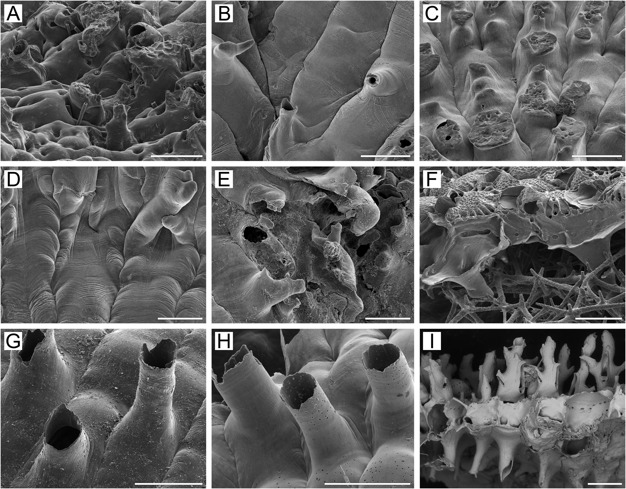
Description. Colonies encrusting, multiserial, unilaminar ( Fig. 2A View FIGURE 2 ), more or less circular, attaining 12 × 15 mm in size, pink when dry. Zooids large, hexagonal to oval, arranged in regular rows in checkered pattern, demarcated by fine sutures between lateral and transverse walls ( Fig. 2A–D View FIGURE 2 ); sutures occluded by secondary calcification in older parts of colony.
Frontal shield umbonuloid ( Fig. 2A–D, I View FIGURE 2 ), thick, convex, pustulose, with numerous, large, elongate areolae along zooidal margins, separated by narrow, high interareolar ridges connected with cystid of suboral avicularium. Some proximal ridges fusing with each other. Thickening of frontal shield resulting in smaller areolae. Umbonuloid component extensive, occupying about 70% of length of frontal shield (72% in one measured zooid), with fine parallel lineation and accretionary banding ( Fig. 2I, L View FIGURE 2 ). Ring scar forming boundary between umbonuloid exterior wall microstructure and extra-umbonuloid calcification clearly visible but not as single discrete line ( Fig. 2L View FIGURE 2 ).
Primary orifice ( Fig. 2A, B, J View FIGURE 2 ) broadly semicircular to bell-shaped or oval; its distal and lateral margins formed by upper terminal part of distal transverse wall ( Fig. 2A, B, E View FIGURE 2 ) and bearing low blunt condyles proximolaterally ( Fig. 2J View FIGURE 2 ). Distal margin of orifice round, proximal margin with low, wide median prominence and broadly rounded proximolateral corners. No oral spines.
Secondary orifice ( Fig. 2A–E, G View FIGURE 2 ) irregularly oval, cormidial, formed proximally by avicularian cystid connected with thin vertical wall surrounding primary orifice ( Fig. 2A–D View FIGURE 2 ). Distally and distolaterally, secondary orifice restricted by vertical walls of distal and lateral zooids. With increasing secondary calcification, outline of secondary orifice often changing, acquiring trapezoidal shape ( Fig. 2E, G View FIGURE 2 ) in ovicellate zooids.
Suboral avicularium with cystid large, bulging, broad, occupying most of frontal shield, with 6–9 communication pores connecting avicularian and hypostegal coelomic cavities ( Fig. 2A, C, D View FIGURE 2 ); surface finely granulated when young, pustulose in older zooids. Avicularian frontal surface (rostral/postmandibular areas) situated asymmetrically with respect to zooidal orifice, on right or left slope of avicularian cystid, normally crossed by zooidal midline, facing distolaterally or laterally, tilted frontodistally at angle of 60–90°. Rostrum semicircular, blunt, directed proximomedially upwards. Palate short, semicircular to rounded-triangular. Palatal foramen repeating shape of palate, opesia oval or semielliptical, surrounded by cryptocystal shelf. Crossbar complete.
Large adventitious avicularia developing by frontal budding in older parts of colony, often associated with ooecia. Zooidal orifices deeply immersed in areas where avicularia are numerous. Avicularian cystid broad, with finely granulated surface ( Fig. 2E, G, H View FIGURE 2 ). Frontal surface of avicularium facing obliquely upwards. Rostrum elongate-oval or elongate-triangular, blunt, oriented distolaterally to proximolaterally. Palate elongate-triangular or spatulate. Palatal foramen heart-shaped, with extensive distal cryptocystal shelf. Opesia oval or semielliptical, cryptocyst narrow. Crossbar complete, with small ligula. In older parts of colony, adventitious avicularia cover most of free space, together with thick secondary calcification, strongly changing appearance of colony (compare Fig. 2A, C, D View FIGURE 2 with Fig. 2E, G View FIGURE 2 ).
Ovicells initially hyperstomial, rapidly becoming subimmersed as ooecium is covered by thick secondary calcification proceeding from daughter and neighbouring zooids; surface finely granular, often with 1–2 sutures demarcating calcification (often bearing interareolar ridges) from different zooids ( Fig. 2E, F, G View FIGURE 2 ). In older parts of colony, secondary calcification covering half to two-thirds of ooecium; ovicell appearing endozooidal when only proximal triangular or semicircular area of ectooecium with pseudopores is visible ( Fig. 2G View FIGURE 2 ). Ooecium formed by distal autozooid around distinctive curved slit ( Fig. 2A, C, D View FIGURE 2 ) in slope of proximal vertical wall of distal zooid contributing to secondary orifice, immediately distal to margin of primary orifice of maternal zooid; this slit leading to communication canal connecting ooecial and visceral coeloms, opening on underside of frontal shield of distal zooid as small, straight, slit-like communication pore at mid-distance between transverse wall and ring-scar ( Fig. 2I View FIGURE 2 ). Ooecium spherical, with straight or slightly concave proximal margin often bearing very low, central convexity ( Fig. 2F View FIGURE 2 ). Ectooecium smooth, with oval, irregular or often slit-like pseudopores arranged in compressed arch around оne large or several small central pseudopores, oval or irregular. Ooecial base surrounded by oval to elongate areolae separated by short, narrow ridges ( Fig. 2F, G View FIGURE 2 ).
Zooids interconnected by two mural pore chambers ( Fig. 2M View FIGURE 2 ) in each distolateral wall, and by one wide, horizontal or 2‒3 (usually 2) compact multiporous septula in basal half of transverse wall. Distally transverse wall can bear up to three buttressed recesses (each with multiporous septula) resembling basal pore chambers ( Fig. 2A View FIGURE 2 ).
Basal surface of zooids ( Figs 2K, M View FIGURE 2 , 30B View FIGURE 30 ) fully calcified, convex, smooth, with numerous tubular protuberances (0.04–0.27 mm in diameter) having fine parallel folds on surface. Boundaries between zooids recognizable basally by deep undulating incisions. Sparse white spots (presumably weakly calcified areas) visible in semitransparent basal wall under light microscope.
Ancestrula and early astogeny not observed.
Remarks. In the general appearance of the zooids, as well as in having: 1) a bulging, broad oval suboral avicularium with semicircular palate, and 2) ooecia with a narrow arch of slit-like pseudopores, R. commandorica n. sp. resembles R. pacifica (see below), but differs from the latter as follows: 1) the primary orifice is submerged in zooids of R. commandorica n. sp. at all stages of zooidal development, whereas it is clearly visible in non-ovicellate zooids and surrounded by a tubular peristome in ovicellate zooids of R. pacifica ; 2) the palatal foramen of the adventitious avicularia is heart-shaped in R. commandorica n. sp., but Y-shaped in R. pacifica ; 3) the two known colonies of R. commandorica n. sp. are both encrusting, whereas colonies of R. pacifica are encrusting only initially, but rapidly grow into erect bilamellar ruffled expansions.
Ecology. Rhamphostomella commandorica n. sp. has been found at depths of 10–250 m, on mixed bottoms (silt, pebbles, boulders). The original substrata for our specimens are unknown.
Distribution. Presently known only from Poludennaya Bight off Medny Island, Commander Islands, and Adak Island, Andreanof Islands, Aleutian Islands, R. commandorica n. sp. can be categorized as a Pacific high-boreal, sublittoral species.

| RV |
Collection of Leptospira Strains |
No known copyright restrictions apply. See Agosti, D., Egloff, W., 2009. Taxonomic information exchange and copyright: the Plazi approach. BMC Research Notes 2009, 2:53 for further explanation.
|
Kingdom |
|
|
Phylum |
|
|
Class |
|
|
Order |
|
|
SubOrder |
Flustrina |
|
SuperFamily |
Lepralielloidea |
|
Family |
|
|
Genus |