
Rhamphostomella cristata ( Hincks, 1889 )
|
publication ID |
https://doi.org/ 10.11646/zootaxa.5131.1.1 |
|
publication LSID |
lsid:zoobank.org:pub:CF550031-D6A9-48A3-A953-A1BD40C72F5E |
|
DOI |
https://doi.org/10.5281/zenodo.6520679 |
|
persistent identifier |
https://treatment.plazi.org/id/03892374-0B25-3328-FF73-ADE11B7AFCCE |
|
treatment provided by |
Plazi |
|
scientific name |
Rhamphostomella cristata ( Hincks, 1889 ) |
| status |
|
Rhamphostomella cristata ( Hincks, 1889) View in CoL
( Figs 4 View FIGURE 4 , 30C View FIGURE 30 , 31J, K View FIGURE 31 )
Rhamphostomella costata var. cristata Hincks, 1889, p. 426 View in CoL , pl. 21, fig. 6.
Rhamphostomella costata var. cristata: Osburn 1912b, p. 286 View in CoL ; 1932, p. 14; Kluge 1962, p. 539, fig. 376; 1975, p. 656, fig. 376; Androsova 1977, p. 202; Denisenko 1988, p. 13; 1990, p. 39; Gontar & Denisenko 1989, p. 359.
Rhamphostomella costata cristata: Tarasova 1983, p. 25 View in CoL , fig. 32; Gontar 1994a, p. 146; 2010, p. 153; Grischenko 1997, p. 174; Gontar et al. 2001, p. 195.
Rhamphostomella cristata: Grischenko 2002, p. 115 View in CoL ; 2003b, p. 237; Denisenko 2011, p. 14; 2013, p. 184.
Rhamphostomella fortissima Bidenkap, 1900a, p. 524 , pl. 19, fig. 8.
Rhamphostomella fortissima: Osburn 1952, p. 427 , pl. 50, figs 1, 2; 1955, p. 38; Gontar 2010, p. 153; Foster 2010, p. 57.
Discopora scabra var. fortissima: Nordgaard 1918, p. 78 ; 1929, p. 7.
Material examined. Neotype: NHMUK 1911.10.1.1576A, one colony fragment, A.M. Norman Collection, Gulf of St Lawrence , Atlantic Ocean, collector J. Whiteaves.
NHMUK 2013.10.21.7a, one colony encrusting oyster shell, RV Norseman , Stn AST–2, 16 July 2011, reef in middle of Boot Bay , coastal waters of Adak Island , Andreanof Islands, Aleutian Islands, Pacific Ocean, 51°44.4ʹ N, 176°30.3ʹ W, depth 10–12 m GoogleMaps , SCUBA, collector P. Kuklinski. ZIRAS 2 /50110, two colony fragments , KIENM Collection , Stn 142, 28 July 1992, Cape Lebyazhy , coastal waters of Medny Island, Commander Islands, Pacific Ocean, 54°35.9ʹ N, 167°52.1ʹ E, depth 30 m GoogleMaps , SCUBA, collector V. V. Oshurkov .
Additional material. 64 specimens. IMB Collection (1972) Stns 6/16, 34/104; (1973) Stns 110/290, 110/292, 158/393, 233/591; KIENM Collection (1991) Stns 215, 221; (1992) Stns 4, 20, 28, 34, 38, 39, 99, 125, 128, 137, 142, 145, 146; KamchatNIRO Collection (2013) Stn 189 (see Appendix 1 for details).
Measurements. ZIRAS 2/50110, Medny Island, Commander Islands, Pacific Ocean ( Figs 4A–M View FIGURE 4 , 30C View FIGURE 30 ). ZL, 0.79–1.33 (1.01 ± 0.14). ZW, 0.47–0.65 (0.56 ± 0.04). ZD, 0.57–0.70 (n = 2). OrL, 0.25–0.33 (0.29 ± 0.02). OrW, 0.27–0.38 (0.31 ± 0.02). OeL, 0.30–0.40 (0.35 ± 0.03). OeW, 0.39–0.55 (0.48 ± 0.03). Av(s)L, 0.15–0.31 (0.24 ± 0.04). Av(ad)L, 0.17–0.33 (0.26 ± 0.04). P(m)N, 14–23 (18). P(oe)N, 11–32 (20) (n = 10).
NHMUK 2013.10.21.7a, Adak Island, Aleutian Islands, Pacific Ocean. ZL, 0.56–1.04 (0.83 ± 0.12). ZW, 0.32– 0.53 (0.43 ± 0.05). ZD, 0.49–0.58 (n = 2). OrL, 0.19–0.27 (0.23 ± 0.01). OrW, 0.21–0.28 (0.24 ± 0.02). OeL, 0.28–0.36 (0.34 ± 0.02) (n = 10). OeW, 0.38–0.47 (0.43 ± 0.02) (n = 10). Av(s)L, 0.13–0.31 (0.21 ± 0.05) (n = 23). P(m)N, 6–16 (12) (n = 10). P(oe)N, 17–25 (20) (n = 10).
Description. Colonies encrusting, multiserial, unilaminar ( Fig. 4A View FIGURE 4 ), more or less circular, attaining up to 51 mm in any one direction; bright red when alive, orange to light pink when dry. Zooids large, oval, hexagonal or pyriform, widest in distal half, arranged in regular rows with checkerboard pattern, demarcated by fine sutures between lateral and transverse walls, visible predominantly in young parts of colony.
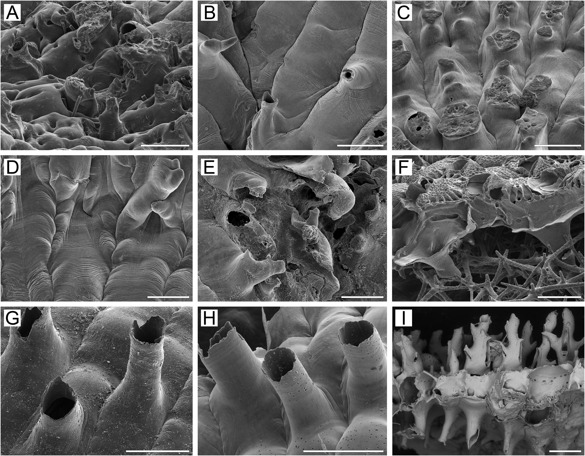
Frontal shield umbonuloid ( Fig. 4A, E, I View FIGURE 4 ), thickened, convex, finely granulated, with numerous, comparatively small round to elongated areolae along zooidal margins, separated by long, narrow, prominent interareolar ridges, arranged centripetally and continuing to apex of suboral avicularium, giving very characteristic striated pattern to frontal shield ( Fig. 4D, E View FIGURE 4 ). With age, size and number of areolae reduced as frontal-shield calcification thickens. Umbonuloid component extensive, occupying about 60% of length of frontal shield (63% in one measured zooid), with fine parallel lineation and accretionary banding ( Fig. 4I, L View FIGURE 4 ). Ring scar discrete ( Fig. 4L View FIGURE 4 ), forming regular boundary between exterior and interior wall microstructure.
Primary orifice ( Fig. 4A, B, J View FIGURE 4 ) roundly quadrangular or bell-shaped; distal and lateral margins formed by upper terminal part of distal transverse wall sometimes forming narrow shelf ( Fig. 4A View FIGURE 4 ). Distal margin of orifice round, proximal margin with broad, low median prominence and broadly rounded proximolateral corners. Condyles absent. Oral spines absent in majority of zooids, but two ephemeral oral spines occasionally evident at distal margin of orifice on marginal zooids.
Secondary orifice ( Fig. 4E–G View FIGURE 4 ) round to semicircular and trapezoidal, cormidial, formed proximally by distal part of frontal shield incorporating avicularian cystid medially and having two tall, symmetrical, triangular lappets laterally. Secondary orifice distally and distolaterally restricted by elevated vertical walls of distal and lateral zooids. In ovicellate zooids, lateral lappets connecting to proximolateral corners of ooecium ( Fig. 4F, G View FIGURE 4 ), conferring incomplete tubular peristomial form to secondary orifice.
Сystid of suboral avicularium large, broad, conical, strongly elevated, occupying most of frontal shield, with finely granulated surface and 4–7 communication pores ( Fig. 4A, D, E View FIGURE 4 ). Frontal surface of avicularium (rostral/ postmandibular areas) situated on left or right side of avicularian cystid, often out of zooidal midline, facing distolaterally. Rostrum elongate-triangular, pointed or sometimes blunt, directed proximomedially upwards, with short terminal hook. Palate elongate-triangular with pointed end. Palatal foramen triangular with rounded distal end, surrounded by very narrow cryptocystal shelf, opesia semicircular. Crossbar complete.
Adventitious avicularia developing in older parts of colony, occupying proximal half of frontal shield proximal to suboral avicularium. Avicularian cystid strongly elevated vertically, with finely granulated surface ( Fig. 4F–H View FIGURE 4 ). Avicularian frontal surface facing obliquely frontodistally to distolaterally. Rostrum elongate-triangular, pointed, directed obliquely frontalwards, with a short terminal hook. Palatal foramen elongate-triangular or oval-triangular with narrow cryptocystal shelf distally, opesia rounded or elliptical, surrounded by narrow cryptocyst. Crossbar complete. In older parts of colony, adventitious avicularia and thick secondary calcification covering most of frontal shield, strongly changing appearance of zooids (compare Fig. 4E View FIGURE 4 with Fig. 4G View FIGURE 4 ).
Ovicells hyperstomial. Ooecium formed by distal autozooid around slit-like concavity with communication pore at bottom, situated in proximalmost part of frontal shield just immediate to distal margin of maternal primary orifice ( Fig. 4E View FIGURE 4 ). Ooecium broader than long ( Fig. 4F–H View FIGURE 4 ), with straight or slightly concave proximal margin. Ectooecium smooth, with round, oval or slit-like pseudopores having radial arrangement. In most cases, ooecia not overgrown by secondary calcification, even in older zooids, or peripheral part surrounded by narrow rim ( Fig. 4G View FIGURE 4 ). In older zooids, ovicells becoming less prominent relative to colony surface because of thickening of frontal shields in surrounding zooids and development of adventitious avicularia; ooecia mostly free of secondary calcification, however.
Zooids interconnected by two mural pore chambers in each distolateral wall ( Fig. 4M View FIGURE 4 ) and usually two multiporous septula in basal half of transverse walls (corresponding to two recesses sometimes with medial buttress between them on distal side, Fig. 4A View FIGURE 4 ). Some such septula were complex, consisting of three to several small pore groups. Up to three “overlapping” septula of various sizes formed wide horizontal “band”.
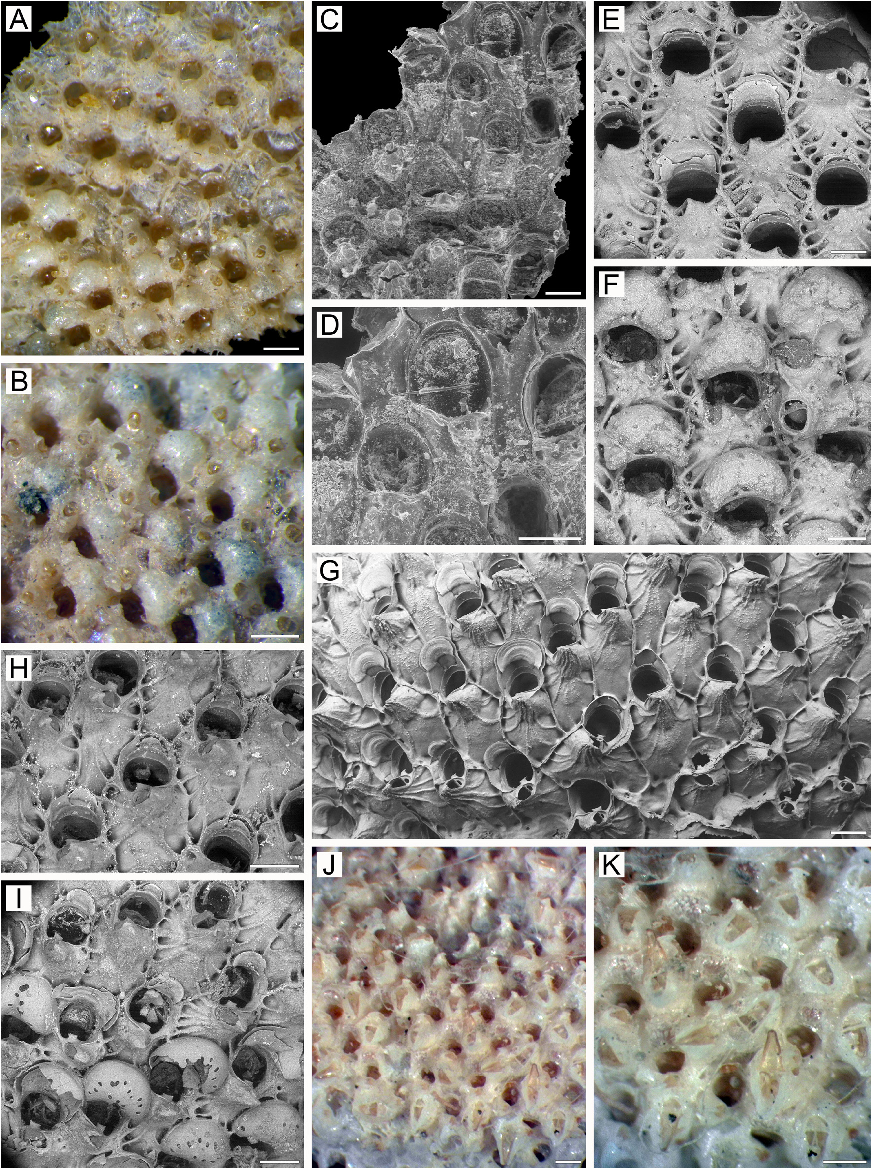
Basal surface of zooids ( Figs 4K View FIGURE 4 , 30C View FIGURE 30 ) fully calcified, convex, smooth, with numerous columnar protuberances (0.14–0.59 mm in diameter), flattened and broadened terminally. Boundaries between zooids clearly recognizable basally by deep, meandering incisions. Abundant white spots (presumably less-calcified areas) visible in semitransparent basal wall under light microscope.
Ancestrula and early astogeny not observed.
Remarks. According to Kluge (1962, 1975), zooids of R. cristata have a small oblique process or prominence on the proximal margin of the primary orifice, and our study confirms this ( Fig. 4J View FIGURE 4 ). Some colonies have zooids with a broad, very low process along the proximal margin, in contrast to the central, clearly prominent and discrete process typical of R. costata .
Kluge (1962, 1975) mentioned a couple of ephemeral oral spines on the distal rim of the orifice in zooids at the colony margin. While most colonies we examined have a few marginal zooids with two oral spines, one colony (NHMUK 2013.10.21.7a) from the shallow waters of Adak Island, Aleutians, has a rather broad zone of marginal zooids, up to three generations deep, all of which have two or three short oral spines.
Some authors have considered R. cristata to be a subspecies or variety of R. costata ( Hincks 1889; Osburn 1912b, 1932; Kluge 1962, 1975; Androsova 1977; Denisenko 1988, 1990; Gontar & Denisenko 1989; Gontar 1994a; Grischenko 1997). However, based on the set of characters described above (see Remarks for R. costata ), R. cristata clearly differs from R. costata and we thus consider it to be a distinct species.
Because we could not locate Hincks’s material, we have selected a neotype for this species based on a specimen collected by J. Whiteaves – similarly to Hincks (1889) – in the Gulf of St Lawrence , North Atlantic, and residing in the Natural History Museum, London .
Ecology. Rhamphostomella cristata has been recorded over a depth range of 10–197 m, mainly on hard bottoms and rock faces (including crevices), boulders, blocks and gravel, and sometimes on silty bottoms overlain with pebbles. In addition to rocky surfaces, colonies encrust serpulid tubes, hydroids, other bryozoans (e.g. Dendrobeania murrayana ) and shells of bivalve molluscs. Some colonies were a component of the cryptic community inhabiting internal cavities formed by the crustose coralline alga Clathromorphum nereostratum .
Distribution. R. cristata is a high-boreal-Arctic, sublittoral species ( Kluge 1962, 1975; Gontar & Denisenko 1989). Arctic records include the Barents Sea ( Bidenkap 1900a; Kluge 1962, 1975; Denisenko 1988, 1990), Kara Sea ( Gontar & Denisenko 1989; Denisenko 2021), East-Siberian Sea ( Nordgaard 1929; Gontar 1994a; Denisenko 2011), Chukchi Sea ( Gontar 2010), Point Barrow, Alaska, Beaufort Sea ( Osburn 1952, 1955), Canadian Arctic Archipelago ( Nordgaard 1929), Hudson Bay ( Osburn 1932), Labrador ( Osburn 1912b), eastern Greenland ( Kluge 1962, 1975; Denisenko & Blicher 2021), Greenland Sea ( Gontar & Denisenko 1989), Spitsbergen ( Kluge 1962, 1975; Gontar et al. 2001), Franz-Josef Land ( Kluge 1962, 1975; Denisenko 1990) and northern Norway ( Nordgaard 1918). In the northwest Atlantic it has been reported from St Lawrence Gulf and Newfoundland ( Hincks 1889; Whiteaves 1901; Gontar & Denisenko 1989). In the northwestern Pacific R. cristata is known from the Bering Sea near the Commander Islands ( Grischenko 1997, 2002, 2003b), from the western Kamchatka shelf of the Sea of Okhotsk (our data) and along northern Primorye, Sea of Japan ( Tarasova 1983). Northeastern Pacific localities include Cook Inlet, Gulf of Alaska ( Foster 2010) and the coastal waters of Adak Island, Andreanof Islands, Aleutian Islands, Pacific Ocean (our data).

| NHMUK |
Natural History Museum, London |
| RV |
Collection of Leptospira Strains |
| V |
Royal British Columbia Museum - Herbarium |
No known copyright restrictions apply. See Agosti, D., Egloff, W., 2009. Taxonomic information exchange and copyright: the Plazi approach. BMC Research Notes 2009, 2:53 for further explanation.
|
Kingdom |
|
|
Phylum |
|
|
Class |
|
|
Order |
|
|
Family |
|
|
Genus |
Rhamphostomella cristata ( Hincks, 1889 )
| Grischenko, Andrei V., Gordon, Dennis P., Taylor, Paul D., Kuklinski, Piotr, Denisenko, Nina V., Spencer-Jones, Mary E. & Ostrovsky, Andrew N. 2022 |
Rhamphostomella cristata:
| Denisenko, N. V. 2011: 14 |
Rhamphostomella costata cristata:
| Gontar, V. I. & Hop, H. & Voronkov, A. Yu. 2001: 195 |
| Grischenko, A. V. 1997: 174 |
| Gontar, V. I. 1994: 146 |
| Tarasova, N. A. 1983: 25 |
Rhamphostomella fortissima:
| Gontar, V. I. 2010: 153 |
| Foster, N. R. 2010: 57 |
| Osburn, R. C. 1952: 427 |
Discopora scabra var. fortissima:
| Nordgaard, O. 1918: 78 |
Rhamphostomella costata var. cristata:
| Gontar, V. I. & Denisenko, N. V. 1989: 359 |
| Denisenko, N. V. 1988: 13 |
| Androsova, E. I. 1977: 202 |
| Kluge, G. A. 1962: 539 |
| Osburn, R. C. 1912: 286 |
Rhamphostomella fortissima
| Bidenkap, O. 1900: 524 |
Rhamphostomella costata var. cristata
| Hincks, T. 1889: 426 |