
Rhamphostomella aleutica, Grischenko & Gordon & Taylor & Kuklinski & Denisenko & Spencer-Jones & Ostrovsky, 2022
|
publication ID |
https://doi.org/ 10.11646/zootaxa.5131.1.1 |
|
publication LSID |
lsid:zoobank.org:pub:CF550031-D6A9-48A3-A953-A1BD40C72F5E |
|
DOI |
https://doi.org/10.5281/zenodo.6520687 |
|
persistent identifier |
https://treatment.plazi.org/id/03892374-0B15-3319-FF73-AB381D9DF971 |
|
treatment provided by |
Plazi |
|
scientific name |
Rhamphostomella aleutica |
| status |
sp. nov. |
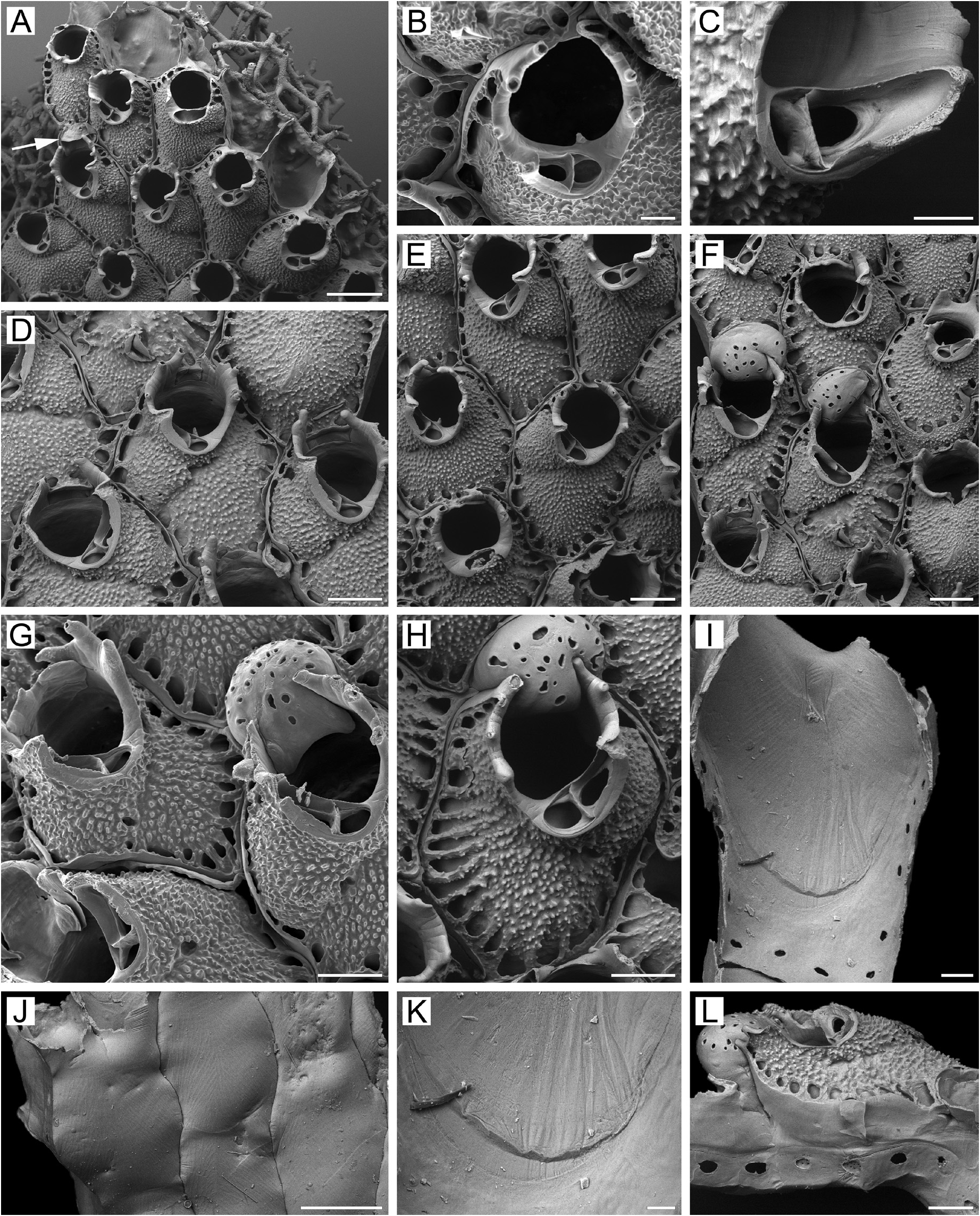
Rhamphostomella aleutica n. sp.
( Fig. 9 View FIGURE 9 )
Diagnosis. Colony encrusting, multiserial. Zooids large, hexagonal. Frontal shield ventricose, finely tuberculated. Marginal areolae deep, separated by short interareolar ridges. Umbonuloid component extensive. Primary orifice submerged, irregularly circular, often with small median process. Condyles absent. No oral spines. Secondary orifice subcircular to irregularly oval, cormidial, defined by tubular peristome. Peristomial rim with acute or blunt-tipped spine-like tubular projections, 2–4 on each side, slightly tilted inwards. Suboral avicularian cystid asymmetrically situated proximolaterally to orifice, with coarsely granular surface. Rostrum elongate triangular with narrowing hooked tip. Crossbar complete. No adventitious avicularia. Ovicells hyperstomial. Ectooecium smooth, lacking secondary calcification, with small pseudopores. Two pore chambers in distolateral wall and 1‒2 multiporous septula in transverse walls. Basal surface of zooids fully calcified, lacking protuberances.
Material examined. Holotype: NHMUK 2010.4.21.1 , one colony encrusting a sponge, Alaska Fisheries Science Center and National Marine Fisheries Service Collection , FV Sea Storm, Haul 190, Stn 114–21, 23 July 2004, coastal waters of Amchitka Island , Rat Islands , western Aleutian Islands , Bering Sea , 51°51.6ʹ N, 178°27.8ʹ E, depth 224– 235 m, collector M.H. Dick. GoogleMaps
Etymology. The species name alludes to the type locality in the Aleutian Islands.
Type locality. Coastal waters of Amchitka Island , Rat Islands , western Aleutian Islands, Bering Sea, 51°51.6ʹ N, 178°27.8ʹ E, depth 224–235 m. GoogleMaps
Measurements. NHMUK 2010.4.21.1, Amchitka Island, Aleutian Islands, Bering Sea ( Fig. 9A–L View FIGURE 9 ). ZL, 0.81– 1.14 (0.93 ± 0.07). ZW, 0.54–0.87 (0.73 ± 0.08). ZD, 0.44–0.62 (n = 2). OrL, 0.27–0.40 (0.32 ± 0.03). OrW, 0.24– 0.40 (0.33 ± 0.03). OeL, 0.34–0.37 (0.35 ± 0.01) (n = 3). OeW, 0.45–0.52 (0.48 ± 0.03) (n = 3). Av(s)L, 0.24–0.39 (0.32 ± 0.03). P(m)N, 15–20 (17) (n = 10). P(oe)N, 16–18 (17) (n = 4).
Description. Colony encrusting, multiserial, unilaminar ( Fig. 9A View FIGURE 9 ), irregular in outline, about 8 × 7 mm in size, deep red when dry. Zooids large, hexagonal, broadly oval, pyriform or irregular ( Fig. 9A, D–H View FIGURE 9 ), arranged in quincunx, demarcated by fine sutures between lateral and transverse walls; sutures visible in both young and old parts of colony.
Frontal shield umbonuloid ( Fig. 9A, I View FIGURE 9 ), moderately convex to strongly inflated, entirely covered by tiny pointed tubercles that enlarge in size and become coarse in older zooids ( Fig. 9B–H View FIGURE 9 ). Frontal shield with deep areolae along margins ( Fig. 9A, D–H View FIGURE 9 ), separated by short narrow interareolar ridges. Umbonuloid component extensive, occupying about 80% of length of frontal shield (77% in one measured zooid), with fine parallel lineation and accretionary banding ( Fig. 9I, K View FIGURE 9 ). Ring scar discrete, forming regular boundary between umbonuloid exterior wall and extra-umbonuloid interior wall microstructure.
Primary orifice ( Fig. 9A View FIGURE 9 ) submerged, irregularly circular, visible only in young zooids; distal and lateral margins formed by upper terminal part of distal transverse wall. Distal margin of orifice round, proximal margin shallowly concave, with small, narrow, pointed median process ( Fig. 9B View FIGURE 9 ) that is barely evident or is absent in some zooids. Condyles absent. No oral spines.
Secondary orifice subcircular to irregularly oval, cormidial, defined by tubular peristome; distally formed by a slightly elevated proximal margin of daughter-zooid frontal shield ( Fig. 9A–B, D View FIGURE 9 ); in older zooids becoming prominent vertical outgrowth ( Fig. 9G View FIGURE 9 ). Laterally and proximally, secondary orifice comprising terminal part of flared peristome, incorporating avicularian cystid on one side. Peristomial rim with spine-like tubular projections, acute or blunt-tipped, 2–4 on each side ( Fig. 9E–H View FIGURE 9 ), slightly tilted inwards. In ovicellate zooids, left and right sides of peristome encroaching onto surface of ooecium ( Fig. 9F–H View FIGURE 9 ), occasionally pressing into it and causing partial ooecial deformation ( Fig. 9F, H View FIGURE 9 ).
Cystid of suboral avicularium occupying one-quarter to one-third of frontal shield, asymmetrically situated proximolaterally to left or right of peristome, broad-based, bulging, with coarsely granulate surface, bearing 2– 4 communication pores connecting avicularian and hypostegal coeloms; postmandibular area of frontal surface often crossing zooidal midline, facing obliquely in frontal direction ( Fig. 9A–H View FIGURE 9 ). Rostrum elongate triangular, with narrowing hooked tip, incorporated into flared peristome, slightly curved distolaterally, directed distolaterally and frontally. Palatal foramen drop-shaped, cryptocystal shelf extensive distally, tapering proximally; opesia oval, surrounded by narrow cryptocyst. Crossbar complete, narrow, vertically deep ( Fig. 9A–H View FIGURE 9 ).
No adventitious avicularia.
Ovicells hyperstomial ( Fig. 9F–H View FIGURE 9 ). Ooecium formed by distal autozooid at colony periphery ( Fig. 9A, L View FIGURE 9 ). Ectooecium smooth, free of secondary calcification, with numerous small, irregular, circular, oval to slit-like pseudopores; proximal margin of ooecium slightly concave, frontal area appressed to distalmost flanks of peristome ( Fig. 9 F–H, L View FIGURE 9 ).
Zooids interconnected by two mural pore chambers in each distolateral wall ( Fig. 9L View FIGURE 9 ) and 1‒2 multiporous septula in basal half of transverse walls. In some zooids, transverse walls have two shallow recesses separated by medial buttress.
Basal surface of zooids ( Fig. 9J View FIGURE 9 ) fully calcified, smooth, thin. Numerous white spots (presumably weakly calcified areas) visible in semitransparent basal wall using light microscopy. Boundaries between zooids indicated basally by broadly sinuous incisions.
Ancestrula and early astogeny not observed.
Remarks. The elevated, subtubular peristome, with 2–4 spinous projections along each lateral margin, makes R. aleutica n. sp. unique in the genus, clearly distinguishable from congeners.
In the primary orifice with a pointed process, the shape and position of the suboral avicularium, and the umbonuloid shield, this species resembles species in Drepanophora (Lepraliellidae) . Considering the pseudoporous ooecium in R. aleutica n. sp., however, these are probably convergent similarities.
Ecology. The sole colony of Rhamphostomella aleutica n. sp. was found encrusting a sponge from 224–235 m depth.
Distribution. Currently known only from the type locality in the coastal waters of Amchitka Island, Rat Islands, western Aleutian Islands, Bering Sea, R. aleutica n. sp. is a Pacific high-boreal, sublittoral species.
| NHMUK |
NHMUK |
No known copyright restrictions apply. See Agosti, D., Egloff, W., 2009. Taxonomic information exchange and copyright: the Plazi approach. BMC Research Notes 2009, 2:53 for further explanation.
|
Kingdom |
|
|
Phylum |
|
|
Class |
|
|
Order |
|
|
Family |
|
|
Genus |