
Rhamphostomella alutacea Gontar, 1993
|
publication ID |
https://doi.org/ 10.11646/zootaxa.5131.1.1 |
|
publication LSID |
lsid:zoobank.org:pub:CF550031-D6A9-48A3-A953-A1BD40C72F5E |
|
DOI |
https://doi.org/10.5281/zenodo.6520689 |
|
persistent identifier |
https://treatment.plazi.org/id/03892374-0B13-331A-FF73-A8531BF2FD5D |
|
treatment provided by |
Plazi |
|
scientific name |
Rhamphostomella alutacea Gontar, 1993 |
| status |
|
Rhamphostomella alutacea Gontar, 1993 View in CoL
( Figs 10 View FIGURE 10 , 32C–E View FIGURE 32 )
Rhamphostomella alutacea Gontar, 1993a, p. 12 View in CoL , fig. 7.
Rhamphostomella alutacea: Denisenko 2013, p. 184 View in CoL .
Material examined. Holotype: ZIRAS 1 /44569, colony encrusting skeleton of sea urchin, Kuril-Sakhalin Expedition of Zoological Institute ( ZIRAS) and Pacific Institute of Fisheries and Oceanography ( TINRO), 15 September 1949, Krabovaya Bight, Shikotan Island, Lesser Kuril Ridge, Pacific Ocean, depth 55 m, shell, rock dredge, collector E.F. Guryanova. Paratype: ZIRAS 2 /50123, five fragments of single colony detached from broken shell of the bivalve Chlamys sp ., MFRT Rodino, 12 September 1992, about 32 km from Cape Hayryuzova, western Kamchatka shelf, Sea of Okhotsk , 57°36.2ʹ N, 156°09.0ʹ E, depth 78–81 m, crab trap, collector A GoogleMaps . V. Grischenko .
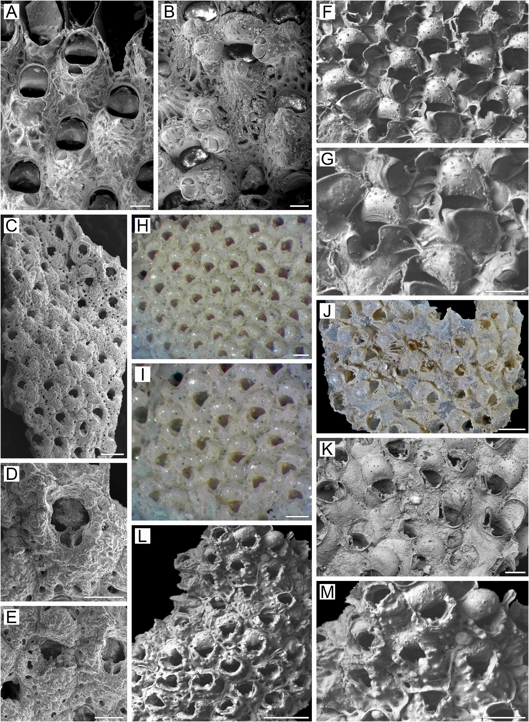
Measurements. ZIRAS 2/50123, western Kamchatka, Sea of Okhotsk ( Fig. 10A–L View FIGURE 10 ). ZL, 0.60–0.93 (0.71 ± 0.09). ZW, 0.35–0.55 (0.44 ± 0.05). ZD, 0.59–0.65 (n = 2). OrL, 0.15–0.25 (0.19 ± 0.03). OrW, 0.20–0.27 (0.22 ± 0.02). OeL, 0.17–0.27 (0.24 ± 0.03). OeW, 0.24–0.35 (0.30 ± 0.03). Av(s)L, 0.07–0.14 (0.10 ± 0.01). P(m)N, 16–26 (19). P(oe)N, 1–10 (6).
Description. Colonies encrusting, multiserial, unilaminar ( Fig. 10A View FIGURE 10 ), irregular in form, attaining 33 mm in maximal dimension; bright red to orange when alive, dark red to pink when dry. Zooids of medium size, hexagonal, oval to pyriform or irregular, arranged in quincunx, demarcated by fine, meandering sutures between lateral and transverse walls ( Fig. 10A, D–H View FIGURE 10 ); boundaries less distinct in older parts of colony.
Frontal shield umbonuloid ( Fig. 10A, D, I View FIGURE 10 ), thickened, pustulose, convex in young zooids, flattened and smoother in older ones owing to its thickening. Large, deep, round to oval areolae along margins ( Fig. 10D–H View FIGURE 10 ), separated by short narrow interareolar ridges that may reach suboral avicularian cystid in distal part of frontal shield, mainly in young zooids. Thickening of frontal shield in older parts of colony resulting in considerable change in appearance, with some areolae reduced in size and obliterated, and others partitioned, resulting in “migration” of openings toward central part of shield and possibly fusing (compare Fig. 10A, D, E View FIGURE 10 with Fig. 10F–H View FIGURE 10 ). Umbonuloid component extensive, occupying about 60% of length of frontal shield (63% in one measured zooid). Ring scar discrete ( Fig. 10I, K View FIGURE 10 ), forming regular boundary between umbonuloid exterior wall and extra-umbonuloid interior wall microstructure.
Primary orifice ( Fig. 10A, B View FIGURE 10 ) submerged, broadly circular or oval; distal and lateral margins formed by upper terminal part of distal transverse wall, with ill-defined rim ( Fig. 10A, B, D, E View FIGURE 10 ). Distal margin of orifice rounded; proximal margin concave, with elongate to short, blunt or bifurcate median lyrula and two small acute or blunt processes situated proximolaterally. Sometimes one or both processes strongly reduced or absent ( Fig. 10A, D–F View FIGURE 10 ). No condyles or oral spines.
Secondary orifice ( Fig. 10E–H View FIGURE 10 ) round to oval, cormidial, formed distally by slightly elevated vertical walls of distal zooid and occasionally by lateral walls of neighbouring zooids ( Fig. 10D, E, G View FIGURE 10 ); proximally restricted by thin walls of peristome, comprising two relatively tall, symmetrical lappets derived from frontal shield and incorporating avicularian cystid medially; lappets connecting to proximolateral corners of ooecium in ovicellate zooids ( Fig. 10F, H View FIGURE 10 ).
Cystid of suboral avicularium small, occupying less than one-quarter of zooid frontal shield; low in young zooids ( Fig. 10A, D, E View FIGURE 10 ), moderately to strongly elevated and conical in some older zooids; situated medially or (more often) lying transversely to left or right of median axis. Frontal surface (rostral/postmandibular areas) crossing zooidal midline. Rostral frontal surface at strong oblique angle to postmandibular area, overall facing laterally and obliquely frontally; this concave aspect of avicularium, as well as position in peristome, gives impression of pseudosinus in secondary orifice. Rostrum short, blunt, semioval, directed laterally to distolaterally and upwards ( Fig. 10C View FIGURE 10 ); palatal foramen conforming to shape of rostrum, no cryptocystal shelf; opesia semicircular or lingulate, sometimes of same size as foramen. Crossbar complete.
No adventitious avicularia.
Ovicells initially hyperstomial, rapidly becoming subimmersed and even appearing endozooidal ( Fig. 10F–H, L View FIGURE 10 ) through overgrowth by secondary calcification from adjoining daughter and neighbouring zooids that cover most of ooecium except for central pseudoporous part. Ooecium formed by distal autozooid. Ooecial and visceral coelomic cavities connected via communication canal opening on underside of frontal shield of distal zooid as large, angular communication slit near proximalmost areolae ( Fig. 10I View FIGURE 10 ). Ooecium with straight or slightly concave proximal margin. Ectooecium smooth, with pseudopores varying markedly in number, size, form (round, oval, elongate, irregular) and pattern of arrangement.
Zooids interconnected by one mural pore chamber in each distolateral wall ( Fig. 10L View FIGURE 10 ). Predominantly two (occasionally one) multiporous septula sometimes added by individual pores in basal half of transverse walls. In some zooids, transverse wall has two shallow recesses separated by medial buttress.
Basal surface of zooids ( Fig. 10J View FIGURE 10 ) fully calcified, convex, smooth or with coarse transverse lineation; without evident protuberances. Boundaries between zooids recognizable basally by deep, sinuous incisions.
Ancestrula and early astogeny not observed.
Remarks. Rhamphostomella alutacea differs from congeners in the strictly transverse position of the suboral avicularium, which is strongly angled upon itself to appear concave, with the rostrum facing obliquely frontally. The only congener with a similar transverse placement of the suboral avicularium, R. radiatula , has a narrow, U-shaped pseudosinus, with the avicularian rostrum often hidden or barely visible frontally ( Fig. 18D–H View FIGURE 18 ).
In her original description, Gontar (1993a) mentioned and illustrated ovicells as having an ooecium with a single triangular or slit-like central pseudopore. In fact, the number, shape, and arrangement of the pseudopores can vary markedly ( Fig. 10F – H View FIGURE 10 ). Contrary to the original description of the frontal shield as flattened, we observed that it can be convex in young zooids.
Ecology. Rhamphostomella alutacea was recorded from 55–81 m depth on sandy and silty bottoms overlain with gravel, where colonies encrust broken shells of the bivalve Chlamys sp. and sea urchin tests.
Distribution. This species was originally described from Krabovaya Bight, Shikotan Island, Lesser Kuril Ridge, northwestern Pacific; we report it also from the western Kamchatka shelf, Sea of Okhotsk. Thus, R. alutacea is a Pacific Asian boreal, sublittoral species.


| V |
Royal British Columbia Museum - Herbarium |
No known copyright restrictions apply. See Agosti, D., Egloff, W., 2009. Taxonomic information exchange and copyright: the Plazi approach. BMC Research Notes 2009, 2:53 for further explanation.
|
Kingdom |
|
|
Phylum |
|
|
Class |
|
|
Order |
|
|
Family |
|
|
Genus |
Rhamphostomella alutacea Gontar, 1993
| Grischenko, Andrei V., Gordon, Dennis P., Taylor, Paul D., Kuklinski, Piotr, Denisenko, Nina V., Spencer-Jones, Mary E. & Ostrovsky, Andrew N. 2022 |
Rhamphostomella alutacea:
| Denisenko, N. V. 2013: 184 |
Rhamphostomella alutacea
| Gontar, V. I. 1993: 12 |