
Rhamphostomella aspera, Grischenko & Gordon & Taylor & Kuklinski & Denisenko & Spencer-Jones & Ostrovsky, 2022
|
publication ID |
https://doi.org/10.11646/zootaxa.5131.1.1 |
|
publication LSID |
lsid:zoobank.org:pub:CF550031-D6A9-48A3-A953-A1BD40C72F5E |
|
DOI |
https://doi.org/10.5281/zenodo.6520691 |
|
persistent identifier |
https://treatment.plazi.org/id/03892374-0B10-3317-FF73-AC7F1D20FE8D |
|
treatment provided by |
Plazi |
|
scientific name |
Rhamphostomella aspera |
| status |
sp. nov. |
Rhamphostomella aspera n. sp.
( Fig. 11 View FIGURE 11 )
Diagnosis. Colony encrusting, multiserial. Zooids very large, broadly hexagonal. Frontal shield thickened, strongly convex, roughly granulated, with shaggy appearance. Marginal areolae separated by prominent radially arranged interareolar ridges continuing to apex of suboral avicularium, giving striated appearance to zooid. Umbonuloid component extensive. Primary orifice roughly quadrangular with round angles and ill-defined rounded lateral condyles. Proximal margin straight with broad low prominence. Pair of short, ephemeral, tubular oral spines often present. Secondary orifice oval or quadrangular, cormidial. Suboral avicularian cystid very large, occupying one-third to half of frontal shield, elevated, with acute conical apex, partially overhanging orifice, covered by interareolar ridges; frontal surface facing laterally to proximolaterally, occasionally proximally. Rostrum semioval, blunt. Crossbar complete, with small ligula. No adventitious avicularia. Ovicells hyperstomial, wider than long. Ectooecium smooth with sparse pseudopores; no secondary calcification. Pore chambers in distolateral wall and 1‒2 multiporous septula in transverse walls. Basal surface of zooids fully calcified, tightly cemented to substratum.
Material examined. Holotype: ZIRAS 1/50542 , colony encrusting sponge, IMB Collection, RV Akademik Oparin , 41st Expedition, Stn 63/ 53, 29 July 2011, northward from Iturup Island, South Kuril Islands, Sea of Okhotsk, 45°45.1ʹ N, 148°33.2ʹ E – 45°44.5ʹ N, 148°33.5ʹ E, depth 264– 274 m, Sigsbee trawl, collectors A.P. Tsurpalo and A.V. Chernyshev. GoogleMaps
Etymology. Latin, asper, rough, shaggy, alluding to the coarsely granular surface of the zooids that contrast strongly with the smooth spherical ooecia.
Type locality. Northward from Iturup Island , South Kuril Islands, Sea of Okhotsk, 45°45.1ʹ N, 148°33.2ʹ E – 45°44.5ʹ N, 148°33.5ʹ E, depth 264– 274 m. GoogleMaps
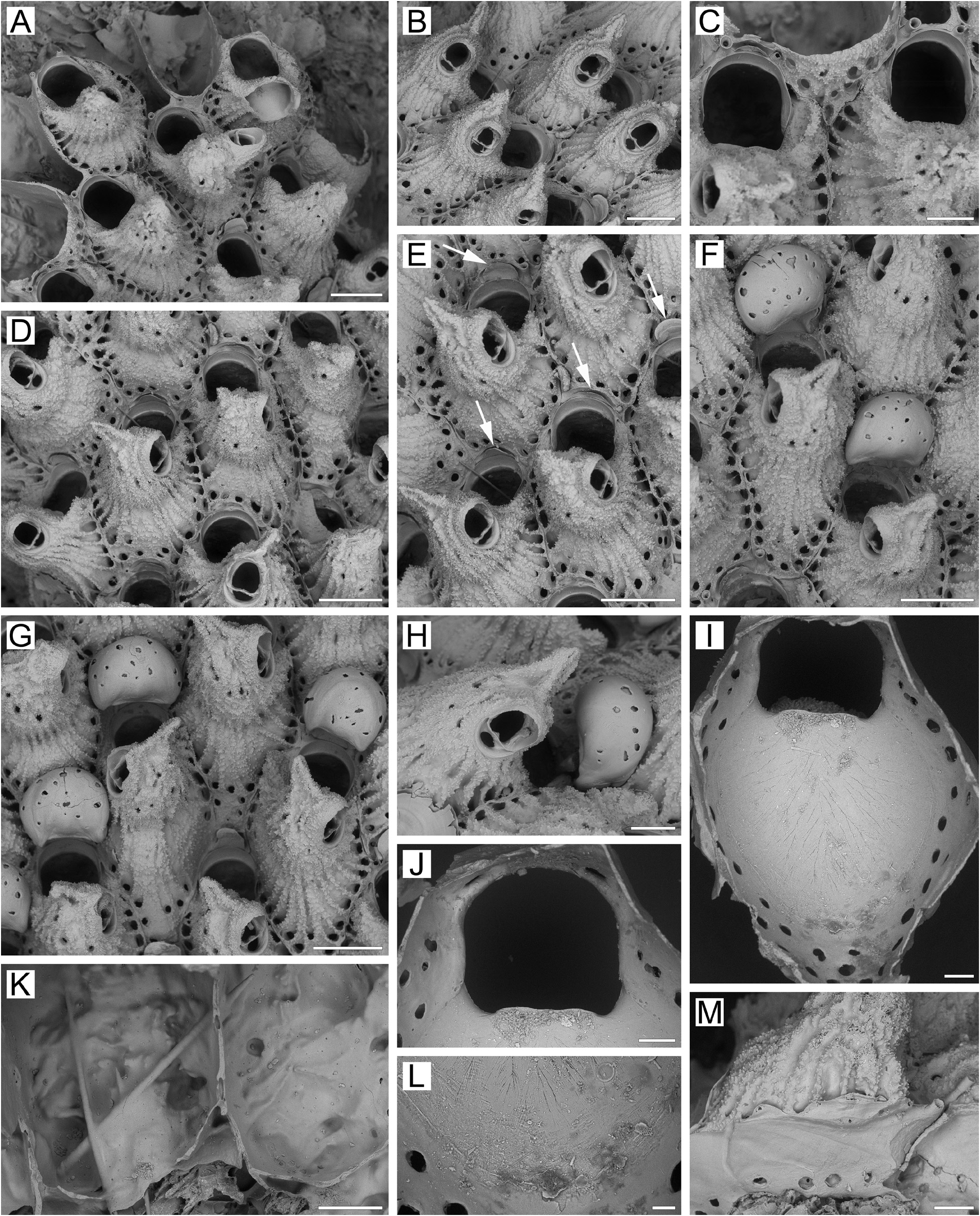
Measurements. ZIRAS 1/50542, Iturup Island, Kuril Islands, Sea of Okhotsk ( Fig. 11A–M View FIGURE 11 ). ZL, 1.33–1.95 (1.58 ± 0.17). ZW, 0.85–1.30 (1.01 ± 0.09). ZD, 1.13–1.19 (n = 2). OrL, 0.40–0.51 (0.44 ± 0.03). OrW, 0.39–0.46 (0.43 ± 0.02). OeL, 0.42–0.55 (0.49 ± 0.04) (n = 10). OeW, 0.57–0.68 (0.62 ± 0.04) (n = 10). Av(s)L, 0.33–0.43 (0.38 ± 0.02). P(m)N, 19–31 (25). P(oe)N, 9–18 (12) (n = 10).
Description. Colony encrusting, multiserial, unilaminar ( Fig. 11A View FIGURE 11 ), patch-like, about 11 × 16 mm in size, yellow when dry. Zooids very large, broadly hexagonal to oval, widest in midlength, arranged in checkered pattern, demarcated by fine sutures between lateral and transverse walls. Boundaries between zooids clearly visible in both young and old parts of colony.
Frontal shield umbonuloid ( Fig. 11A, I View FIGURE 11 ), thickened, strongly convex, with rough surface composed of pointed granules conferring a shaggy appearance, with circular areolae along raised margins (sometimes forming double rows for a short distance) ( Fig. 11B–F View FIGURE 11 ), separated by elongate, prominent, radially arranged interareolar ridges connected with cystid of suboral avicularium, frequently continuing to apex and thus giving a striated appearance to zooid. Areolae diminishing in size in older parts of colony. Umbonuloid component extensive, occupying about 80% of length of frontal shield (78% in one measured zooid), with fine parallel lineation and accretionary banding ( Fig. 11I, L View FIGURE 11 ). Ring scar indistinct ( Fig. 11I, L View FIGURE 11 ), nevertheless forming regular boundary between umbonuloid exterior wall and extra-umbonuloid interior wall microstructure.
Primary orifice ( Fig. 11C, I, J View FIGURE 11 ) quadrangular with round angles to hoof-shaped, often longer than wide; distal and lateral margins formed by upper terminal part of distal transverse wall, with prominent shelf distally ( Fig.11A, C View FIGURE 11 ); low, ill-defined rounded condyles laterally ( Fig. 11C, I, J View FIGURE 11 ). Distal margin of orifice rounded, proximal margin straight with broad, low prominence having shallow median concavity and broadly rounded proximolateral corners. Pair of short, ephemeral, tubular oral spines ( Fig. 11A, C, E, M View FIGURE 11 ) often present in marginal and non-ovicellate zooids and occasionally in some ovicellate zooids.
Secondary orifice ( Fig. 11D, E View FIGURE 11 ) oval or quadrangular, cormidial, distolateral curvature formed by vertical walls of distal and lateral zooids, proximally restricted by thickened distal margin of frontal shield that medially incorporates cystid of suboral avicularium ( Fig. 11B, D View FIGURE 11 ).
Suboral avicularian cystid very large, occupying distal one-third to half of frontal shield, bulbous to conical, strongly elevated, with roughly tuberculate surface, and with 3–7 minute frontal communication pores ( Fig. 11A–H, M View FIGURE 11 ); gently tilted distally, partially overhanging orifice, covered by interareolar ridges that continue to acute conical projection on top, which is directed frontally or inclined laterally.Avicularian frontal surface (rostral/postmandibular areas) crossing zooidal midline or not, facing laterally to proximolaterally, occasionally proximally. Rostrum semioval, blunt, with acute projection at the apex of the cystid directed distolaterally upwards; palatal foramen semioval or rounded triangular, laterally bordered by narrow cryptocystal shelf, opesia oval, semiround or ellipsoid. Crossbar complete, with small ligula.
No adventitious avicularia.
Ovicells hyperstomial ( Fig. 11F–H View FIGURE 11 ). Ooecium wider than long, formed by distal autozooid around shallow, arch-like concavity with communication pore at bottom, situated in proximalmost part of frontal shield just distal to distal margin of maternal primary orifice ( Fig. 11A, B, D, E View FIGURE 11 ). Ectooecium smooth, sharply contrasting with shaggy, coarsely granular surface of frontal shield, with circular to slit-like sparse pseudopores. Ovicells remain prominent, with ooecium uncovered by secondary calcification.
Zooids interconnected by four mural pore chambers in each distolateral wall of zooid ( Fig. 11M View FIGURE 11 ) and 1‒2 small multiporous septula in basal half of transverse walls, sometimes accompanied by individual pores.
Basal surface of zooids ( Fig. 11A, K View FIGURE 11 ) fully calcified, inflated, tightly cemented to substratum.
Ancestrula and early astogeny not observed.
Remarks. The orientation of the frontal surface of the suboral avicularium varies greatly in this species. While in the majority of zooids it is situated on the lateral slope of the avicularian cystid and faces laterally, in others it is on the proximal slope, with corresponding orientation ( Fig. 11D View FIGURE 11 , lowest zooid), and occasionally faces frontally ( Fig. 11A View FIGURE 11 , central zooid).
Rhamphostomella aspera n. sp. most resembles R. obliqua n. sp. in the general appearance of zooids and in having: 1) a primary orifice of similar shape, 2) a bulbous to conical, strongly elevated suboral avicularian cystid tilted distally and overhanging the orifice, with a lingulate mandible; and 3) hyperstomial ovicells, with the ooecium free of secondary calcification. However, these species differ as follows: 1) the primary orifice has a broad, low, median prominence in R. aspera n. sp. and straight in R. obliqua n. sp.; 2) the suboral avicularian cystid has a pointed, conical apex that is sometimes tilted laterally in R. aspera n. sp., but a blunt apex in R. obliqua n. sp.; 3) the opesial area of the suboral avicularium faces mostly laterally to proximolaterally or occasionally proximally in R. aspera n. sp., but distolaterally to laterally in R. obliqua n. sp.; 4) the interareolar ridges are long, prominent and solid, and continue to the avicularian apex in R. aspera n. sp., but are generally shorter and lower, reaching only the sides of the avicularian cystid in the distal half of zooids in R. obliqua n. sp.; 5) the entire frontal surface is evenly coarse, giving a shaggy appearance to R. aspera n. sp., but smooth to finely granular in R. obliqua n. sp., except for the coarsely granulated surface of the avicularian cystid; 6) colonies of R. aspera n. sp. are yellow, those R. obliqua n. sp. are either saturated-brown or light-brown; 7) the orifice in R. aspera n. sp. (0.40–0.51 × 0.39–0.46 mm) is larger than that in R. obliqua n. sp. (0.34–0.41 × 0.33–0.39 mm); 8) the ooecium in R. aspera n. sp. (0.42–0.55 × 0.57–0.68 mm) is larger than that in R. obliqua n. sp. (0.33–0.39 × 0.45–0.55 mm), with non-overlapping ranges.
Ecology. The only specimen of Rhamphostomella aspera n. sp. was encrusting a sponge from 264–274 m depth.
Distribution. This species is known from only a single locality north of Iturup Island, South Kuril Islands, Sea of Okhotsk, and can be categorized as a Pacific Asian high-boreal, sublittoral species.
| ZIRAS |
ZIRAS |
| IMB |
IMB |
No known copyright restrictions apply. See Agosti, D., Egloff, W., 2009. Taxonomic information exchange and copyright: the Plazi approach. BMC Research Notes 2009, 2:53 for further explanation.
|
Kingdom |
|
|
Phylum |
|
|
Class |
|
|
Order |
|
|
SubOrder |
Flustrina |
|
SuperFamily |
Lepralielloidea |
|
Family |
|
|
Genus |