
Rhamphostomella plicata (Smitt, 1868)
|
publication ID |
https://doi.org/10.11646/zootaxa.5131.1.1 |
|
publication LSID |
lsid:zoobank.org:pub:CF550031-D6A9-48A3-A953-A1BD40C72F5E |
|
DOI |
https://doi.org/10.5281/zenodo.7626602 |
|
persistent identifier |
https://treatment.plazi.org/id/03892374-0B0E-3306-FF73-AECA1DF9F82A |
|
treatment provided by |
Plazi |
|
scientific name |
Rhamphostomella plicata (Smitt, 1868) |
| status |
|
Rhamphostomella plicata (Smitt, 1868) View in CoL
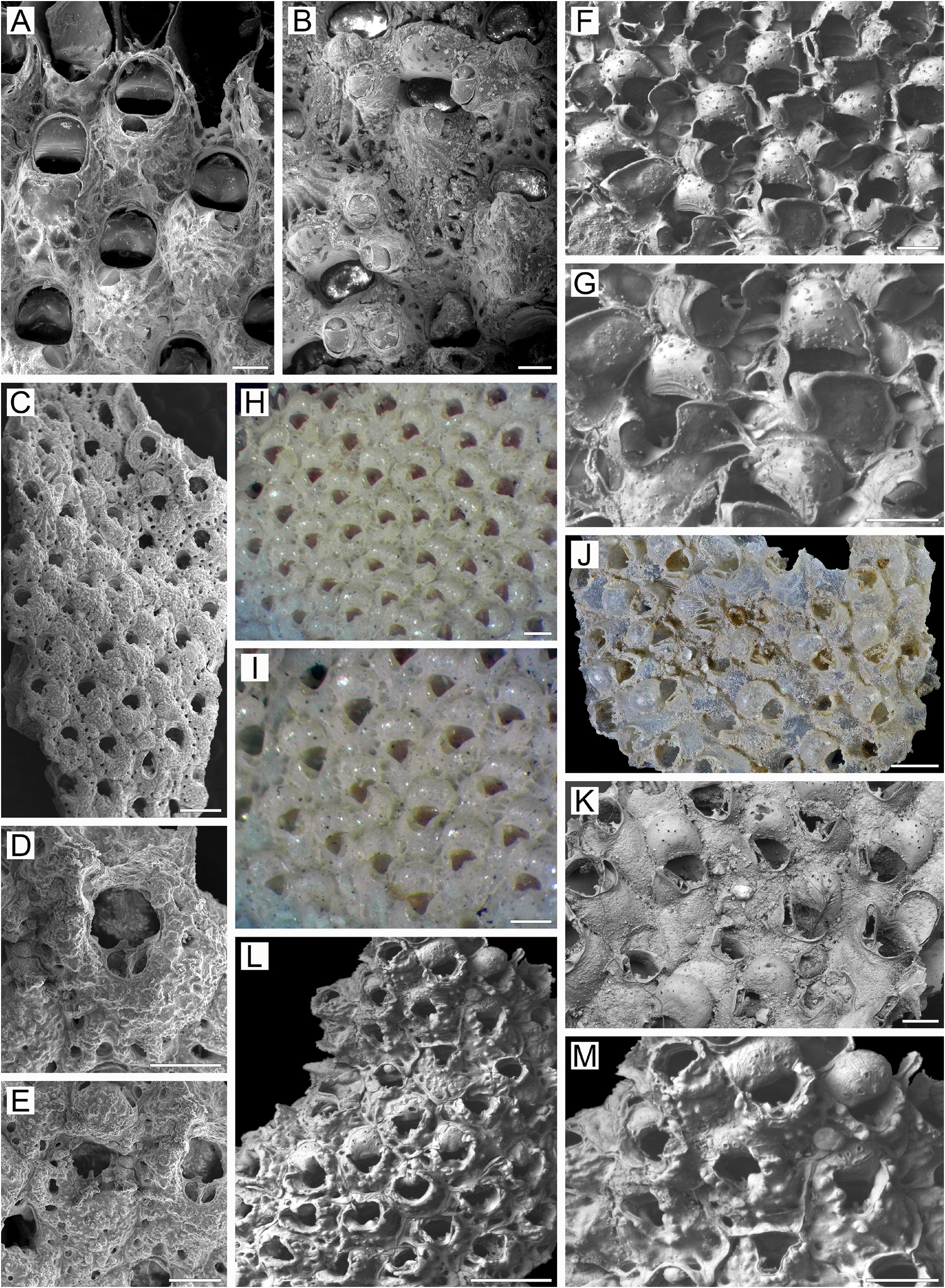
( Figs 17 View FIGURE 17 , 32J, K View FIGURE 32 )
Cellepora plicata Smitt, 1868a, p. 30 , 31 (part), pl. 28, figs 189, 190 (?).
Cellepora plicata: Smitt 1868b, p. 484 , 485.
Discopora plicata :? Smitt 1878a, p. 31;?1878b, p. 24; Nordgaard 1918, p. 78.
Rhamphostomella plicata: Lorenz 1886, p. 12 View in CoL , 13; Nordgaard 1905, p. 171, pl. 5, figs 14, 15; 1906, p. 30, 41, pl. 4, figs 49, 50; Gostilovskaya 1978, p. 230, fig. 146; Kluge 1962, p. 544, fig. 381; 1975, p. 662, fig. 381; Powell 1968a, p. 2312, fig. 10, pl. 13b; Winston & Hayward 2012, p. 121, fig. 78.
Rhamphostomella lorenzi Kluge, 1907, p. 188 .
Rhamphostomella lorenzi: Kluge 1915, p. 386 .
Additional references. Rhamphostomella plicata: Osburn 1936, p. 542 View in CoL ; Gostilovskaya 1957, p. 456; 1964, p. 220; Kluge 1961, p. 142; Denisenko 1984, p. 76; 1988, p. 13; 1990, p. 39; 2008, p. 188; 2013, p. 184; Gontar & Denisenko 1989, p. 357; Gontar et al. 2001, p. 195; Grischenko 2002, p. 115; Kuklinski 2002b, p. 203; Denisenko & Kuklinski 2008, p. 48; Kuklinski & Taylor 2009, p. 497; Gontar 2010, p. 153; Denisenko et al. 2016, p. 366.
Material examined. Lectotype: SMNH-Type-1696, two fragments of one colony, Swedish Arctic Expedition , August 1861, Waygat Islands, Hinlopen Strait, Svalbard and Jan Mayen, 79°10.0ʹ N, 19°00.0ʹ E, depth 110–146 m, mud. GoogleMaps
SMNH- 132277 , colony on bivalve shell, Sandeberg Expedition, Stn 28, 1877, off Waideguba, Kola Peninsula, Barents Sea, Russia, depth 73–100 m, gravel and shells. NHMUK 1911.10.1.1590 About NHMUK , five colonies encrusting hydroid stolons and spirorbid tubes, ex Copenhagen Museum Collection, from G.M. R. Levinsen, A.M. Norman Collection, Greenland. USNM (no inventory number), two colony fragments, Thacher Island, Gulf of Maine, Massachusetts, USA, northwestern Atlantic Ocean .
Measurements. NHMUK 1911.10.1.1590, Greenland ( Fig. 17A, D–G, I–J, L–M View FIGURE 17 ). ZL, 0.82–1.20 (0.99 ± 0.11). ZW, 0.37–0.60 (0.47 ± 0.05). ZD, 0.35–0.41 ( n = 2). OrL, 0.21–0.28 (0.24 ± 0.03). OrW, 0.25–0.33 (0.31 ± 0.02). OeL, 0.25–0.38 (0.33 ± 0.03). OeW, 0.32–0.45 (0.39 ± 0.03). Av(s)L, 0.15–0.28 (0.23 ± 0.04). P(m)N, 5–10 (7). P(oe)N, 11–19 (16) ( n = 10).
Description. Colonies encrusting, multiserial, unilaminar ( Fig. 17A View FIGURE 17 ), small, irregular in form, attaining about 14 mm in maximal size, beige to greyish when dry. Zooids large, oblong-rectangular to oval, or trapezoidal and irregular, in shape, often swollen and broadened distally, flattened and tapering proximally, arranged in quincunx, demarcated by fine sutures between lateral and transverse walls; sutures less visible in older parts of colony.
Frontal shield umbonuloid ( Fig. 17A, I View FIGURE 17 ), thin, moderately convex, lower towards proximal margin, smooth in developing zooids ( Fig. 17A View FIGURE 17 ), finely dimpled to wrinkled or reticulated in fully-formed zooids ( Fig. 17D–H View FIGURE 17 ). Areolae sparse, elongate to triangular, separated by short interareolar ridges; ridges generally developed in distal half of zooid ( Fig. 17D–G View FIGURE 17 ), may be strongly reduced or absent in young zooids ( Fig. 17A, D, E View FIGURE 17 ). Interior of frontal shield ( Fig. 17I, L View FIGURE 17 ) showing distinct or indistinct ring scar ( Fig. 17I, L View FIGURE 17 ). Umbonuloid component occupying about 40% of length of frontal shield (38% in one measured zooid), with fine parallel lineation and accretionary banding.
Primary orifice ( Fig. 17B, J View FIGURE 17 ) submerged, inversely pyriform, subcircular to oval; distal and lateral margins formed by upper terminal part of distal transverse wall. Distal margin of orifice rounded, proximal margin concave, tapering, with small median lyrula having straight, bifurcate or alate apex, and two small, short processes lateroproximally with rounded or pointed tips; processes sometimes ill-defined ( Fig. 17F View FIGURE 17 ) or lacking ( Fig. 17I View FIGURE 17 ). No condyles.
Secondary orifice ( Fig. 17D–H View FIGURE 17 ) circular or broadly triangular in outline, cormidial; in contrast with most species studied, distally and distolaterally restricted by flared, thickened upper part of transverse wall of maternal zooid. Proximal and lateral walls of daughter and distolateral zooids adjoining this wall ( Fig. 17D, E View FIGURE 17 ). Secondary orifice formed laterally and proximally by thin-walled, flared peristome of two triangular lappets from frontal shield, one lappet incorporating or virtually replaced by cystid of suboral avicularium; flared lappet on opposite side of peristome, plus avicularian cystid, defining broad, deep V- to U-shaped pseudosinus in secondary orifice ( Fig. 17D–G View FIGURE 17 ). Distally, lappets inclining to vertical walls of distolateral zooids, abutting to proximolateral corners of ooecia in ovicellate zooids. No oral spines.
Cystid of suboral avicularium small, quite elevated, with dimpled surface and one communication pore, asymmetrically placed proximal to orifice on left or right ( Fig. 17A–H View FIGURE 17 ). Avicularian frontal surface (rostral/ postmandibular areas) to one side of midline, but sometimes crossing it, facing obliquely frontally. Rostrum elongatelingulate, narrowing, with blunt tip, extending somewhat over orifice, directed distolaterally and frontally. Palate elongate, triangular, with rounded distal end; foramen elongate triangular or elongate oval, with cryptocystal shelf distally; opesia round triangular ( Fig. 17A–C, F–H View FIGURE 17 ). Crossbar complete. Suboral avicularia lacking in some zooids.
No adventitious avicularia.
Ovicells hyperstomial ( Fig. 17F–H View FIGURE 17 ), ooecium free of secondary calcification except for very narrow basal rim visible in some ooecia ( Fig. 17G, H View FIGURE 17 ). Ooecium formed by distal autozooid close to colony periphery. Ooecial coelomic cavity connecting to visceral coelom via communication canal opening on underside of proximal part of frontal shield as oval slit-like communication pore situated at apex of triangular area (ovicell floor) or halfway between transverse wall and ring scar ( Fig. 17I View FIGURE 17 ). Ooecium smooth, with straight or insignificantly concave proximal margin and small, scattered, circular or irregular pseudopores.
Zooids interconnected by two mural pore chambers in each distolateral wall ( Fig. 17M View FIGURE 17 ). In transverse walls, communication pores as horizontal “band” or two multiporous septula.
Basal wall of zooids fully calcified, smooth, flattened or slightly convex, with tubular protuberances (up to 0.60 mm long, 0.20–0.33 mm in diameter), textured by fine parallel lineation on surface ( Fig. 17K View FIGURE 17 ). Numerous white spots (presumably less-calcified areas) visible in semitransparent basal wall by light microscopy. Boundaries between zooids recognizable basally by sinuous sutures.
Ancestrula and early astogeny not observed.
Remarks. In describing the new species Cellepora plicata, Smitt (1868a) indicated figures 189–196 (pl. 28) as illustrations. Comparing them with his own specimens, Lorenz (1886) distinguished three species ( Rhamphostomella spinigera , R. radiatula and R. plicata ) and indicated that only figures 189–191 and 195 of Smitt represent R. plicata . Nonetheless, only figure 189 unambiguously illustrates this species (figure 190 shows the colony basal surface). As to the other illustrations, figure 191 presumably shows R. bilaminata ; figure 192, R. spinigera ; figure 193, R. radiatula ; and figures 195, 196, R. hincksi (see additional discussion in the Remarks elsewhere in the text).
Nordgaard (1906, p. 31) wrote of R. plicata , “This species is distinguishable from the next one ( R. hincksi ) by the circumstance that the proximal margin of the oral aperture is more rounded, the aperture has not so marked a triangular shape as is the case with hincksi . The most conspicuous difference, however, is that plicata has a distinct median denticle [lyrula] that is absent in hincksi ”. We can also add the strong difference in the frontal shield relief in these species.
Ecology. Rhamphostomella plicata is known from 12–146 m depth on mixed bottoms (sand, shell, gravel), where colonies encrust mollusk shells, ascidians and colonies of other bryozoans.
Distribution. This is a boreal-Arctic, circumpolar, sublittoral species. Arctic records include the Barents Sea ( Smitt 1868 a, 1868b, 1879 b; Bidenkap 1900a; Nordgaard 1905; Kuznetsov 1941; Kluge 1962, 1975; Denisenko 1984, 1988, 1990), White Sea ( Kluge 1907; Gostilovskaya 1957, 1978), Kara Sea ( Nordgaard 1912; Kluge, 1962, 1975), Laptev Sea ( Gontar & Denisenko 1989), Chukchi Sea ( Kluge 1962, 1975; Denisenko 2008; Denisenko & Kuklinski 2008; Gontar 2010), Canadian Arctic Archipelago ( Nordgaard 1906; Osburn 1936), Baffin Bay ( Gontar & Denisenko 1989), Davis Strait ( Kluge 1962, 1975), western Greenland ( Norman 1906; Levinsen 1914; Osburn 1919, 1936; Denisenko & Blicher 2021), eastern Greenland ( Levinsen 1916; Denisenko & Blicher 2021) [ Smitt (1868b) just mentioned Greenland], Iceland ( Nordgaard 1924), Franz Josef Land ( Denisenko 1990), Spitsbergen ( Smitt 1868b; Gontar et al. 2001; Kuklinski 2002b; Kuklinski & Taylor 2009) and northern Norway ( Nordgaard 1905, 1918). In the northwestern Atlantic, R. plicata has been reported from St Lawrence Gulf ( Whiteaves 1901) and in the Gulf of Maine near Thacher Island ( Winston & Hayward 2012; our data). The only record from the northeastern Atlantic is from the Faroe Islands ( Denisenko et al. 2016). In the northwestern Pacific, it is documented in the Bering Sea between St Lawrence Island and Chukotskiy Cape ( Kluge 1961; Grischenko 2002), and along the eastern coastal waters of Sakhalin Island, Sea of Okhotsk ( Kluge et al. 1959; Kluge 1961).

| R |
Departamento de Geologia, Universidad de Chile |
| USNM |
Smithsonian Institution, National Museum of Natural History |
No known copyright restrictions apply. See Agosti, D., Egloff, W., 2009. Taxonomic information exchange and copyright: the Plazi approach. BMC Research Notes 2009, 2:53 for further explanation.
|
Kingdom |
|
|
Phylum |
|
|
Class |
|
|
Order |
|
|
SubOrder |
Flustrina |
|
SuperFamily |
Lepralielloidea |
|
Family |
|
|
Genus |
Rhamphostomella plicata (Smitt, 1868)
| Grischenko, Andrei V., Gordon, Dennis P., Taylor, Paul D., Kuklinski, Piotr, Denisenko, Nina V., Spencer-Jones, Mary E. & Ostrovsky, Andrew N. 2022 |
Rhamphostomella lorenzi :
| Kluge, G. A. 1915: 386 |
Rhamphostomella lorenzi
| Kluge, G. A. 1907: 188 |
Rhamphostomella plicata :
| Winston, J. E. & Hayward, P. J. 2012: 121 |
| Gostilovskaya, M. G. 1978: 230 |
| Powell, N. A. 1968: 2312 |
| Kluge, G. A. 1962: 544 |
| Nordgaard, O. 1905: 171 |
| Lorenz, L. von 1886: 12 |
Discopora plicata
| Nordgaard, O. 1918: 78 |
| Smitt, F. A. 1878: 31 |
Cellepora plicata Smitt, 1868a , p. 30
| Smitt, F. A. 1868: 30 |
Cellepora plicata : Smitt 1868b , p. 484
| Smitt, F. A. 1868: 484 |