
Rhamphostomella radiatula ( Hincks, 1877 )
|
publication ID |
https://doi.org/ 10.11646/zootaxa.5131.1.1 |
|
publication LSID |
lsid:zoobank.org:pub:CF550031-D6A9-48A3-A953-A1BD40C72F5E |
|
DOI |
https://doi.org/10.5281/zenodo.7628944 |
|
persistent identifier |
https://treatment.plazi.org/id/03892374-0B0B-3302-FF73-AFF61AA4FE3D |
|
treatment provided by |
Plazi |
|
scientific name |
Rhamphostomella radiatula ( Hincks, 1877 ) |
| status |
|
Rhamphostomella radiatula ( Hincks, 1877) View in CoL
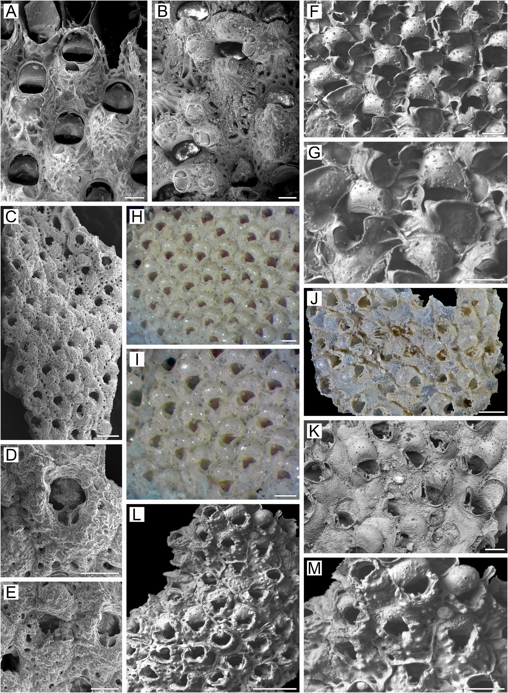
( Figs 18 View FIGURE 18 , 32L, M View FIGURE 32 )
Cellepora plicata: Smitt 1868a, p. 30 , 31 (part), pl. 28, fig. 193.
Lepralia radiatula Hincks, 1877, p. 104 , pl. 10, figs 9–14.
Rhamphostomella radiatula: Lorenz 1886, p. 13 View in CoL , pl. 7, fig. 9 (mentioned as fig. 10 in the text); Nordgaard 1905, p. 172, pl. 5, figs. 16, 17; Kluge 1962, p. 543, fig. 380; 1975, p. 660, fig. 380; Gostilovskaya 1978, p. 229, fig. 145; Androsova 1958, p. 173, fig. 104; Winston & Hayward 2012, p. 124, fig. 79; Taylor 2021, p. 76, fig. 5a–e.
Discopora radiatula: Nordgaard 1918, p. 78 .
Additional references. Rhamphostomella radiatula: Nordgaard 1906, p. 32 View in CoL , 41; 1924, p. 9; Kluge 1907, p. 196; 1908a, p. 534; 1928, p. 257; 1961, p. 142; 1964, p. 190; Gostilovskaya 1957, p. 455; Kluge et al. 1959, p. 213; Hansen 1962, p. 40; Gontar 1980, p. 13; 1992, p. 198; 1993b, p. 202; Mawatari & Mawatari 1981, p. 56; Denisenko 1988, p. 13; 1990, p. 39; 2013, p. 184; Gontar & Denisenko 1989, p. 354; Bennike et al. 1994, p. 199; Grishankov 1995, p. 48; Grischenko 1997, p. 175; 2002, p. 115; 2003b, p. 237; Gontar et al. 2001, p. 195; Kuklinski 2002b, p. 203; Ostrovsky 2009, p. 206, fig. 78b; 2013, p. 8, fig. 2.41b.
Material examined. Neotype: NHMUK 1911.10 View Materials .1.1592, two fragments from one colony, ex Copenhagen Museum Collection, from G.M. R. Levinsen, A.M. Norman Collection, Iceland.
NHMW 72987 View Materials , one colony fragment, L. Lorenz Collection, II Austro-Hungarian Polar Expedition , 1882–1883, Jan Mayen, depth 20–130 m, collector F. Fischer. NHMUK 1899.5 View Materials .1.878, six colony fragments, no locality given, T. Hincks Collection . ZIRAS 19 /50114, two colony fragments , KIENM Collection , Stn 132, 23 July 1992, Rock Sivuchy Kamen , coastal waters of Medny Island , Commander Islands, Bering Sea, 54°47.4ʹ N, 167°39.3ʹ E, depth 10 m GoogleMaps , SCUBA, collector V.I. Shalukhanov .
Additional material. 157 colonies and colony fragments. IMB Collection (1973) Stns 149/384, 229/587; (2011) Stn 27/22; PIBOC Collection (1991) Stns 14, 17, 18, 19, 20, 41; KIENM Collection (1992) Stns 3, 5, 7, 20, 27, 28, 29, 30, 32, 34, 38, 43, 44, 46, 56, 58, 61, 63, 65, 66, 67, 68, 69, 70, 72, 75, 79, 81, 88, 94, 96, 97, 99, 110, 111, 116, 119, 121, 128, 129, 130, 132, 136, 144, 145, 146, 147, 148; A. V. Grischenko Collection (1992) Stns 7, 8 (see Appendix 1 for details).
Measurements. ZIRAS 19/50114, Medny Island, Commander Islands, Bering Sea ( Fig. 18A–M View FIGURE 18 ). ZL, 0.48– 0.77 (0.61 ± 0.07). ZW, 0.35–0.53 (0.41 ± 0.05). ZD, 0.41–0.58 (n = 2). OrL, 0.15–0.27 (0.21 ± 0.03). OrW, 0.18– 0.28 (0.22 ± 0.02). OeL, 0.20–0.25 (0.22 ± 0.01). OeW, 0.22–0.35 (0.28 ± 0.04). Av(s)L, 0.05–0.09 (0.07 ± 0.01) (n = 10). P(m)N, 12–19 (17) (n = 20). P(oe)N, 4–10 (8) (n = 20).
Description. Colonies encrusting, multiserial, unilaminar ( Fig. 18A View FIGURE 18 ), more or less circular, attaining 14 mm in maximal dimension, bright pink-reddish to brown-yellow when alive, light brown to pink when dry. Zooids small, hexagonal, trapezoid, oval to pyriform, arranged in checkered pattern, demarcated by fine sinuous sutures between lateral and transverse walls; sutures less visible in older parts of colony.
Frontal shield umbonuloid ( Fig. 18A, D–G, I View FIGURE 18 ), strongly thickened, convex, smooth in youngest zooids, normally with numerous tubercles of differing form and size, concentrated around secondary orifice and also distributed over entire frontal surface ( Fig. 18D–H View FIGURE 18 ). Large, deep circular areolae along zooidal margins, separated by short, narrow (in young zooids) to thick (in older zooids) interareolar ridges. Secondary calcification may be strongly developed, resulting in general thickening of frontal shield and enlargement of tubercles ( Fig. 18E View FIGURE 18 ). Umbonuloid component occupying about 60% of length of frontal shield (62% in one measured zooid), with accretionary banding ( Fig. 18I, J, L View FIGURE 18 ). Ring scar discrete ( Fig. 18I, L View FIGURE 18 ).
Primary orifice ( Fig. 18B, I View FIGURE 18 ) deeply submerged, circular to oval; distal and lateral margins formed by upper, terminal part of distal transverse wall ( Fig. 18A View FIGURE 18 ). Distal margin of orifice rounded, proximal margin concave, tapering, with prominent, conical, acute median lyrula, curving in frontal direction ( Fig. 18A, B, G, I, J View FIGURE 18 ). Condyles absent.
Secondary orifice ( Fig. 18D, G View FIGURE 18 ) irregularly oval or broadly triangular in outline, with narrowly sinuate, Ushaped proximal margin, cormidial; distally restricted by vertical thickening of proximal wall of daughter zooid ( Fig. 18D, E View FIGURE 18 ); proximally formed by thin-walled, low peristome of two lappets from frontal shield, one of which incorporates cystid of suboral avicularium (on left or right side). In ovicellate zooids, peristomial lappets connecting with ooecium, overgrowing its proximal surface toward each other and forming incomplete circle ( Fig. 18F–H View FIGURE 18 ). No oral spines.
Cystid of suboral avicularium small, with tubercular surface and one communication pore ( Fig. 18D, F View FIGURE 18 ), situated medially and transversely relative to zooidal midline, within pseudosinus of secondary orifice. Avicularian frontal surface (rostral/postmandibular areas) concave, crossing zooidal midline, facing obliquely distally. Rostrum short, curved, with tip hooked in profile, directed laterally to distolaterally and upwards ( Fig. 18C, D, F–I View FIGURE 18 ), sometimes concealed within peristome and barely visible frontally. Palate short, lingulate to broadly triangular, foramen elongate oval; palatal face at right angle to postmandibular area, with semicircular opesia. Crossbar complete.
No adventitious avicularia.
Ovicells cleithral, initially hyperstomial, soon becoming subimmersed and later appearing endozooidal when only frontal area of ooecium remains visible ( Fig. 18F, G, M View FIGURE 18 ), being sunken in secondary calcification originating from frontal shields of distal and distolateral zooids. Ooecium formed by distal autozooid, ooecial fold arising on colony periphery concurrently with formation of frontal shield of distal zooid ( Fig. 18A View FIGURE 18 ). Ooecium with straight proximal margin and scattered circular, oval to slit-like pseudopores, some occluded by secondary calcification.
Zooids interconnected by three mural pore chambers ( Fig. 18M View FIGURE 18 ) in each distolateral wall and multiporous septula in transverse walls, sometimes with buttressed recesses.
Basal surface of zooids ( Fig. 18K View FIGURE 18 ) fully calcified, inflated, rough, textured by irregular lineation, without protuberances. Boundaries between zooids unrecognizable basally.
Ancestrula and early astogeny not observed.
Remarks. Smitt (1868a, pl. 28, fig. 193) initially illustrated R. radiatula under the name Cellepora plicata , together with three other species. Hincks (1877) described R. radiatula as a separate species (in Lepralia ) from Iceland and Labrador, and Lorenz (1886) moved it to Rhamphostomella . Although we found six fragments of this species on a slide in the T. Hincks Collection housed in the Natural History Museum, London (NHMUK 1899.5.1.878, with the inscription “? Part of type material”), they are in poor condition and cannot be used for identification or comparison. Accordingly, we selected a neotype from Iceland from the A.M. Norman Collection, also in Natural History Museum, London.
The following combination of characters distinguishes R. radiatula from congeners: 1) suboral and frontal tubercles present, 2) small zooidal size, and 3) the right-angled aspect of the avicularian frontal surface.As discussed above, the only congener having a similarly right-angled frontal face to the suboral avicularium is R. alutacea . That species has a broad pseudosinus in the secondary orifice, in contrast to the narrow pseudosinus in R. radiatula , and the frontal surface of the avicularium is well visible in frontal view compared to R. radiatula , in which the avicularian frontal surface is barely visible frontally.
Kluge (1962, 1975) described the primary orifice of Arctic R. radiatula as having a pair of small triangular points on the median lyrula. We did not observe these in our material.
Kluge (1962, 1975) indicated that colonies of R. radiatula loosely overgrow the substratum. In contrast, our observations of a large number of specimens show that colonies are tightly cemented to a variety of hard substrata (barnacles, shells of molluscs, stones, etc.). In most cases, the basal wall of colonies conforms to irregularities in the substratum microtopography, resulting in very uneven patterning of the basal surface ( Fig. 18K View FIGURE 18 ).
Ecology. Rhamphostomella radiatula has been found at depths of 2–280 m, predominantly on hard bottoms— rocks (including vertical surfaces and crevices), boulders, blocks, and gravel—and scattered on mixed seabeds with sand or silt overlain with broken shells. In addition to rocky surfaces, colonies encrust bivalve mollusc shells, ascidians, colonies of other bryozoans ( Flustrellidra gigantea , Tricellaria beringia , Celleporina robertsoniae ), polychaete tubes, sponges, barnacles and occasionally holdfasts of brown and red algae, and exists as a part of cryptic community inhabiting the cavities formed by the crustose coralline alga Clathromorphum nereostratum .
Distribution. This is a boreal-Arctic, circumpolar, sublittoral species. In the Arctic R. radiatula has been reported from the Barents Sea ( Nordgaard 1896, 1918; Bidenkap 1900a, 1900b; Kluge 1906, 1915, 1962, 1975; Denisenko 1988, 1990), White Sea ( Gostilovskaya 1957, 1978; Grishankov 1995; Ostrovsky 2009, 2013), Kara Sea ( Nordgaard 1912; Kluge 1962, 1975; Denisenko 2021), Canadian Arctic Archipelago ( Nordgaard 1906; Osburn 1932), Baffin Bay ( Hansen 1962), Davis Strait ( Hansen 1962; Kluge 1962, 1975), Labrador ( Hincks 1877; Gontar & Denisenko 1989), western Greenland ( Levinsen 1914; Denisenko & Blicher 2021), eastern Greenland ( Levinsen 1914, 1916; Denisenko & Blicher 2021), Jan Mayen Island ( Lorenz 1886; Nordgaard 1907a), Iceland ( Hincks 1877; Nordgaard 1924), Franz Josef Land ( Denisenko 1990), Spitsbergen ( Gontar et al. 2001; Kuklinski 2002b) and northern Norway ( Smitt 1868a; Nordgaard 1905, 1918). In the northwestern Atlantic it has been reported from St Lawrence Gulf ( Kluge 1962, 1975) and in the Gulf of Maine ( Winston & Hayward 2012). In the northwestern Pacific there are records from Africa Cape, eastern Kamchatka ( Kluge 1961; Grischenko 2002), Commander Islands, Bering Sea ( Kluge 1961; Grischenko 1997, 2002, 2003b; our data); Urup, Chiproy and Simushir Islands of the middle Kuril Islands, Sea of Okhotsk ( Gontar 1980, 1993b; our data); western coastal waters of southern Sakhalin Island ( Androsova 1958; Kluge et al. 1959) and Moneron Island in the Sea of Japan ( Kluge 1961), and the Pacific coast of Hokkaido ( Mawatari & Mawatari 1981). In the northeastern Pacific it occurs in the Gulf of Alaska near Kodiak Island (our data).
Bennike et al. (1994) documented R. radiatula from middle Pleistocene deposits in central western Greenland. Taylor (2021) reported this species from Pleistocene deposits of Scotland.

| R |
Departamento de Geologia, Universidad de Chile |
| T |
Tavera, Department of Geology and Geophysics |
| V |
Royal British Columbia Museum - Herbarium |
No known copyright restrictions apply. See Agosti, D., Egloff, W., 2009. Taxonomic information exchange and copyright: the Plazi approach. BMC Research Notes 2009, 2:53 for further explanation.
|
Kingdom |
|
|
Phylum |
|
|
Class |
|
|
Order |
|
|
Family |
|
|
Genus |
Rhamphostomella radiatula ( Hincks, 1877 )
| Grischenko, Andrei V., Gordon, Dennis P., Taylor, Paul D., Kuklinski, Piotr, Denisenko, Nina V., Spencer-Jones, Mary E. & Ostrovsky, Andrew N. 2022 |
Rhamphostomella radiatula:
| Nordgaard, O. 1905: 172 |
| Lorenz, L. von 1886: 13 |
Lepralia radiatula
| Hincks, T. 1877: 104 |
Cellepora plicata: Smitt 1868a , p. 30
| Smitt, F. A. 1868: 30 |