
Rhamphostomella hincksi Nordgaard, 1906
|
publication ID |
https://doi.org/ 10.11646/zootaxa.5131.1.1 |
|
publication LSID |
lsid:zoobank.org:pub:CF550031-D6A9-48A3-A953-A1BD40C72F5E |
|
DOI |
https://doi.org/10.5281/zenodo.7626600 |
|
persistent identifier |
https://treatment.plazi.org/id/03892374-0B04-330B-FF73-ACCF1997FC7D |
|
treatment provided by |
Plazi |
|
scientific name |
Rhamphostomella hincksi Nordgaard, 1906 |
| status |
|
Rhamphostomella hincksi Nordgaard, 1906 View in CoL
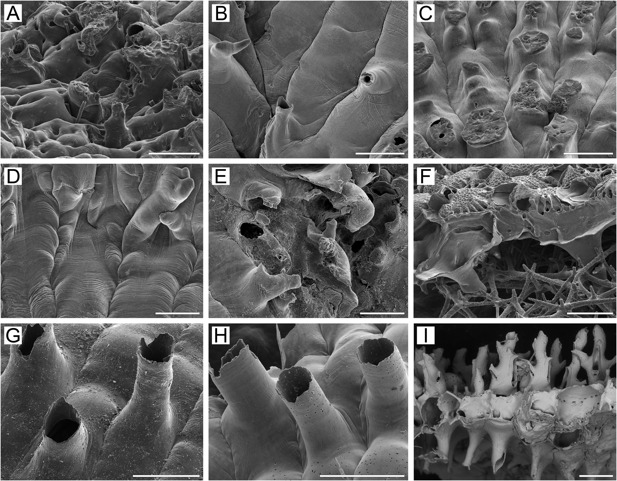
( Figs 15 View FIGURE 15 , 30G View FIGURE 30 , 32H, I View FIGURE 32 )
? Cellepora plicata Smitt, 1868a, p. 30 , 31 (part), pl. 28, figs 195, 196.
Cellepora plicata: Hincks 1877, p. 106 , pl. 11, figs 3, 4.
Ramphostomella [sic] hincksi Nordgaard, 1906, p. 31 , 41, pl. 4, fig. 51.
Rhamphostomella hincksi: Kluge 1962, p. 541 View in CoL , fig. 378; 1975, p. 658, fig. 378; Powell 1968a, p. 2311, fig. 10, pl. 13a; Hayami 1970, p. 332, pl. 36, fig. 1.
Additional references. Rhamphostomella hincksi: Osburn 1955, p. 38 View in CoL ; Hansen 1962, p. 40; Hayami 1975, p. 89; Sakagami et al. 1980, p. 330; Gontar 1980, p. 18; 1990, p. 133; 2010, p. 153; 2013, p. 184; Gontar & Denisenko 1989, p. 357; Denisenko 1990, p. 39; 2008, p. 187; Kuklinski 2002b, p. 203; Denisenko & Kuklinski 2008, p. 48; Foster 2010, p. 57.
Material examined. Neotype: NHMUK 1976.8.6.39pt, three fragments from one colony, RV Ernest Holt , Stn 41, 74°25.0ʹ N, 18°02.0ʹ E (about 22 km westwards from Medvezhii Island, western Barents Sea), depth 128 m. GoogleMaps
NHMUK 68.3 About NHMUK .13.46, one colony, 1858, Spitsbergen , collectors O. Sorella and N. Nordenskjold. NHMUK 1877.11 About NHMUK .28.112, two colonies encrusting pieces of the same bivalve shell, A.M. Norman Collection, HMS Valorous, 1875, Davis Strait . NHMUK 1899.5 About NHMUK .1.876, two colony fragments , T. Hincks Collection , Labrador . NHMUK 1963.2 About NHMUK .12.244, three colony fragments, no locality given, Dundee Collection . NHMW 72986 View Materials , one colony, 1884, L. Lorenz Collection, II Austro-Hungarian Polar Expedition , 1882–1883, Jan Mayen, depth 160–180 m, collector F. Fischer. NHMW 92534 View Materials (=1884.II.48), one colony fragment, L. Lorenz Collection, II Austro-Hungarian Polar Expedition , 1882–1883, Jan Mayen, depth 160–180 m, collector F. Fischer. USNM 11130 About USNM , nine colony fragments, Arctic Research Laboratory Collection ,? August 1948, Point Barrow, Alaska, Beaufort Sea, depth 55.5 m, collector G.E. MacGinitie. ZIRAS 7 /50119, two colony fragments detached from broken shells of bivalve mollusc Chlamys sp ., MFRT Rodino , 12 September 1992, about 32 km from Cape Hayryuzova , western Kamchatka shelf, Sea of Okhotsk, 57°36.2ʹ N, 156°09.0ʹ E, depth 78–81 m, crab trap, collector A GoogleMaps . V. Grischenko .
Measurements. ZIRAS 7/50119, western Kamchatka, Sea of Okhotsk ( Fig. 15A–G, I, K View FIGURE 15 ). ZL, 0.77–1.35 (1.00 ± 0.14). ZW, 0.37–0.60 (0.50 ± 0.06). ZD, 0.43–0.55 (n = 2). OrL, 0.15–0.28 (0.22 ± 0.03). OrW, 0.22–0.35 (0.29 ± 0.04). OeL, 0.28–0.32 (0.31 ± 0.01). OeW, 0.33–0.40 (0.37 ± 0.02). Av(s)L, 0.15–0.27 (0.20 ± 0.03). P(m)N, 7–13 (10). P(oe)N, 18–26 (25) (n = 10).
Description. Colonies encrusting, multiserial, unilaminar ( Fig. 15A View FIGURE 15 ), more or less circular, attaining 16 mm in maximal dimension, reddish or burgundy when alive, pink when dry. Zooids large, hexagonal ( Fig. 15D View FIGURE 15 ), widest at midlength, arranged in regular, straight rows, packed in quincunx; demarcated by fine, undulating sutures between lateral and transverse walls; sutures visible in both young and old parts of colony.
Frontal shield umbonuloid ( Fig. 15D, E, I View FIGURE 15 ), inflated or moderately convex, smooth to weakly dimpled centrally, with series of deep areolae along zooidal margins ( Fig. 15D–G View FIGURE 15 ) separated by radially arranged interareolar ridges; in younger zooids, ridges relatively short, low, some connecting with cystid of suboral avicularium ( Fig. 15A, D, E View FIGURE 15 ). In older zooids, ridges tall, thickened, elongate, often joining along zooid midline and connecting to peristomial lappet and avicularian cystid, giving strongly costate appearance to frontal shield ( Fig. 15F, G View FIGURE 15 ). Interior of frontal shield ( Fig. 15I View FIGURE 15 ) with discrete ring scar ( Fig. 15K View FIGURE 15 ). Umbonuloid component occupying about 40% of length of frontal shield (44% in one measured zooid), with fine parallel lineation and accretionary banding.
Primary orifice submerged, irregularly round; rounded distally, sinuate or bisinuate ( Fig. 15B View FIGURE 15 ) proximally ( Fig. 15A, B, I View FIGURE 15 ); if bisinuate, with small process ( Fig. 15B View FIGURE 15 ). Distal and lateral margins of primary orifice formed by upper terminal part of distal transverse wall.
Secondary orifice ( Fig. 15C–E View FIGURE 15 ) broadly triangular in outline, cormidial, distally and distolaterally restricted by thickening of vertical walls of distal and distolateral zooids, laterally and proximally formed by avicularian cystid (often with small distal lappet on its rostrum) on one side and high lappet of frontal shield on opposite side; lappet triangular, straight, slightly concave or sinuous in profile, together with avicularium forming proximally broad deep V-shaped pseudosinus in secondary orifice ( Fig. 15C–H View FIGURE 15 ). Distally, lappets connect with lateral walls of distolateral zooids; in ovicellate zooids, lappets not fused with proximolateral corners of ooecium. No oral spines.
Cystid of suboral avicularium ( Fig. 15A–H View FIGURE 15 ) relatively small but distinct, bulbous, strongly elevated, with coarsely dimpled surface, and 1–3 (normally 2) communication pores connecting avicularian and hypostegal coeloms, asymmetrically placed to left or right side of proximal peristomial rim. Inclined frontal surface (rostral/ postmandibular areas) of avicularium converging toward or crossing zooidal midline, facing obliquely frontally. Rostrum oblong-oval, weakly curving inward, with small, hooked tip directed laterally to distolaterally and upwards, extending somewhat over orifice ( Fig. 15C, H View FIGURE 15 ). Palate semielliptical to triangular, with rounded distal end; palatal foramen elongate-oval or triangular, with rounded angles; opesia semicircular. Crossbar complete.
No adventitious avicularia.
Ovicells initially hyperstomial ( Fig. 15H View FIGURE 15 ), but ooecia rapidly becoming subimmersed by peripheral overgrowth of secondarily thickened lateral and proximal walls of distolateral and daughter zooids ( Fig. 15F, G View FIGURE 15 ); thickened lateral walls plugging gaps between distal margins of peristomial lappets and proximal corners of ooecium, thus completing secondary orifice in ovicellate zooids ( Fig. 15F, G View FIGURE 15 ). Ooecium formed by distal autozooid; ooecial fold arises on colony periphery concurrently with frontal shield of distal zooid. Ooecial coelomic cavity connected with visceral coelom via communication canal opening on underside of proximal part of frontal shield as small, curved slit-like communication pore close to transverse wall ( Fig. 15I View FIGURE 15 ). Ooecium with slightly concave proximal margin and numerous small, scattered circular and oval (sometimes irregular) pseudopores.
Zooids interconnecting by two mural pore chambers in each distolateral wall ( Fig. 15L View FIGURE 15 ). Communication pores in basal part of transverse walls arranged either as horizontal “band” or forming two multiporous septula.
Basal wall of zooids ( Figs 15J View FIGURE 15 , 30G View FIGURE 30 ) fully calcified, smooth, slightly convex, with tubular protuberances (up to 0.47 mm long, up to 0.28 mm in diameter). Boundaries between zooids indicated basally by gently sinuous incisions.
Ancestrula and early astogeny not observed.
Remarks. Described and illustrated by Hincks (1877) as Cellepora plicata from Iceland, R. hincksi was recognized and redescribed as a separate species by Nordgaard (1906) based on specimen from the Barents Sea. Still, it is rather possible that figures 195 and 196 of Smitt (1868a, pl. 28) show the same species. Regrettably, only a tiny, poorly preserved fragment of the presumed R. hincksi survived in Nordgaard’s collection in the Natural History Museum, University of Oslo (E. Di Martino, pers. comm., 2020). To correct this situation, we have selected a neotype for this species based on a specimen collected in the Barents Sea from the RV Ernest Holt. Three fragments of one colony are deposited at the Natural History Museum, London.
In having a sinuate, elevated secondary orifice formed by an asymmetrically set avicularian cystid on one side and a high triangular lappet on the opposite side, and spherical ooecia with small, evenly distributed pseudopores, R. hincksi strongly resembles R. plicata (Smitt, 1868) . Historically, this resemblance led to some misidentifications.
The differences between these species are as follows: 1) the frontal shield has a series of deep marginal areolae separated by tall, radially arranged ridges along the entire lateral wall in R. hincksi , but only a few areolae separated by short ridges along the distal half of the zooid in R. plicata ; 2) the palatal foramen of the suboral avicularium is gently curved in R. hincksi but straight in R. plicata ; 3) ooecia are rapidly surrounded by growing and thickening vertical walls of neighbouring zooids in R. hincksi , but not in R. plicata ; 4) the primary orifice lacks a lyrula in R. hincksi but may occasionally bear a very small denticle ( Fig. 15B View FIGURE 15 ) (see also Nordgaard 1906; Osburn 1952; Kluge 1962, 1975), whereas a distinct lyrula is always present in R. plicata .
Ecology. Rhamphostomella hincksi has been recorded from depts of 10–270 m, predominantly on mixed bottoms, including silt, sand and gravel overlain with broken mollusc shells. Colonies encrust mollusc shells and colonies of other bryozoans.
Distribution. This is a boreal-Arctic, circumpolar, sublittoral species. In the Arctic R. hincksi has been recorded in the Barents Sea (? Smitt 1868a; Nordgaard 1896; Bidenkap 1900a; Waters 1900; Andersson 1902; Norman 1903; Kluge 1962, 1975; Denisenko 1990), Kara Sea ( Kluge 1962, 1975; Denisenko 2021), Laptev Sea ( Kluge 1962, 1975; Gontar 1990), Chukchi Sea ( Kluge 1962, 1975; Denisenko 2008; Denisenko & Kuklinski 2008; Gontar 2010), Point Barrow, Alaska, Beaufort Sea ( Osburn 1955), Canadian Arctic Archipelago ( Nordgaard 1906), Baffin Bay ( Hansen 1962), Davis Strait ( Hansen 1962; Kluge 1962, 1975), Hudson Bay ( Gontar & Denisenko 1989), western Greenland ( Norman 1876; Kluge 1908b; Levinsen 1914; Osburn 1919, 1936; Denisenko & Blicher 2021), eastern Greenland ( Levinsen 1916; Denisenko & Blicher 2021), Iceland ( Hincks 1877; Gontar & Denisenko 1989), Jan Mayen Island ( Lorenz 1886), Franz Josef Land ( Denisenko 1990), and Spitsbergen ( Kuklinski 2002b). In the northern Atlantic, it is known from St Lawrence Gulf ( Whiteaves 1901). Northwestern Pacific records are from the Sea of Okhotsk, including the eastern shore of southern Sakhalin Island ( Kluge 1961; Kluge et al. 1959), the western Kamchatka shelf (our data), coastal waters of Iturup and Shikotan Islands and south Kuril Islands ( Kluge 1961; Kluge et al. 1959; Gontar 1980). The only known locality in the northeastern Pacific is Cook Inlet, Gulf of Alaska ( Foster 2010).
R. hincksi has also been reported from Miocene and Neogene deposits in northern Japan ( Hayami 1970, 1975).

| NHMUK |
Natural History Museum, London |
| RV |
Collection of Leptospira Strains |
| T |
Tavera, Department of Geology and Geophysics |
| V |
Royal British Columbia Museum - Herbarium |
No known copyright restrictions apply. See Agosti, D., Egloff, W., 2009. Taxonomic information exchange and copyright: the Plazi approach. BMC Research Notes 2009, 2:53 for further explanation.
|
Kingdom |
|
|
Phylum |
|
|
Class |
|
|
Order |
|
|
Family |
|
|
Genus |
Rhamphostomella hincksi Nordgaard, 1906
| Grischenko, Andrei V., Gordon, Dennis P., Taylor, Paul D., Kuklinski, Piotr, Denisenko, Nina V., Spencer-Jones, Mary E. & Ostrovsky, Andrew N. 2022 |
Rhamphostomella hincksi:
| Hayami, T. 1970: 332 |
| Powell, N. A. 1968: 2311 |
| Kluge, G. A. 1962: 541 |
Ramphostomella [sic] hincksi
| Nordgaard, O. 1906: 31 |
Cellepora plicata: Hincks 1877 , p. 106
| Hincks, T. 1877: 106 |
Cellepora plicata Smitt, 1868a , p. 30
| Smitt, F. A. 1868: 30 |