
Rhamphostomella obliqua, Grischenko & Gordon & Taylor & Kuklinski & Denisenko & Spencer-Jones & Ostrovsky, 2022
|
publication ID |
https://doi.org/ 10.11646/zootaxa.5131.1.1 |
|
publication LSID |
lsid:zoobank.org:pub:CF550031-D6A9-48A3-A953-A1BD40C72F5E |
|
DOI |
https://doi.org/10.5281/zenodo.6520697 |
|
persistent identifier |
https://treatment.plazi.org/id/03892374-0B01-3304-FF73-AB5F1B52FEF4 |
|
treatment provided by |
Plazi |
|
scientific name |
Rhamphostomella obliqua |
| status |
sp. nov. |
Rhamphostomella obliqua n. sp.
( Fig. 16 View FIGURE 16 )
Diagnosis. Colony encrusting, multiserial. Zooids very large, broadly hexagonal. Frontal shield thin-walled, moderately convex, finely granular. Interareolar ridges low, short, reaching sides of suboral avicularian cystid in distal half of zooid. Umbonuloid component large. Primary orifice bell-shaped, slightly longer than wide, with blunt, ill-defined, lateral condyles; proximal margin straight. Secondary orifice conforming to shape of primary orifice, cormidial, with low, thin-walled proximal peristome. No oral spines. Suboral avicularian cystid strongly elevated, with blunt apex, occupying one-quarter to one-third of frontal shield symmetrically, bulbous to conical, coarsely granular, strongly tilted distally, overhanging orifice, facing distolaterally to laterally. Rostrum elongate oval. Crossbar complete. No adventitious avicularia. Ovicells hyperstomial. Ectooecium smooth with sparse circular to slit-like pseudopores, no secondary calcification. Two mural pore chambers in distolateral wall and two multiporous septula in transverse walls. Basal surface of zooids fully calcified, flat, smooth.
Material examined. Holotype: ZIRAS 1/50541 , colony encrusting internal surface of broken shell of Chlamys sp. , IMB Collection, RV Akademik Oparin , 41st Expedition, Stn 31/26, 17 July 2011, eastward from Simushir Island , middle Kuril Islands, Pacific Ocean, 47°02.9ʹ N, 152°13.6ʹ E – 47°03.4ʹ N, 152°14.7ʹ E, depth 82– 115 m, Sigsbee trawl, collectors A.P. Tsurpalo and A.V. Chernyshev. GoogleMaps
NHMUK 2013.10.21.2 , one colony, RV Norseman , Stn AS–1, 17 July 2011, coastal waters of Adak Island , Andreanof Islands, Aleutian Islands, Pacific Ocean, 51°46.2ʹ N, 176°25.6ʹ W, depth 10 m, SCUBA, collector P. Kuklinski. GoogleMaps NHMUK 2013.10.21.8a, one colony, RV Norseman , Stn AS–1, 17 July 2011, coastal waters of Adak Island , Andreanof Islands, Aleutian Islands, Pacific Ocean, 51°46.2ʹ N, 176°25.6ʹ W, depth 10 m GoogleMaps , SCUBA, collector P. Kuklinski.
Additional material. Three specimens. IMB Collection (2011) Stns 31/26, 64/54 (see Appendix 1 for details).
Etymology. The species name refers to the oblique position of the large subavicularian cystid, which is strongly angled over the orifice.
Type locality. Eastward from Simushir Island , middle Kuril Islands, Pacific Ocean, 47°02.9ʹ N, 152°13.6ʹ E – 47°03.4ʹ N, 152°14.7ʹ E, depth 82–115 m. GoogleMaps
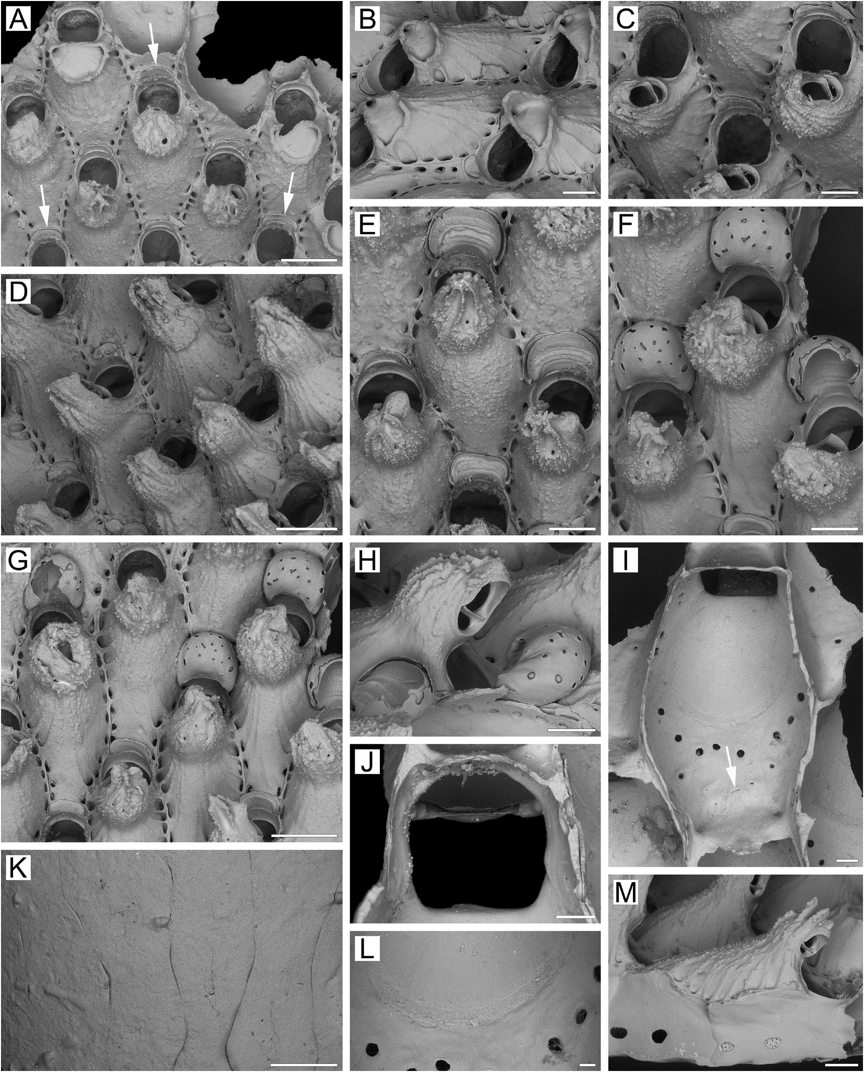
Measurements. ZIRAS 1/50541, Simushir Island, Kuril Islands, Pacific Ocean ( Fig. 16A–M View FIGURE 16 ). ZL, 1.15–1.92 (1.50 ± 0.15). ZW, 0.65–0.95 (0.81 ± 0.08). ZD, 0.73–0.81 (n = 2). OrL, 0.34–0.41 (0.38 ± 0.02). OrW, 0.33–0.39 (0.35 ± 0.02). OeL, 0.38–0.48 (0.43 ± 0.03) (n = 15). OeW, 0.45–0.55 (0.48 ± 0.03) (n = 15). Av(s)L, 0.21–0.43 (0.33 ± 0.05). P(m)N, 17–27 (22). P(oe)N, 13–21 (18) (n = 10).
Description. Colonies encrusting, multiserial, unilaminar ( Fig. 16A View FIGURE 16 ), subcircular, deep brown to light brown when dry; maximal size observed 18 × 20 mm. Zooids very large, broadly hexagonal, widest in midline, rarely elongate oval and tapering proximally, arranged in checkered pattern, demarcated by fine sutures between lateral and transverse zooidal walls; sutures less visible in older parts of colony.
Frontal shield umbonuloid ( Fig. 16A, I View FIGURE 16 ), thin-walled, fragile, moderately convex, finely granulated, with single row of mostly elongate areolae along raised margins, separated by low, interareolar ridges; ridges normally less prominent in young zooids ( Fig. 16A View FIGURE 16 ), though sometimes evident in them too ( Fig. 16B View FIGURE 16 ). In older parts of colony, these ridges, when developed, often connecting to cystid of suboral avicularium and continuing to its apex ( Fig. 16D, M View FIGURE 16 ), but in some instances not very prominent on avicularium ( Fig. 16E–G View FIGURE 16 ). Interior of frontal shield ( Fig. 16I View FIGURE 16 ) with very fine ring scar ( Fig. 16I, L View FIGURE 16 ). Umbonuloid component occupying about 70% of length of frontal shield (68% in one measured zooid), with fine parallel lineation and accretionary banding. In cleaned specimens, semicircle of large pores (lower openings of areolar canals) evident proximal to ring scar.
Primary orifice ( Fig. 16C, J View FIGURE 16 ) bell-shaped, sometimes semioval, slightly longer than wide; distal and lateral margins formed by upper terminal part of distal transverse wall bearing prominent shelf distally ( Fig. 16A–C, E, F View FIGURE 16 ) and forming blunt, ill-defined condyles laterally ( Fig. 16J View FIGURE 16 ). Distal margin of orifice rounded, proximal margin straight, with broadly rounded proximolateral corners.
Secondary orifice ( Fig. 16A–E View FIGURE 16 ) conforming to shape of primary orifice, cormidial; distolateral curvature restricted by thickening of proximal and lateral vertical walls of daughter and neighbouring zooids; proximally bounded by low, thin-walled peristome formed by frontal shield and centrally incorporating cystid of suboral avicularium ( Fig. 16D, F, H View FIGURE 16 ). In ovicellate zooids, peristome usually reaching proximolateral corners of ooecium. No oral spines.
Сystid of suboral avicularium occupying distal one-quarter to one-third of zooidal frontal shield, situated mostly symmetrically relative to orifice, bulbous to conical, strongly elevated, coarsely and irregularly thickened on top; surface coarsely granulated, many granules look like sharp spinules, thus contrasting with rest of frontal shield, with 1–5 minute communication pores ( Fig. 16A–H, M View FIGURE 16 ). Avicularian cystid strongly angled distally, overhanging orifice in older parts of colony ( Fig. 16H View FIGURE 16 ), gradually tapering terminally, with raised vertical ridges uniting into conical tip. Avicularian frontal surface (rostral/postmandibular areas) situated on distolateral slope of cystid, usually crossing zooidal midline, sometimes to one side of it, facing distolaterally to laterally. Rostrum elongate oval, directed proximomedially to proximolaterally and frontally; palate lingulate, foramen elongate oval, bordered by narrow cryptocystal shelf; opesia more or less semicircular. Crossbar complete.
No adventitious avicularia.
Ovicells hyperstomial in all parts of colony, ooecium never overgrown by secondary calcification. Ooecium formed by distal autozooid around crescentic slit with communication pore at bottom, situated in proximalmost part of frontal shield very close to distal margin of maternal primary orifice. Ooecial coelomic cavity connected to visceral coelom via communication canal opening on underside of proximal part of frontal shield of distal zooid as small, straight, slit-like communication pore situated halfway between transverse wall and ring scar ( Fig. 16I View FIGURE 16 ). Ooecium with concave proximal margin. Ectooecium smooth, with sparse, circular to slit-like pseudopores.
Zooids interconnected by two mural pore chambers ( Fig. 16M View FIGURE 16 ) in each distolateral wall. Communication pores spread across basal part of transverse walls either as horizontal “band” or forming two multiporous septula. In some zooids, transverse walls distally with two shallow recesses separated by median buttress.
Basal surface of zooids ( Fig. 16K View FIGURE 16 ) fully calcified, flat, smooth. Boundaries between some zooids recognizable by intermittent fine incisions.
Ancestrula and early astogeny not observed.
Remarks. R. obliqua n. sp. is similar to R. aspera n. sp., but differs from the latter in at least seven characters, described above (see Remarks for R. aspera n. sp.). This species also superficially resembles R. scabra in the conical, prominent suboral avicularian mucro, laterally facing palatal foramen, and lingulate mandible, but differs from the latter in lacking additional adventitious avicularia.
Ecology. Rhamphostomella obliqua n. sp. is known from depths of 10–435 m on pebbles and broken shells of the bivalve mollusc Chlamys sp.
Distribution. The known distribution is based on three records, including one from the Sea of Okhotsk side of the middle to southern Kuril Islands, another from their Pacific side, and the third from the Pacific side of Adak Island, Andreanof Islands, Aleutian Islands. Rhamphostomella obliqua n. sp. is thus a Pacific high-boreal, sublittoral to upper bathyal species.
| ZIRAS |
ZIRAS |
| IMB |
IMB |
| NHMUK |
NHMUK |
No known copyright restrictions apply. See Agosti, D., Egloff, W., 2009. Taxonomic information exchange and copyright: the Plazi approach. BMC Research Notes 2009, 2:53 for further explanation.
|
Kingdom |
|
|
Phylum |
|
|
Class |
|
|
Order |
|
|
SubOrder |
Flustrina |
|
SuperFamily |
Lepralielloidea |
|
Family |
|
|
Genus |