
Pholcus wonju Lee & Lee, 2024
|
publication ID |
https://doi.org/10.11646/zootaxa.5432.2.3 |
|
publication LSID |
lsid:zoobank.org:pub:8B157EDE-7ADB-4AC0-8C98-66D68D8EB27B |
|
DOI |
https://doi.org/10.5281/zenodo.10899097 |
|
persistent identifier |
https://treatment.plazi.org/id/0387A476-7D1D-B042-FF29-236E7266FDF4 |
|
treatment provided by |
Plazi |
|
scientific name |
Pholcus wonju Lee & Lee |
| status |
sp. nov. |
Pholcus wonju Lee & Lee , sp. nov.
Figs 1E–F View FIGURE 1 , 2E–F View FIGURE 2 , 5, 14, 17G, 18
Type material. Holotype: ♂, SOUTH KOREA: Gangwon-do: Wonju-si, Sillim-myeon, near Sangwonsa Temple in Mt. Chiaksan (37˚17'25"N, 128˚04'54"E, 505 m), 12 Aug. 2021, Sam-Kyu Kim leg. ( NIBR) . Paratypes: 1♀, same data as for holotype (KNU-Ar 20230050) ; 2♂♂, same data as for holotype (KNU-Ar 20230051–52) ; 3♀♀, same data as for holotype (KNU-Ar 20230053–55) ; 2♂♂ 2♀♀, same data as for holotype (KNU-Ar 20230056) .
Other material examined. 2♂♂ 1♀, Wonju-si , Gwirae-myeon, near Cheoneunsa Temple (37˚13'44"N, 127˚53'54"E, 353 m), 30 Aug. 2016, Jun-Gi Lee & Jun-Ho Lee leg. ( KNU) ; 2♂♂ 3♀♀, Wonju-si , Sillim-myeon, Chiak Service Area (37˚15'20"N, 128˚03'00"E, 451 m), 25 May 2013, Doo-Yeong Choi leg. (KNU-kise 218) ; 1♂ 1♀, ditto ( KNU) .
Etymology. The specific epithet is derived from the type locality, Wonju-si. Noun in apposition.
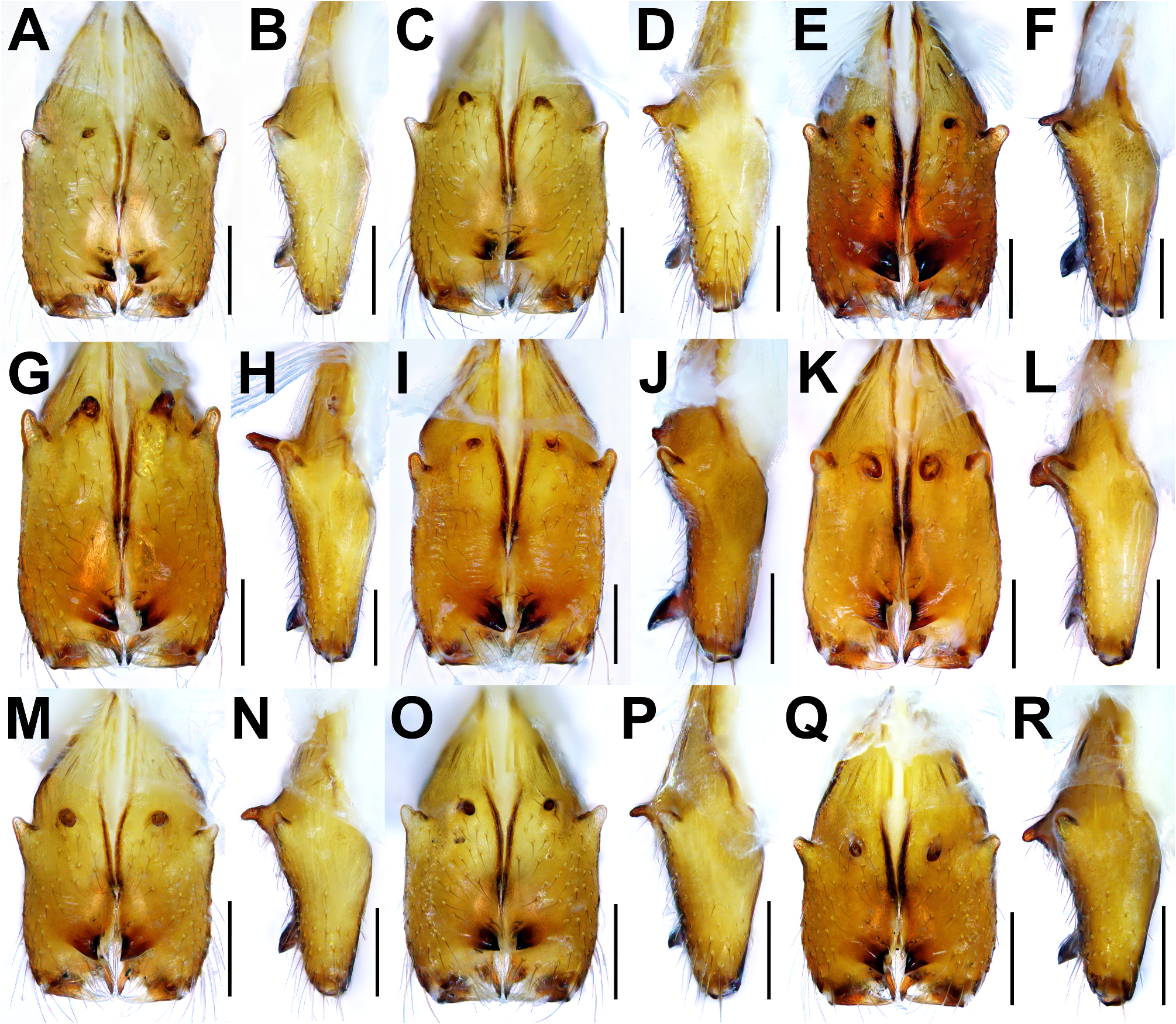
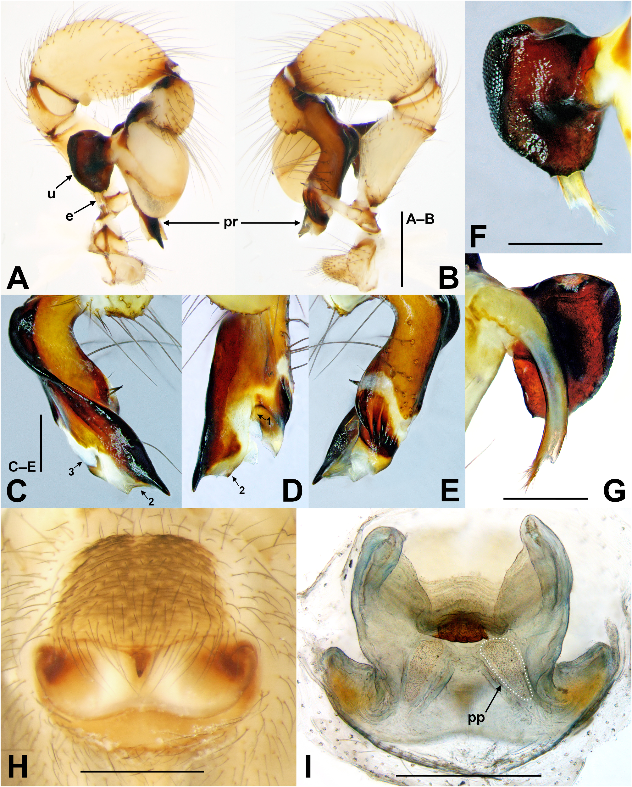
Diagnosis. Males superficially similar to Pholcus crassus Paik, 1978 having prolateral process of procursus with heavily sclerotized outer margin and membranous lobe at inner margin, slender and pointed tip, and short dorsal process ( Fig. 5D View FIGURE 5 ), but can be distinguished by: 1) outer margin of prolateral process of procursus not elongated ( Fig. 5D View FIGURE 5 ) (strongly convex in P. crassus , see Huber 2011: fig. 2293); 2) dorsal process of procursus thin and digitiform ( Fig. 5D View FIGURE 5 ) (stout and triangular in P. crassus , see Huber 2011: fig. 2293); 3) uncus oval, distally with several denticles ( Fig. 5F View FIGURE 5 ) (oblong, distally without several denticles in P. crassus , see Huber 2011: fig. 2292). Females at a glance similar to P. crassus , having epigynal anterior plate much longer than posterior plate, slightly elongated posterior margin of posterior plate ( Fig. 5H View FIGURE 5 ), and long oblong pore plates of internal genitalia ( Fig. 5I View FIGURE 5 ), but can be distinguished by: 1) epigynal anterior plate trapezoid ( Fig. 5H View FIGURE 5 ) (triangular in P. crassus , see Huber 2011: fig. 2169); 2) epigynal posterior plate with pair of sclerotized areas anterolaterally ( Fig. 5H View FIGURE 5 ) (sclerotized areas indistinct in P. crassus , see Huber 2011: fig. 2169); 3) anterolateral portion of internal genitalia strongly elongated, pointing anteriorly ( Fig. 5I View FIGURE 5 ) (slightly elongated, pointing medially in P. crassus , see Huber 2011: figs 2170, 2295).
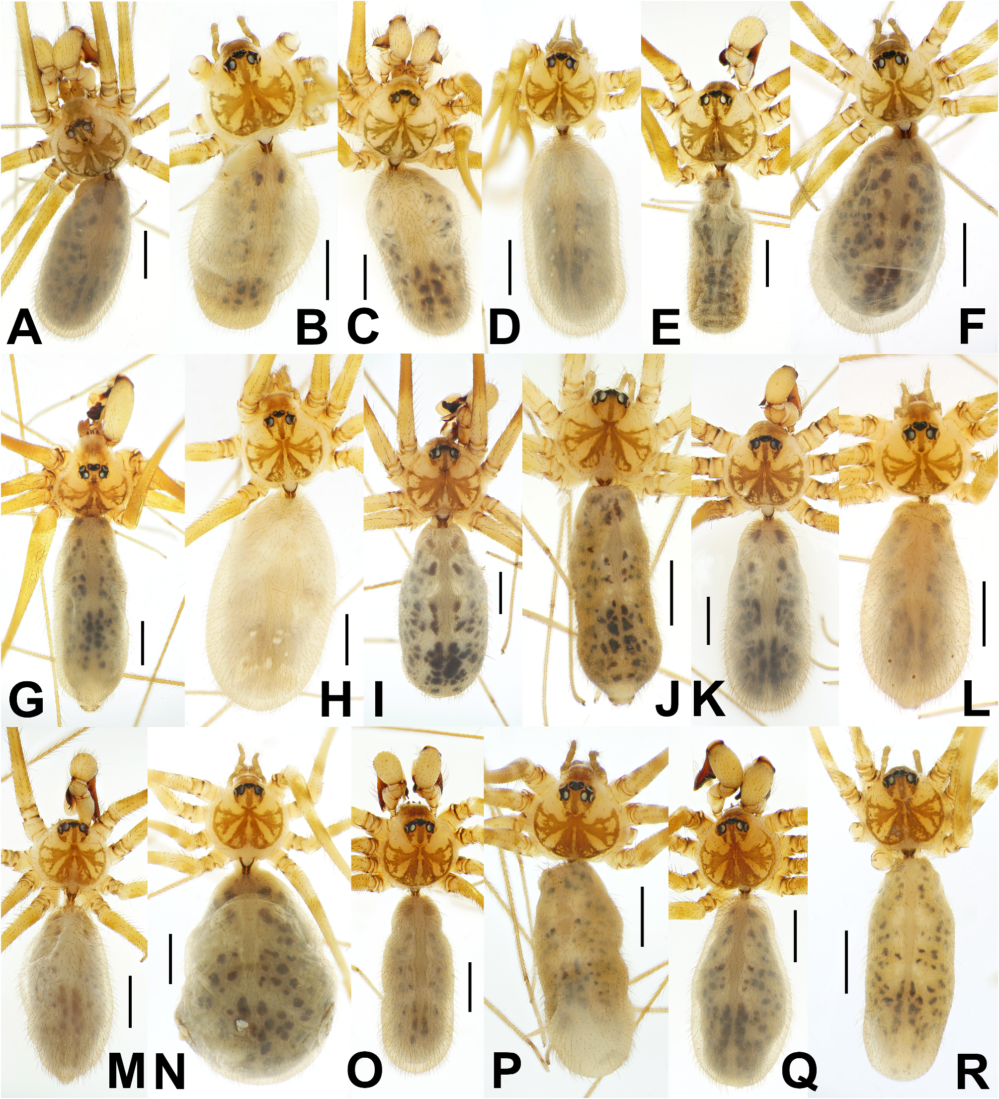
Description. Male ( holotype). Habitus as in Fig. 1E View FIGURE 1 . Total length 5.5. Prosoma 1.9 long, 1.9 wide. Carapace round, pale yellow, with dark brown radial marks and marginal band on thoracic area. Eye area slightly elevated, posteriorly with dark brown marks laterally and medially. Diameter of AME 116 µm, ALE 174 µm, PME 170 µm, PLE 174 µm. AME–AME 67 µm, PME–PME 241 µm, PME–ALE 50 µm. Clypeus with large dark brown mark, without process. Chelicera ( Fig. 2E–F View FIGURE 2 ) with thumb-like proximo-lateral apophysis, slender and straight frontal apophysis, and ventrally curved, notched triangular distal apophysis distinctly longer and larger than proximo-lateral apophysis. Sternum longer than wide, shield-like, pale yellow with brown marks laterally and posteriorly. Opisthosoma 3.5 long, 1.4 wide, cylindrical, greyish yellow, without cuticular patterns, with cardiac pattern and many black granular spots dorsally and laterally. Leg femora and tibiae yellowish brown with two whitish and two dark brown bands distally, two dark brown bands proximally; patellae, metatarsi, tarsi dark brown, without bands; leg Ⅰ femur distinctly darker. Leg Ⅰ 51.6 (13.3 + 0.8 + 13.4 + 21.7 + 2.4), leg II 35.7 (9.9 + 0.8 + 8.9 + 14.4 + 1.7), leg III 24.6 (7.1 + 0.8 + 6.0 + 9.4 + 1.3), leg IV 30.2 (9.3 + 0.6 + 8.1 + 10.6 + 1.6). Ratio of leg Ⅰ (16.0: 1.0: 16.2: 26.2: 2.8), leg II (13.1: 1.0: 11.7: 19.0: 2.2), leg III (9.2: 1.0: 7.8: 12.2: 1.7), leg IV (15.3: 1.0: 13.3: 17.3: 2.6). Leg formula 1243. Tibia Ⅰ L/d 75. Tibiae, metatarsi, tarsi with short vertical setae, tibiae and metatarsi Ⅰ, II with long curved hairs. Tibiae with three trichobothria, except tibia Ⅰ (prolaterally absent). Retrolateral trichobothrium on tibia Ⅰ at 6% proximally. Tarsus I with 27 pseudosegments, mostly irregular. Palp ( Fig. 5A–G View FIGURE 5 ). Trochanter apophysis ( Fig. 5B View FIGURE 5 ) less than half as long as femur, straight, slender, distally blunt, ventro-subdistally with single curved hair, proximo-retrolaterally with tubercle; femur ventrally swollen; tibia with prolatero-ventral tubercle; procursus ( Fig. 5B–E View FIGURE 5 ) dark brown, nearly perpendicularly curved dorsally, with large ventral knee; dorso-subdistally strongly swollen, with two spines near retrolateral ridges ( Fig. 5E View FIGURE 5 ); procursus tip ( Fig. 5C–E View FIGURE 5 ) with three distinct ridges and slightly curved whitish line retrolaterally ( Fig. 5E View FIGURE 5 ), short and slender dorsal process (arrowed 1 in Fig. 5D View FIGURE 5 ), long rectangular prolateral process longitudinally curved dorsally, marginally strongly sclerotized, distally elongated and pointed, entally with membranous lobe (arrowed 2 in Fig. 5C–D View FIGURE 5 ), and short blunt ventral process (arrowed 3 in Fig. 5C View FIGURE 5 ); genital bulb ( Fig. 5A View FIGURE 5 ) oval, pale yellow; uncus ( Fig. 5F View FIGURE 5 ) about 0.7 times as long as genital bulb, dark brown, oval, distally slightly serrated, outer margin with numerous tiny scales; pseudoappendix absent; embolus ( Fig. 5G View FIGURE 5 ) slender, distally fringed, weakly sclerotized, longer than uncus.
Female (one of paratypes, KNU-Ar 20230050). Habitus as in Fig. 1F. View FIGURE 1 Somatic characteristics generally similar to male, but cheliceral apophyses absent, legs slightly shorter. Total length 4.9. Prosoma 1.3 long, 1.3 wide. Diameter of AME 98 µm, ALE 152 µm, PME 134 µm, PLE 137 µm. AME–AME 51 µm, PME–PME 183 µm, PME–ALE 44 µm. Opisthosoma 3.3 long, 2.0 wide. Leg Ⅰ 29.4 (7.1 + 0.6 + 7.4 + 12.2 + 2.2), leg II 20.3 (5.4 + 0.6 + 5.0 + 7.8 + 1.5), leg III 15.2 (4.2 + 0.5 + 3.7 + 5.6 + 1.2), leg IV 19.3 (5.7 + 0.6 + 4.9 + 7.4 + 0.8). Ratio of leg Ⅰ (12.6: 1.0: 13.3: 21.8: 3.9), leg II (9.2: 1.0: 8.4: 13.2: 2.5), leg III (8.1: 1.0: 7.0: 10.8: 2.3), leg IV (9.9: 1.0: 8.6: 13.0: 1.3). Leg formula 1243. Tibia I L/d 50. Epigyne ( Fig. 5H View FIGURE 5 ). Anterior plate slightly longer than wide, trapezoid, dark brown, posteromedial margin slightly extended posteriorly; posterior plate bright ivory, about half as long as anterior plate, posterolaterally slightly extended, with pair of sclerotized areas anterolaterally; epigynal knob dark brown, thick, pointed, half as long as posterior plate; posterior sclerotized cuticle yellowish brown, slightly procurved, very thin; interspace between posterior plate and posterior sclerotized cuticle yellowish brown. Internal genitalia ( Fig. 5I View FIGURE 5 ). Anterior arch short and transverse, medially strongly sclerotized; anterolateral portion strongly extended, blunt lanceolate with distal margin folded once inwardly; genital valve slightly wavy, laterally hidden by anterolateral extensions dorsally; lateral portion strongly curved toward anteriorly and distally concave; pore plates long obovate, close together anteriorly, gradually wide apart posteriorly.
Variation. Males. Prosoma width: 1.5–1.9 (mean 1.7), tibia Ⅰ: 10.5–13.6 (mean 12.1) (n=10).
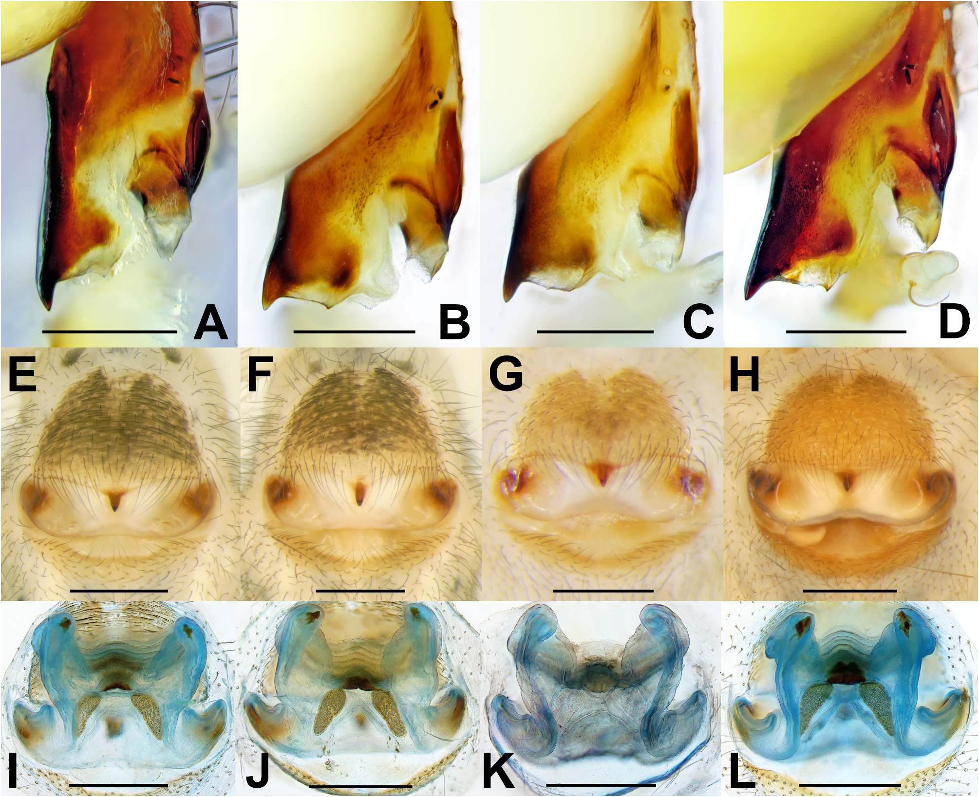
An intrapopulational variation was observed on the shape of inner margin of prolateral process in procursus tip ( Fig. 14A–D View FIGURE 14 ).
Females. Prosoma width: 1.3–1.7 (mean 1.6), tibia Ⅰ: 7.1–9.0 (mean 8.1) (n=10). Intrapopulational variations were observed on the shape of sclerotized mark of epigynal anterior plate ( Fig. 14E–H View FIGURE 14 ), the shape of the distal portion of strongly elongated projection in anterior arch, and the shape and size of pore plates of internal genitalia were also varied ( Fig. 14I–L View FIGURE 14 ).
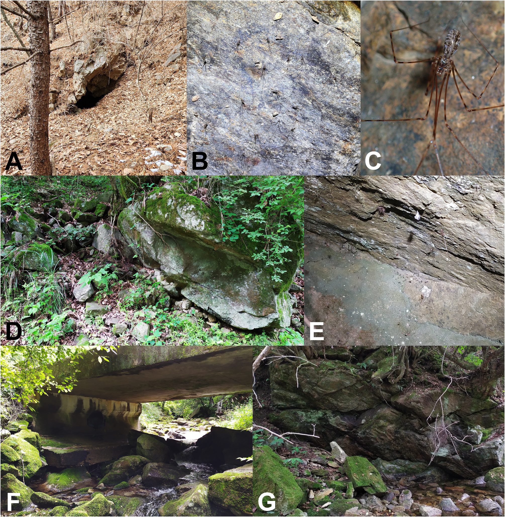
Habitat information. This species was found on rock walls and crevices under rocks in streamside of mountainous regions ( Fig. 17G View FIGURE 17 ). Sympatry is observed between this species with Pholcus chiakensis Seo, 2014 from Chiak Service Area and near Cheoneunsa Temple, Wonju-si, sharing the same habitat together.
Distribution. Korea (Wonju) ( Fig. 18 View FIGURE 18 ).

| NIBR |
National Institute of Biological Resources |
| KNU |
Kyungpook National University |
No known copyright restrictions apply. See Agosti, D., Egloff, W., 2009. Taxonomic information exchange and copyright: the Plazi approach. BMC Research Notes 2009, 2:53 for further explanation.