
Promitobates trapista, Bragagnolo, Cibele & Pinto-Da-Rocha, Ricardo, 2012
|
publication ID |
https://doi.org/ 10.5281/zenodo.280969 |
|
DOI |
https://doi.org/10.5281/zenodo.6174045 |
|
persistent identifier |
https://treatment.plazi.org/id/038687A2-FFD4-C26C-FF3C-5AABFC14FB42 |
|
treatment provided by |
Plazi |
|
scientific name |
Promitobates trapista |
| status |
sp. nov. |
Promitobates trapista View in CoL sp. n.
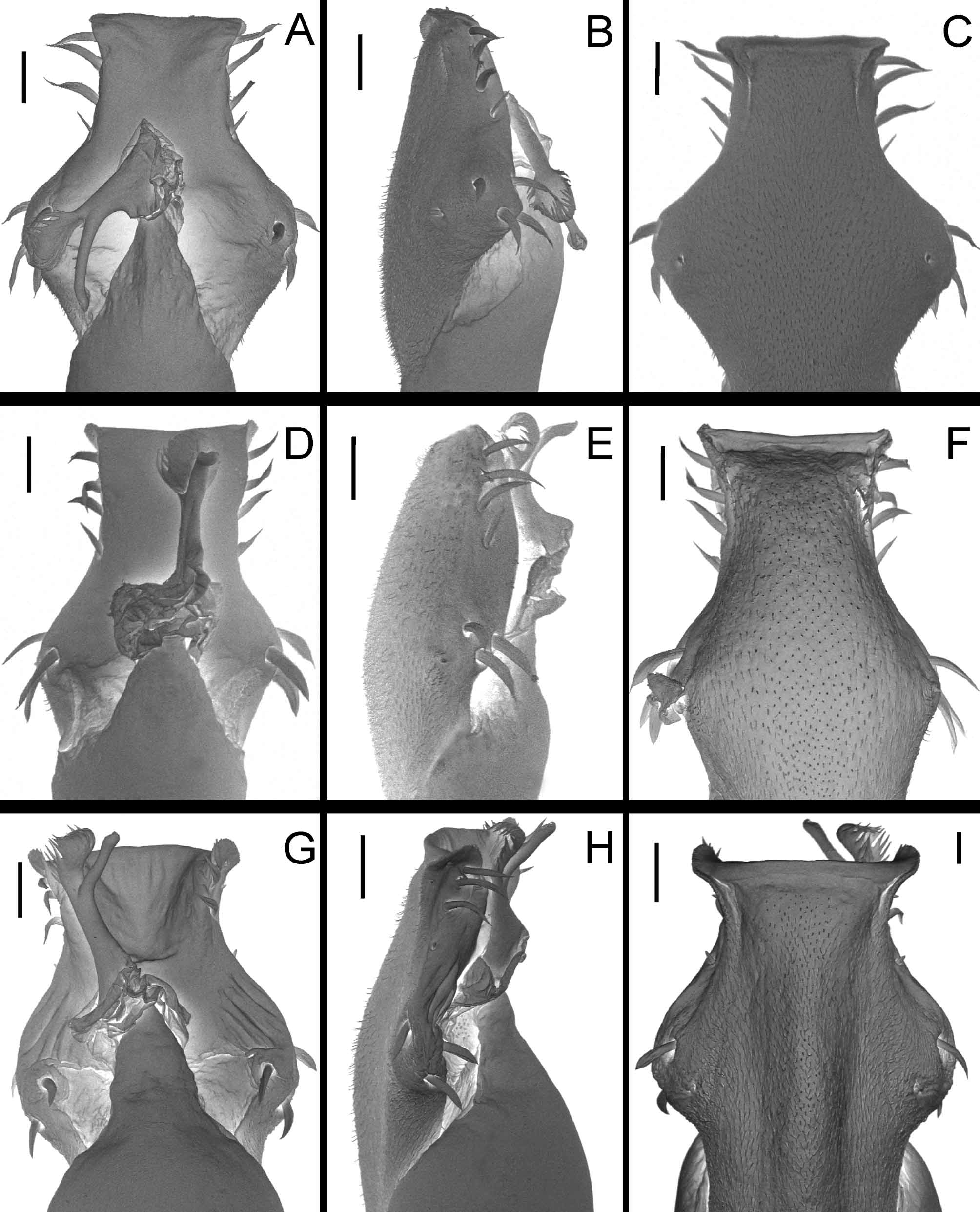
( Figs. 6 View FIGURE 6 B, 8D, 10D, 18G, H, I)
Promitobates View in CoL sp.: Bragagnolo et al. 2007: 393 (cit).
Type material. BRAZIL. São Paulo: Cotia (Reserva Morro Grande), VII.2008, R. Pinto-da-Rocha et al. leg; holotype 3 ( MZSP 31197). Paratypes, same locality as holotype, 15.XII.2002, A.A. Nogueira et al. leg. 2 3 ( MZSP 27406).
Etymology. Trapista is a kind of ale beer, made by Belgian Trappist monks.
Diagnosis. It resembles P. a l e sp. n. by external apophysis of coxa IV short and single-branched; also resembles P. hatschbachi and P. ornatus by the pattern of tubercles on dorsal scutum. It can be distinguished form them by coloration, with dorsal scutum and legs I–III dark-yellow and apice of coxa IV, trochanter IV and femur IV black.
Male description. Measurements (holotype). Dorsal scutum; maximum width: 4.5; total length: 4.2; prosoma length: 1.8. Leg IV; length of external apophysis of coxa: 0.95; femur IV length: 37.6. Tarsal formula: 7; 15; 10; 8.
Dorsum ( Figs. 6 View FIGURE 6 B, 8D). Anterior margin of prosoma with two or three tubercles on corners. Ocularium with two high apophyses and tubercles behind apohphysis. Prosoma with tubercles behind ocularium, arranged in “U”. Area I with a row of small tubercles up to the posterior groove and two enlarged median tubercles. Area II with a row of tubercles up to the posterior groove and small scattered tubercles. Areas III and IV fused, III with two high spiniform apophyses and tubercles on median region. Lateral margin of dorsal scutum with one row of tubercles which is placed from ocularium to area II. Posterior margin with two median spiniform tubercles and one row of rounded tubercles increasing in size to the corners. Free tergites I and II with small scattered tubercles and one enlarged pair of tubercles on the corners; III with a median spiniform tubercle.
Chelicera. Segment I with three setiferous tubercles; II unarmed.
Pedipalp. Trochanter with a small ventral spine. Femur with a small ventral basal spine and one subapical, prolateral spine. Tibia setation: mesal IiIi; ectal IiIi. Tarsus setation: mesal IIi; ectal IIi.
Venter. Coxa I with median row of six setiferous tubercles and two apical tubercles; trochanter I with median tubercle and few granules scattered. Coxa II with median row of eight small setiferous tubercles and two apical tubercles; trochanter II with median tubercle. Coxa III with small setiferous tubercles, distributed roughly in rows, the anterior and posterior one, increasing in size distally; trochanter III with median tubercle. Coxa IV with granules scattered and three tubercles on posterior margin; genital area unarmed; free sternites with row of tubercles; anal operculum with few granules.
Legs. ( Fig. 10 View FIGURE 10 D). Coxa I–II with one anterior dorso basal apophysis and one posterior; coxa III with anterior dorso basal apophysis; coxa IV with high tubercles, external apical apophysis directed ventrally and with one basal rounded tubercle, two small internal spiniform apophysis. Trochanters I–III with small scattered tubercles. Trochanter IV with one dorsal apical and one dorso basal rounded apophyses and one small internal apical spiniform apophysis. Femur more than 10 times longer than dorsal scutum, straight and unarmed.
Coloration. Dorsal scutum, chelicerae, pedipalps and legs I–III dark-yellow. Apex of coxa IV, trochanter IV and femur IV black.
Penis ( Fig. 18 View FIGURE 18 G–I). Ventral plate convex, apex straight and distal margin corner rounded and directed ventrally; three pairs of distal setae, one pair of median setae; three pairs of basal setae, arranged in “V” and one pair of ventral basal setae. Stylus unarmed, apex straight and swollen. Ventral process almost parallel to the stylus, apex spoon-shaped, with serrate margins directed ventrally.
Female. Unknown.
Geographical distribution ( Fig. 3 View FIGURE 3 B). Only known from type locality.


| MZSP |
Sao Paulo, Museu de Zoologia da Universidade de Sao Paulo |
No known copyright restrictions apply. See Agosti, D., Egloff, W., 2009. Taxonomic information exchange and copyright: the Plazi approach. BMC Research Notes 2009, 2:53 for further explanation.
|
Kingdom |
|
|
Phylum |
|
|
Class |
|
|
Order |
|
|
Family |
|
|
SubFamily |
Mitobatinae |
|
Genus |
Promitobates trapista
| Bragagnolo, Cibele & Pinto-Da-Rocha, Ricardo 2012 |
Promitobates
| Bragagnolo 2007: 393 |