
Amblyscarta pinna, Mejdalani & Domahovski & Cavichioli, 2019
|
publication ID |
https://doi.org/ 10.11646/zootaxa.4711.2.9 |
|
publication LSID |
lsid:zoobank.org:pub:E7DCF456-8EC4-4E36-B864-742A065C0ED9 |
|
DOI |
https://doi.org/10.5281/zenodo.5921259 |
|
persistent identifier |
https://treatment.plazi.org/id/E754DBA0-3121-4698-A1F9-4F51E9ECEBFC |
|
taxon LSID |
lsid:zoobank.org:act:E754DBA0-3121-4698-A1F9-4F51E9ECEBFC |
|
treatment provided by |
Plazi |
|
scientific name |
Amblyscarta pinna |
| status |
sp. nov. |
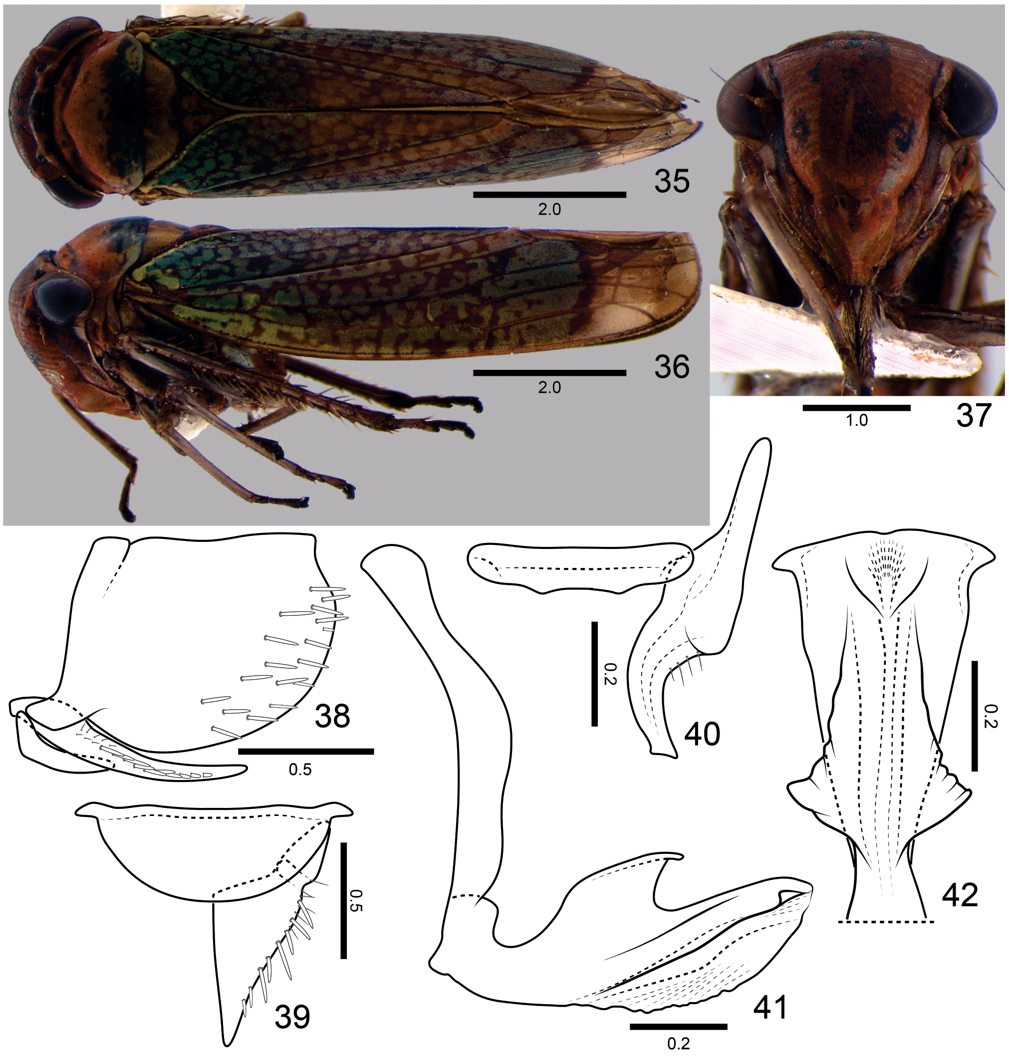
Amblyscarta pinna View in CoL sp. nov.
( Figs 35–50 View FIGURES 35–42 View FIGURES 43–50 )
Total length (mm). Male 9.0–9.3 (holotype 9.0); female 9.7–9.8.
Color ( Figs 35–37 View FIGURES 35–42 ). Ground color of anterior dorsum orange; crown with two dark brown to black median spots, one at transition to frons and another, larger, at posterior margin; posterior portion of pronotum green with dark brown to black spots and vermiculations; mesonotum with dark brown to black markings on scutoscutellar suture and scutellum. Forewing green with dark brown to black vermiculations from base to distal portion of anteapical cells, where a transverse dark brown to black stripe is located; area behind this stripe smoky subhyaline. Face mostly orange. Legs yellow to brown.
Male terminalia. Pygofer ( Fig. 38 View FIGURES 35–42 ), in lateral view, slightly produced posteriorly; posterior margin slightly rounded inferiorly and with emargination on superior portion; without processes; macrosetae distributed mostly on posterior third. Valve ( Fig. 39 View FIGURES 35–42 ), in ventral view, large; posterior margin broadly rounded. Subgenital plate ( Figs 38, 39 View FIGURES 35–42 ), in ventral view, triangular, narrowing gradually towards apex; fused basally to its counterpart; with uniseriate macrosetae along outer margin, microsetae also present; in lateral view, short, not extending as far posteriorly as pygofer apex. Style ( Fig. 40 View FIGURES 35–42 ), in dorsal view, extending much farther posteriorly than connective; without preapical lobe; apical portion slightly curved outwards, inner margin not dilated, with tiny apical tooth; apex distinctly truncate. Aedeagus ( Figs 41, 42 View FIGURES 35–42 ) symmetrical; shaft, in lateral view, with conspicuous, slightly asymmetrical dorsal finlike process; with pair of longitudinal lateral flanges forming apically dentiform process directed outwards; ventral margin with pair of flanges, each one with basal projection; gonopore located apically.
Female terminalia. Sternite VII ( Figs 43, 44 View FIGURES 43–50 ), in ventral view, with strong triangular projection on posterior margin. “Internal” sternite VIII without distinct sclerites. Pygofer ( Figs 43, 44 View FIGURES 43–50 ), in lateral view, moderately produced posteriorly; posterior margin rounded; macrosetae distributed on posterior portion and extending anteriorly along ventral margin. Valvifer I ( Fig. 45 View FIGURES 43–50 ), in lateral view, subquadrangular. Valvula I ( Figs 45, 46 View FIGURES 43–50 ), in lateral view, with acute apex; dorsal sculptured area extending from basal portion to apex of blade, strigate; ventral sculptured area restricted to apical portion, strigate; ventral interlocking device distinct along basiventral half of blade; in ventral view, basal portion of valvula I expanded outwards. Valvula II ( Figs 47–49 View FIGURES 43–50 ), in lateral view, expanded beyond basal curvature; dorsal margin convex, bearing about 20 teeth; basal portion of most teeth projected dorsally, their posterior portion flat, apical teeth triangular; denticles distributed on teeth and on dorsal and ventral apical portions of blade (ventral denticulate apical portion longer than dorsal portion); preapical prominence distinct; apex obtuse; valvula with ducts extending towards teeth and apex. Gonoplac ( Fig. 50 View FIGURES 43–50 ) of the usual Cicadellinae type: in lateral view, with basal half narrow; apical half expanded, gradually narrowing towards apex; latter obtuse; tiny denticuli on apical portion and extending anteriorly along ventral margin.
Material examined. Male holotype: “ Brasil, MT [Mato Grosso], Cláudia, Fazen-\ da Continental , 11.5841°S \ 55.3003°W, 365m, light trap, \ 17-19.vi.2017, RR Cavichioli \ & AC GoogleMaps Domahovski ” ( DZUP). Paratypes: 1♂, 2♀, “ Brasil, MT , Novo Mundo, \ Pq. [Parque] Est. [Estadual] do Cristalino \ 09.4517°S 55.8396°W, \ 240m, sweep, 21- 25.vi. \ 2017, AC GoogleMaps Domahovski ” ( MNRJ, 1♀ DZUP) .
Etymology. The new species name comes from the Latin and refers to the dorsal fin-like process of the aedeagus ( Fig. 41 View FIGURES 35–42 ). It is a noun in apposition.
Remarks. This new species can be promptly recognized by the peculiar fin-like process of the aedeagus ( Fig. 41 View FIGURES 35–42 ), which is unique within the genus. As in the case of the two previous new taxa, the color pattern ( Figs 35, 36 View FIGURES 35–42 ) will readily distinguish A. pinna sp. nov. from those poorly known species that were not included in Young’s (1977) key ( A. cervicula , A. lignea , A. modesta , and A. schaumi ; see digital images in Wilson et al. 1999).

No known copyright restrictions apply. See Agosti, D., Egloff, W., 2009. Taxonomic information exchange and copyright: the Plazi approach. BMC Research Notes 2009, 2:53 for further explanation.
|
Kingdom |
|
|
Phylum |
|
|
Class |
|
|
Order |
|
|
Family |
|
|
Genus |