
Eoplatyrhina bolcensis ( Heckel, 1851 ) Marram̀ & Carneval & Claeso & Naylo & Kriwe, 2020
|
publication ID |
https://doi.org/ 10.1080/14772019.2020.1783380 |
|
publication LSID |
lsid:zoobank.org:pub:B4C7A979-7972-409B-B489-A6DDD5E35FE5 |
|
DOI |
https://doi.org/10.5281/zenodo.10932539 |
|
persistent identifier |
https://treatment.plazi.org/id/0385D508-FFE8-FFC1-90DA-6CFEB2962379 |
|
treatment provided by |
Felipe |
|
scientific name |
Eoplatyrhina bolcensis ( Heckel, 1851 ) |
| status |
comb. nov. |
† Eoplatyrhina bolcensis ( Heckel, 1851) comb. nov.
Figs 2–7 View Figure 2 View Figure 3 View Figure 4 View Figure 5 View Figure 6 View Figure 7
1833–1843 Narcopterus bolcanus Agassiz : vol. 1: 44 (nomen nudum; no description or figure).
1833–1843 Narcopterus bolcanus Agassiz : vol. 3: 382.
1833–1843 Narcopterus bolcanus Agassiz : vol. 4: 38.
1835 Narcopterus bolcanus Agassiz : 14.
1851 Platyrhina bolcensis Heckel : 324 (first occurrence of name and description).
1854 Platyrhina (?) bolcana; Pictet: 277.
1860 Platyrhina bolcensis Heckel ; Molin: 587.
1874 Platyrhina bolcensis Heckel ; De Zigno: 177.
1894 Platyrhina bolcensis (Heckel) Molin ; Jaekel: 106, fig. 18.
1904 Platyrhina bolcensis (Heckel) ; Eastman: 27.
1905 Platyrhina bolcensis (Heckel) ; Eastman: 351.
1922 Platyrhina bolcensis (Agassiz) Heckel ; D’ Erasmo: 12.
1980 Platyrhina bolcensis Heckel ; Blot: 344.
1987 Platyrhina bolcensis Molin, 1860 ; Cappetta: 139.
2004 Platyrhina bolcensis ; Carvalho: 78, fig. 12A, C.
2012 Platyrhina bolcensis Molin, 1860 ; Cappetta: 346.
2014 Platyrhina bolcensis Heckel, 1851 ; Carnevale, Bannikov, Marram̀a, Tyler & Zorzin: 41.
2018c ‘ Platyrhina ’ bolcensis ; Marram̀a, Carnevale, Engelbrecht, Claeson, Zorzin, Fornasiero & Carnevale: 287, fig. 12C.
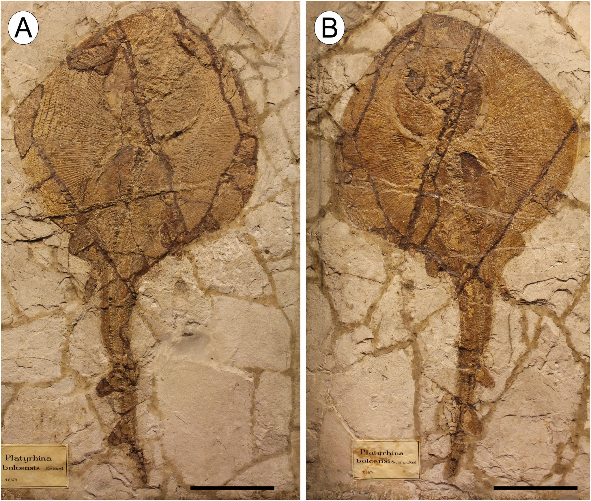
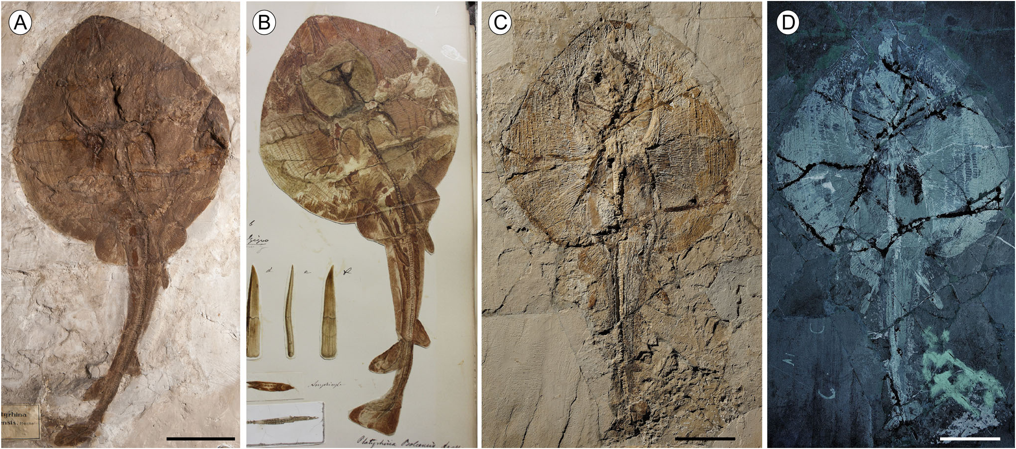
Holotype. MGP-PD 8873 C/8874C, articulated skeleton in part and counterpart, lacking the caudal fin, 338.5 mm disc width ( DW, hereafter; Fig. 2 View Figure 2 ).
Referred material. MGP-PD 26279C/26280C, completely articulated skeleton in part and counterpart, 384.2 mm DW, 840.3 mm TL ( Fig. 3A, B View Figure 3 ); MGGC 7449/7450, articulated skeleton in part and counterpart, lacking dorsal and caudal fins, 379.4 mm DW ( Fig. 3C, D View Figure 3 ).
Type locality and horizon. Monte Postale site, Bolca Konservat-Lagerst¨atte, Italy; early Eocene, Ypresian, middle Cuisian, SBZ 11 ( NP 13, CNE 5); 50.7–48.9 Ma ( Papazzoni et al. 2017).
Diagnosis. As for the genus.
Description
† Eoplatyrhina bolcensis ( Heckel, 1851) comb. nov. is represented by three partially complete articulated specimens in part and counterpart ( Figs 2 View Figure 2 , 3 View Figure 3 ), including the holotype ( MGP-PD 8873C/8874C) and two additional specimens ( MGP-PD 26279C/26280C and MGGC 7449/ 7450). Counts and measurements are listed in the Supplemental material (File 1, Table S1 View Table 1 ). The examined specimens are similar in size. The largest one measures 84 cm TL and 38 cm DW. The pectoral disc of † Eoplatyrhina gen. nov. is notably expanded, ovoid or shovel shaped, slightly longer than wide and reaching its maximum width just posterior to its mid-length. The snout is broad and rounded. The tail is not very stout, slightly longer than disc length, with two dorsal fins inserting posteriorly on the tail. The overall body shape and proportions are similar to those of the extant thornbacks Platyrhina and Platyrhinoidis .
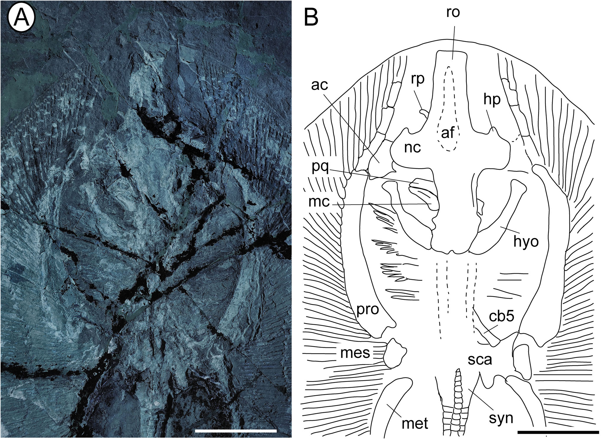
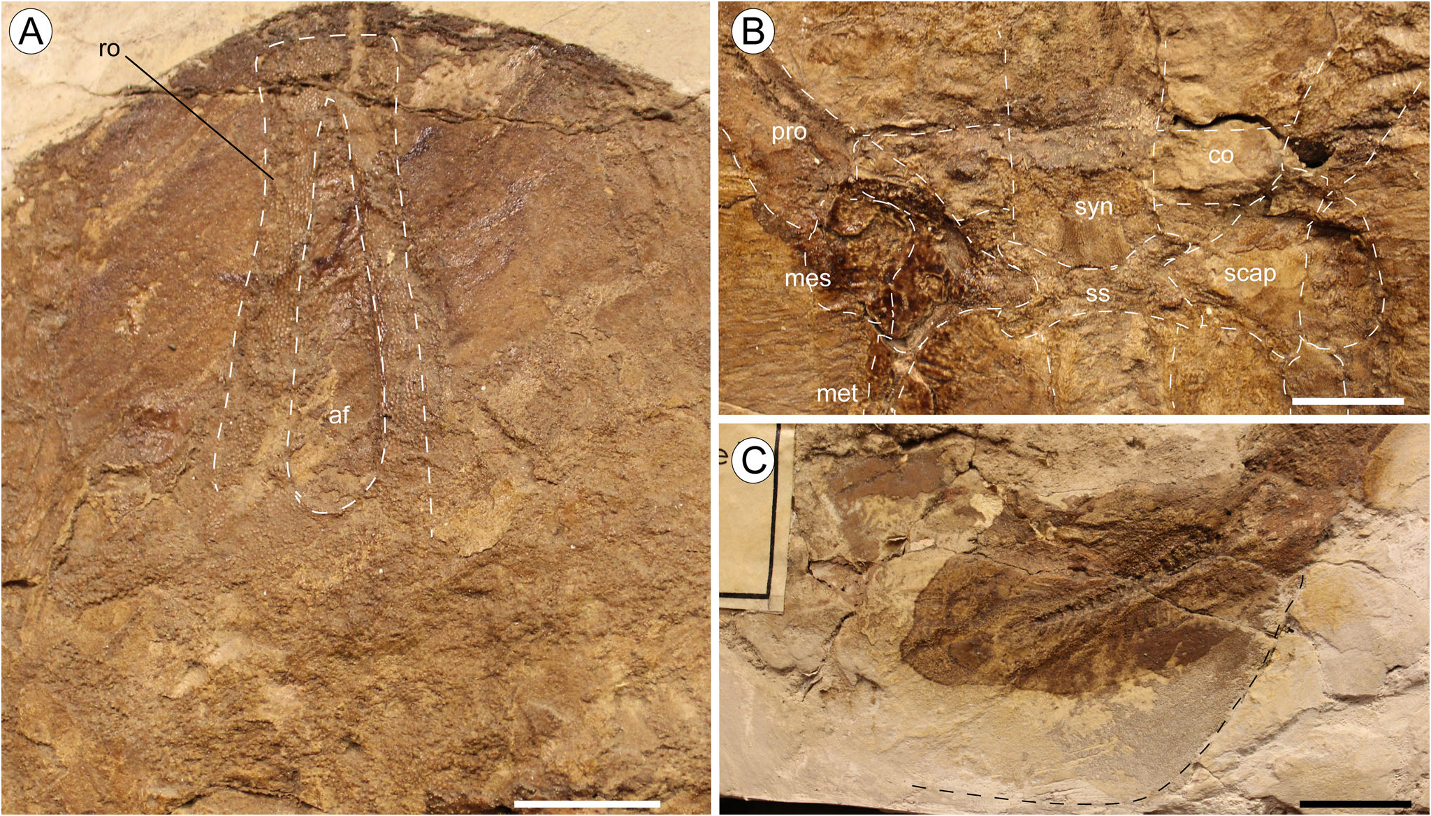
Neurocranium. The rostral cartilage fails to reach the anterior margin of the disc, as in all platyrhinids. This element is long and tapers gradually anteriorly ( Figs 4 View Figure 4 , 5A View Figure 5 ), resembling the condition typical of Platyrhinoidis and † Tethybatis , and differs from the short rostrum observed in † Tingitanius and Platyrhina . Unlike other platyrhinids, the anterior margin of the rostral cartilage is not pointed but trough-shaped, with the rostral node slightly expanded laterally ( Figs 4 View Figure 4 , 5A View Figure 5 ). Rostral appendices at the tip of the rostrum are absent. A small rod-like process lateral to the rostral cartilage and just anterior to the nasal capsule in MGGC 7449/7450 can be interpreted as one of the two rostral processes, which are uniquely present in extant thornbacks. Although McEachran et al. (1996) considered these structures homologous to the rostral appendices of skates and guitarfishes, Carvalho (2004) pointed out that the rostral processes of platyrhinids, originating ventral to the rostral cartilage, might represent outgrowths of the lamina orbitonasalis, unlike the rostral appendices that are secondary chondrifications fused laterally to the rostral node. The nasal capsules are ovoid, laterally expanded, and at right angles to the rostrum, as in † Tethybatis . A single small horn-like process (= tab-like process of Claeson et al. 2013) can be recognized on the anterior margin of each nasal capsule, similar to the extant platyrhinids and † Tingitanius . The antorbital cartilages are well developed and plate-like and have an irregular outline. They project laterally from the postero-lateral margin of the nasal capsules and articulate distally with the propterygia. It is difficult to distinguish the preorbital process or the jugal arch, but a small and narrow postorbital process can be recognized in the otic region, just posterior to the supraorbital crest. The orbital region is longer than wide. The anterior fontanel extends through almost the entire length of the rostral cartilage and resembles an isosceles triangle with a close and concave posterior border, similar to the condition seen in † Tingitanius , and in contrast to the oval-shaped fontanel of Platyrhina , or to the figure-eight shape typical of Platyrhinoidis .
Jaws, hyoid and gill arches. Specimens of † Eoplatyrhina bolcensis comb. nov. are mostly preserved in dorsal view, obscuring the jaws, which are displaced and difficult to describe ( Fig. 4 View Figure 4 ). For the same reason, teeth are not exposed in any specimen, and therefore their morphology remains unknown. It is also unclear whether the labial cartilages are present, as in mature specimens of Platyrhina . The hyomandibulae are stout, robust and slightly arched, with a concave inner margin, narrow at their medial section. They project anterolaterally. As in † Tethybatis , there is a large space between the hyomandibulae and mandibular arch, which is interpreted by Carvalho (2004) as indicative of the presence of a large spiracular opening. In radiographs, this space is not present in Platyrhinoidis or Platyrhina , while it is present in Zanobatus . The distal part of the hyomandibulae appears taphonomically separated from the Meckel’ s cartilage. The fifth ceratobranchials articulate with the anterior margin of the scapulocoracoid, and the remaining gill arches are poorly preserved or missing.
Synarcual and vertebral column. Although the synarcual can be identified as a tubular mineralized structure between the neurocranium and scapulocoracoid, its morphology remains ambiguous. The dorsally exposed specimens obscure the pattern of free centra. In † Tingitanius , the first exposed vertebral centrum of the synarcual is located posterior to the articulation of the suprascapular cartilage with the synarcual. In Platyrhina , the first free centrum is situated at the level of the scapulocoracoid articulation with the synarcual. In Platyrhinoidis , the first free centrum is rostral to the scapulocoracoid articulation with the synarcual. The vertebral column of † Eoplatyrhina bolcensis comb. nov. consists of about 132 vertebral centra, in the most complete specimen MGP-PD 26279C/26280C. There are 20–24 trunk centra (from the first distinguishable centrum to the anterior margin of the puboischiadic bar), and 113–118 from the puboischiadic bar to the tip of the tail (of these, about 23 are caudal). The vertebral centra are highly calcified, sub-rectangular in shape and anteroposteriorly compressed. There are about 15 or 16 pairs of ribs.
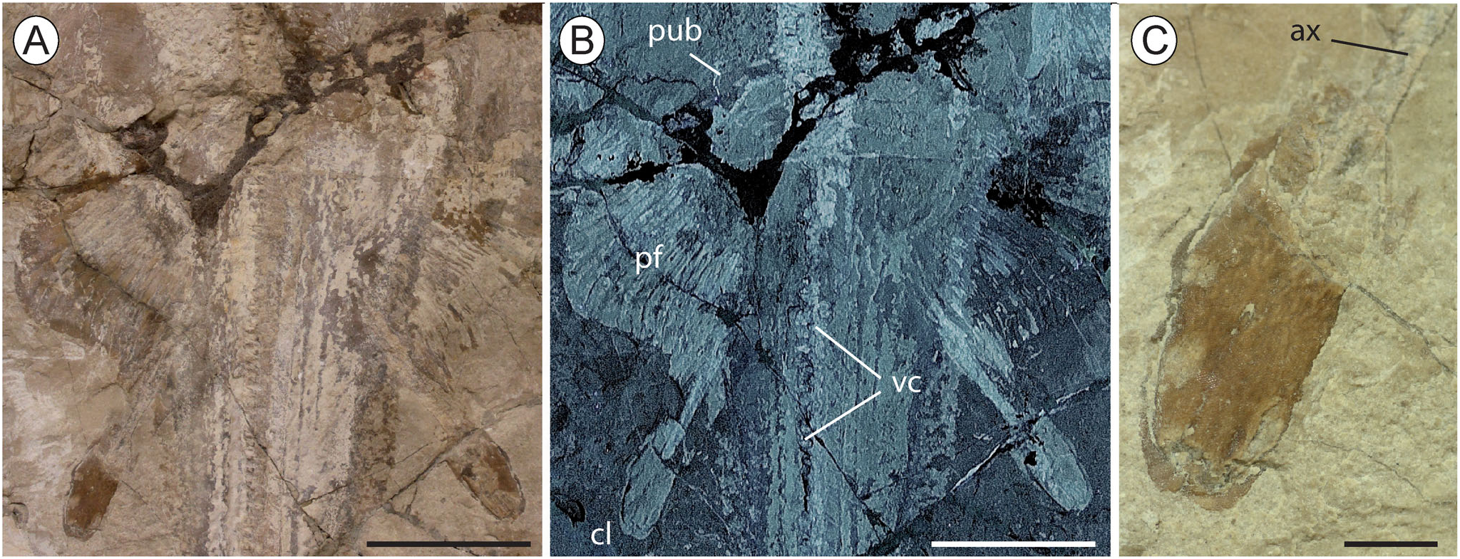
Appendicular skeleton and fins. It is difficult to describe the morphology of the coracoid bar because the specimens are mostly exposed in dorsal view, but the scapular processes of the scapulocoracoid seem to be short in MGP-PD 26279C/80C ( Fig. 5B View Figure 5 ). This specimen shows a small medially fused suprascapular cartilage; this cartilage is hourglass-shaped, with concave anterior and posterior borders, exhibiting deep indentations into which the distal edges of the scapular processes of the scapulocoracoid fit. Laterally, the scapulocoracoid articulates with the proximal portion of the pterygia through equidistant condyles. The propterygium is long and arched, tapers distally and extends to the anterior disc margin ( Fig. 4 View Figure 4 ). The propterygium is segmented, with the first segment lying anterior to the mouth, close to the level of the antorbital cartilage. The proximal section of the propterygium does not extend far posteriorly to the procondyle, and does not articulate with the scapulocoracoid. A single unsegmented mesopterygium seems to be present. The metapterygium is as long and curved as the propterygium, but it is unclear whether it is segmented distally. The pectoral fins are clearly of the plesodic type, with radials reaching the external border of the pectoral disc. All the radials articulate with the pterygia. Each pectoral radial contains 10–12 segments and bifurcates distally only once at about the eighth segment. There are approximately 81–87 pectoral radials, of which 35–38 are propterygial, 8–10 mesopterygial, and 38–41 metapterygial. The pectoral radials of † E. bolcensis comb. nov. are robust, stiff and completely covered by mineralized tissue, forming the so-called ‘crustal calcification’ typical of most of batoids except the benthic stingrays and skates ( Schaefer & Summers 2005).
The puboischiadic bar is partly recognizable in MGGC 7449/7450, where it seems straight or slightly bent, narrow and plate-like ( Fig. 6 View Figure 6 ). It is difficult to recognize the postpelvic processes on the posterior margin of the puboischiadic bar that are typical for living platyrhinids. There are about 18–21 pelvic radials. The structure of the first pelvic radial is unclear but the pelvic condyles seem close together and not separated as in skates. All the specimens show straight and stout claspers, whose length represents about 10% TL ( Fig. 6 View Figure 6 ). As in Platyrhinoidis , their distal extremity does not reach the origin of the first dorsal fin; they differ from those characteristic of Platyrhina , whose clasper tips can extend beyond the first dorsal-fin origin (e.g. Last et al. 2016; White & Last 2016). The clasper glands are almost entirely covered by dermal denticles, and consequently their skeletal morphology is difficult to describe. However, the axial cartilage is rod-like, possibly calcified over most of its length, and extends and inserts over the complete length of the clasper to the ventral terminal cartilage.
Dorsal and caudal fins. † Eoplatyrhina bolcensis comb. nov. possesses two dorsal fins located in the posterior half of the tail. The extent of the fin radial cartilages into the fin web is not precisely ascertainable, but they are possibly aplesodic. The base of the dorsal fins has a length of about 5% TL. No impression of dorsal-fin radials is visible. The caudal fin is only preserved in MGP-PD 26279C/80C ( Fig. 5C View Figure 5 ). It is about 11% TL and contains about 23 vertebrae not reaching the posterior-most border of the caudal fin. There are about 20–25 caudal-fin radials on the ventral and dorsal sides (40–50 in total), which do not reach the external margin of the caudal fin (aplesodic).
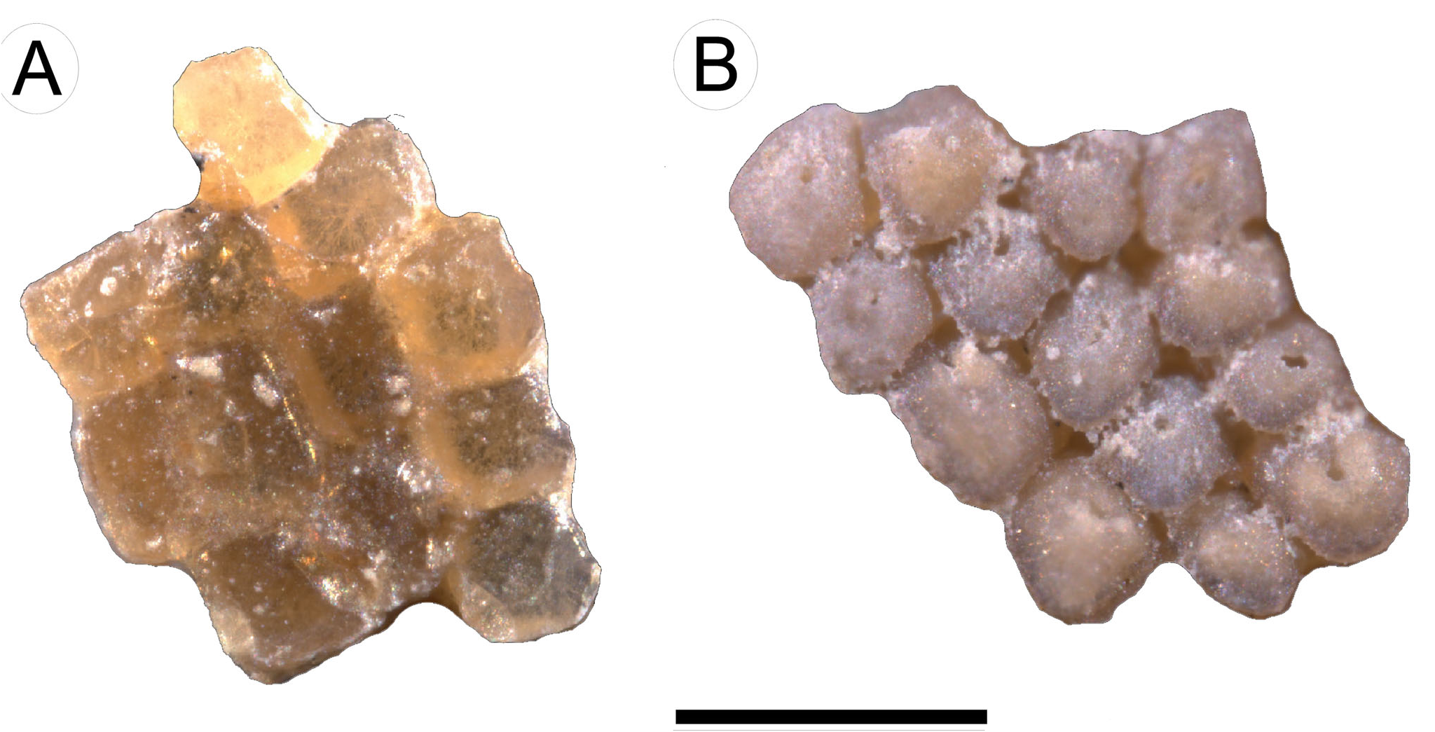
Dermal denticles. As in extant platyrhinids (see Deynat 2005), the entire body of † E. bolcensis comb. nov. is covered with numerous small dermal denticles that form a continuous and regular covering ( Fig. 7 View Figure 7 ). Denticle size is quite uniform across the body. Some denticles were extracted from the dorsal side of the disc of MGP-PD 26279C/80C for a detailed analysis. Their crown is about 200 µm wide and rhomboidal or lozenge-shaped ( Fig. 7 View Figure 7 ). The denticle root is deeper than the crown height and a nutritive foramen can be recognized near the centre. Extant thornbacks and † Tingitanius possess parallel rows of enlarged dermal denticles (thorns) over the posterior part of the disc and tail, a condition that was regarded as diagnostic for platyrhinids. However, this is not the case for † Eoplatyrhina bolcensis comb. nov. and † Tethybatis , in which thorns are completely absent ( Carvalho 2004), possibly representing a feature supporting this sister-group relationship.
No known copyright restrictions apply. See Agosti, D., Egloff, W., 2009. Taxonomic information exchange and copyright: the Plazi approach. BMC Research Notes 2009, 2:53 for further explanation.
|
Kingdom |
|
|
Phylum |
|
|
Class |
|
|
Order |
|
|
Family |
|
|
Genus |
Eoplatyrhina bolcensis ( Heckel, 1851 )
| Marram ̀, Giuseppe, Carneval, Giorgio, Claeso, Kerin M., Naylo, Gavin J. P. & Kriwe, Jurgen 2020 |
Platyrhina bolcensis
| Molin 1860 |
Platyrhina bolcensis
| Molin 1860 |
Platyrhina bolcensis
| Molin 1860 |
Platyrhina bolcensis
| Heckel 1851 |
Platyrhina ’ bolcensis
| Heckel 1851 |