
Pyrochroa coccinea (Linnaeus)
|
publication ID |
https://doi.org/10.11646/zootaxa.4966.3.5 |
|
publication LSID |
lsid:zoobank.org:pub:4159E660-EA53-48E2-9724-DDB48DB6FC43 |
|
DOI |
https://doi.org/10.5281/zenodo.4736771 |
|
persistent identifier |
https://treatment.plazi.org/id/038587AA-CA59-9067-FF4A-4654FAF87914 |
|
treatment provided by |
Plazi |
|
scientific name |
Pyrochroa coccinea (Linnaeus) |
| status |
|
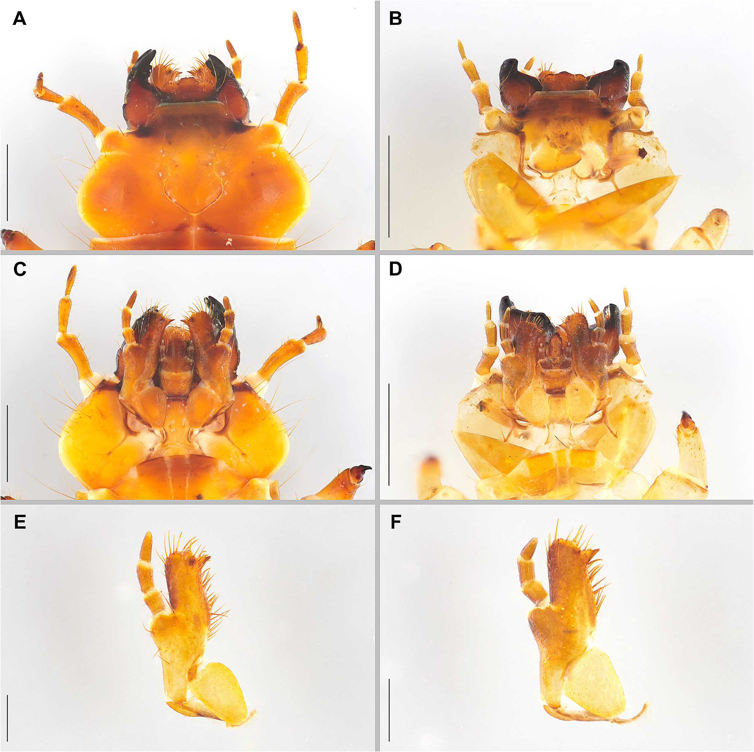
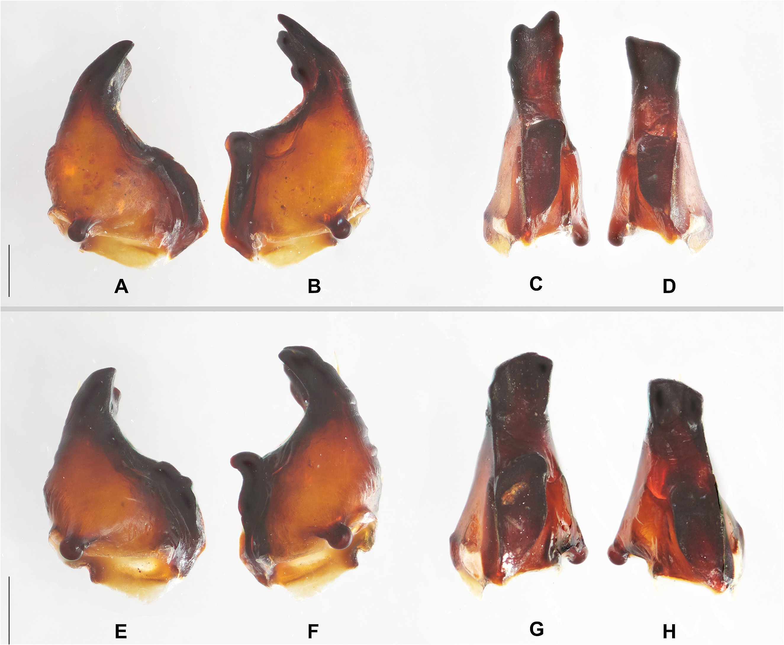
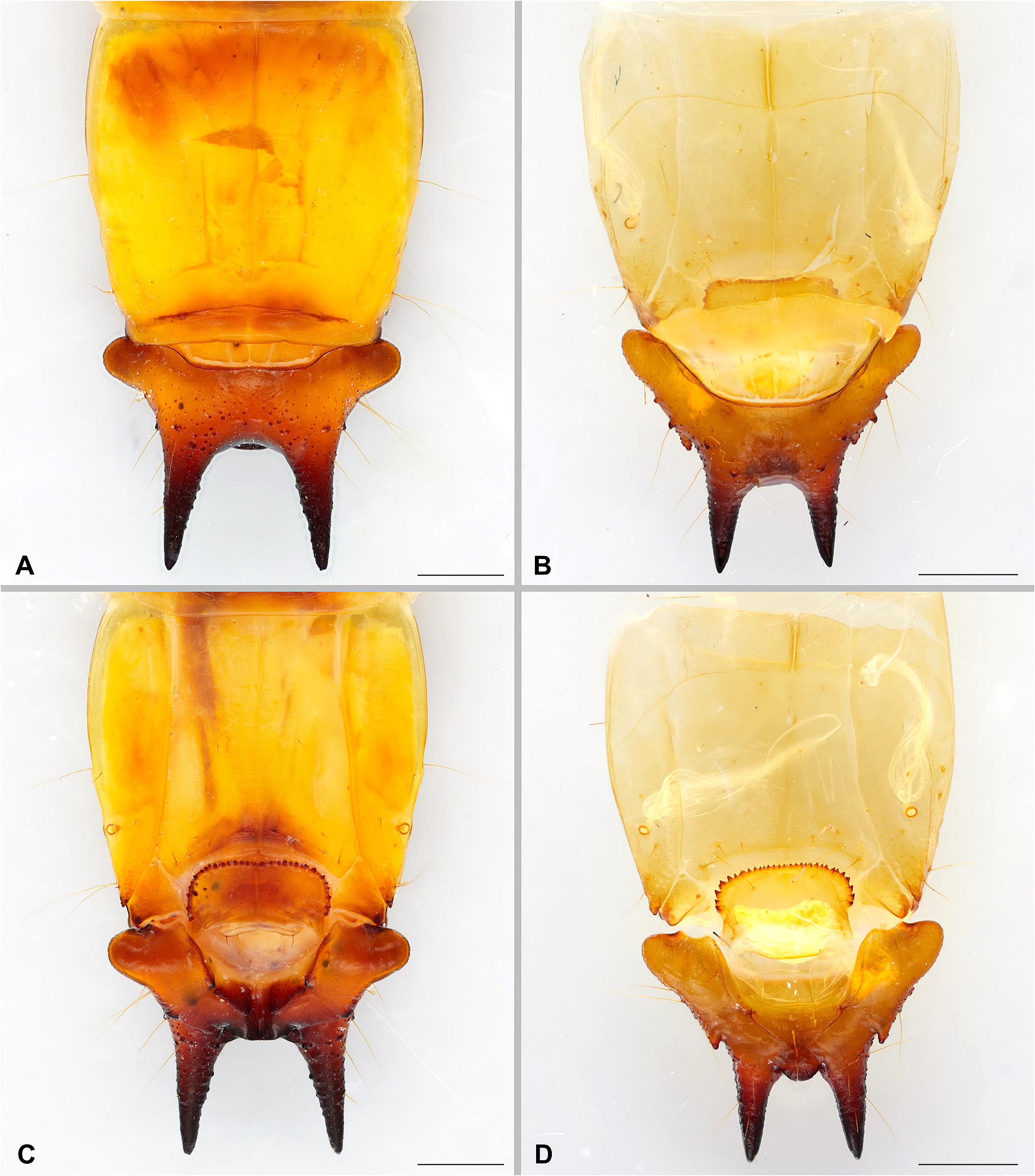
Pyrochroa coccinea (Linnaeus) ( Figs 1A View FIGURE 1 , 2A, C, E View FIGURE 2 , 3 A–D View FIGURE 3 , 4A, C View FIGURE 4 , 5A View FIGURE 5 )
Diagnosis. The most diagnostic characters are structures associated with the highly sclerotized urogomphal plate: lateral lobes rounded; long, slender, straight, subparallel urogomphi; urogomphal lip ventrally furrowed and two urogomphal pits with parallel rugulae. Other diagnostic characters vs. P. serraticornis are shown in Table 1 View TABLE 1 .
Description of mature larva. Body length 3.5 cm (from mesal labral apex to apices of urogomphi) and maximum width 5.1 mm (across widest portion of urite VIII) ( Fig. 1A View FIGURE 1 ). Body orthosomatic with sides subparallel, moderately sclerotized except most of cephalic capsule, mandibles, and urogomphal plate which are more heavily sclerotized; body vestiture consisting of short to moderately elongate, scattered setae. Thoracic and abdominal terga lacking parabasal ridges. Head and body dark yellowish to amber, melanisation much darker in areas of heavy sclerotization such as mandibles, urogomphi, urogomphal lip and urogomphal pits.
Head. Prognathous, flattened, exserted from prothorax, about 4.5 mm width ( Figs 2A, C View FIGURE 2 ). Epicranial suture lyriform with stem short, frontal arms complete nearly to antennal insertions, endocarinae absent ( Fig. 2A View FIGURE 2 ). Free, symmetrical trilobate labrum, central lobe with a median thin notch, lateral lobes with high concentration of stout setae ( Fig. 2A View FIGURE 2 ). Two pairs of stemmata on each lateral side. Antennal insertions fully exposed; antennae moderately long, slender, 3-segmented; antennomere I slightly curved outwards, narrower in the middle area, wider apically; sensorium of segment 2 small, conical; antennomere III narrower than I and II, as long as II, acutely rounded apically; setae distributed only on the inner surface of I, well distributed on all the surface of II and III. Mouthparts retracted ( Figs 2 A, C View FIGURE 2 ). Mandibles heavily sclerotized, movable, asymmetrical, molar area of mandibles well developed; left mandible bigger, bearing a blunt molar tooth, apex rounded bidentate with two smaller subapical teeth, inner surface slightly concave ( Figs 3B, C View FIGURE 3 ); apex of right mandible sub-securiform, inner surface heavily concave so as to make it appear bidentate from a ventral view ( Figs 3A, D View FIGURE 3 ). Maxillae ( Fig. 2E View FIGURE 2 ) each with 1-segmented cardo which is diagonally folded upward upon itself toward the stipes and thus appearing 2-segmented; a well-developed, undivided, pad-like maxillary articulating area; ventral surface of stipes quite glabrous, bearing few scattered setae; galea and lacinia fused to form maxillary mala; mala bearing stout apical and adoral spiniform setae and a welldeveloped pointed uncus at apico-adoral margin; 3-segmented, filiform maxillary palpus, palpomere II about 1.4X length of I, III subequal in length to II; I–II bearing stout long spiniform setae, III tapering distally, acutely rounded apically. Labium with mentum trapezoidal ( Fig. 2C View FIGURE 2 ); submentum with a median pair of stout setae, shape elongate with sub-basal hourglass-shaped narrowing, apical margin slightly more heavily sclerotized ( Fig. 2C View FIGURE 2 ); ligula well developed, elongate, with several pointed setae on each side in the direction of the labial palps; each labial palpus short, 2-segmented, I twice as long as II. Hypostomal rods ( Fig. 2C View FIGURE 2 ) well developed, divergent; gular sutures separate ( Fig. 2C View FIGURE 2 ).
Thorax and Abdomen. Thorax flattened, with sides of prothorax subparallel, meso- and metathorax rounded ( Fig. 1A View FIGURE 1 ); cervicosternum divided into three plates ( Fig. 2C View FIGURE 2 ). Legs well developed, moderately short, 5-segmented including tarsungulus, vestiture consisting of sparse, short setae. Abdomen flattened, with sides slightly converging forward, moderately sclerotized; tergites I–VII subequal in length and width; tergite VIII approximately 2 times as long as others ( Fig. 1A View FIGURE 1 ). Sternite VIII emarginate apically ( Fig. 4C View FIGURE 4 ). Ventrolateral margins of abdominal laterotergite VIII emarginate, with lanceolate shape ending with more sclerotized acute apex ( Fig. 4C View FIGURE 4 ). Tergite IX divided into four plates ( Fig. 4A View FIGURE 4 ), hinged, capable of considerable dorso-longitudinal movement, extending ventrally, thus forming the urogomphal plate, widest basally where it forms well developed rounded lateral lobes ( Figs 4A, C View FIGURE 4 ); surface of urogomphal plate bearing numerous, well-developed, callosities and several setigerous calli, in particular, long setae are associated with three pairs of calli on the dorsolateral surfaces ( Fig. 4A View FIGURE 4 ), one pair of calli at the base of urogomphi and one pair of calli on the ventrolateral inner surface of urogomphi ( Fig. 5A View FIGURE 5 ); urogomphi heavily sclerotized, long, slender, straight, subparallel, tapering and acuminate apically; ventral surface of urogomphal plate sharply excavate basally at articulation with sternites IX and X, excavation narrowing distally to bases of urogomphi and urogomphal lip, almost to form a tube under the urogomphal lip ( Fig. 4C View FIGURE 4 ). Urogomphal plate possessing a heavily sclerotized urogomphal lip ventrally ( Figs 4A, C View FIGURE 4 , 5A View FIGURE 5 ), between the two, heavily sclerotized urogomphal pits ( Fig. 5A View FIGURE 5 ), which, in turn, arise distally between the heavily sclerotized fixed urogomphi, and bear characteristic parallel rugulae. Sternite IX broadly transversely U-shaped ( Fig. 4C View FIGURE 4 ), slightly invaginate in correspondence of a middle, thin, weak unsclerotized line, partially recessed into shallow emargination of sternite VIII, possessing continuous semi-circular arch of approximately 34–36 well-developed asperities along anterior margin; ventrolateral margin heavily sclerotized forming a blunt tooth ( Fig. 4C View FIGURE 4 ). Segment X reduced, transversely ovate, with basal margin rounded, recessed into emarginations of sternite IX, visible ventrally ( Fig. 4C View FIGURE 4 ).
Spiracles. One pair of well-developed ovate thoracic spiracles ventrolaterally positioned on laterotergites along anterior margin of mesothorax. Paired, ovate abdominal spiracles, subequal in size, located on dorsolateral margin of abdominal tergite I ( Fig. 1A View FIGURE 1 ) and ventrolateral margins of abdominal laterotergites II–VII; paired spiracles of abdominal laterotergite VIII annular-ovate, located ventrolaterally at distal 1/3 of its length ( Fig. 4C View FIGURE 4 ).


No known copyright restrictions apply. See Agosti, D., Egloff, W., 2009. Taxonomic information exchange and copyright: the Plazi approach. BMC Research Notes 2009, 2:53 for further explanation.
|
Kingdom |
|
|
Phylum |
|
|
Class |
|
|
Order |
|
|
Family |
|
|
Genus |