
Pliciloricus ukupachaensis, Sørensen & Herranz & Grzelak & Shimabukuro & Kristensen & Zeppilli, 2023
|
publication ID |
https://doi.org/ 10.5852/ejt.2023.879.2169 |
|
publication LSID |
lsid:zoobank.org:pub:BE6ED0BD-4A61-4F50-A8C2-BE84C6F00656 |
|
DOI |
https://doi.org/10.5281/zenodo.8155363 |
|
persistent identifier |
https://treatment.plazi.org/id/AEF3321F-BCFB-4FF2-A8EC-81BF8E929462 |
|
taxon LSID |
lsid:zoobank.org:act:AEF3321F-BCFB-4FF2-A8EC-81BF8E929462 |
|
treatment provided by |
Felipe |
|
scientific name |
Pliciloricus ukupachaensis |
| status |
sp. nov. |
Pliciloricus ukupachaensis View in CoL sp. nov.
urn:lsid:zoobank.org:act:AEF3321F-BCFB-4FF2-A8EC-81BF8E929462
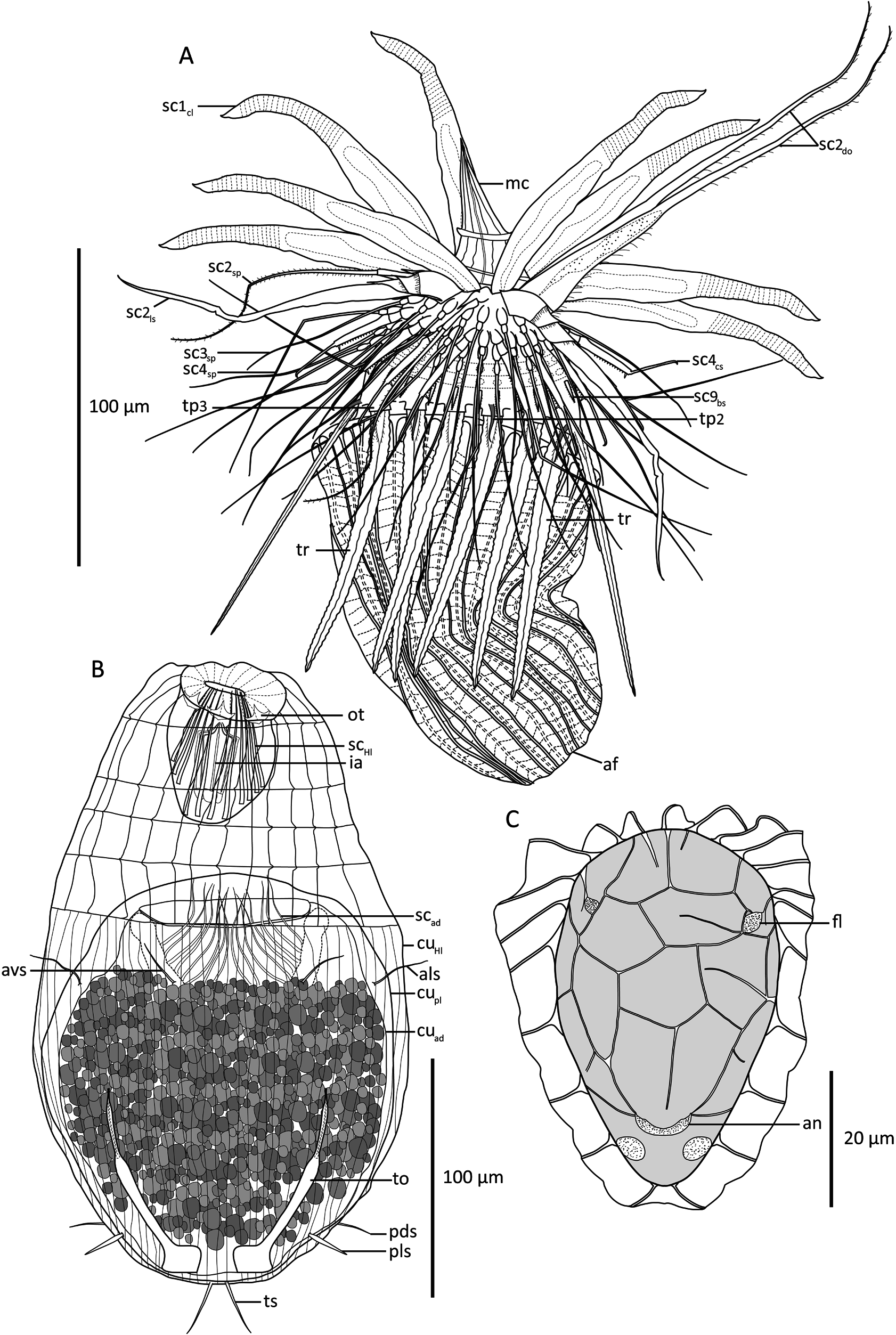
Figs 2–8 View Fig View Fig View Fig View Fig View Fig View Fig View Fig
Diagnosis
Adult Pliciloricus with a mouth cone with narrow basis, but getting conspicuously broader, before gradually narrowing again towards distal end. Introvert Row 1: females with eight regular, club-shaped clavoscalids with 23–25 annulated rings on distal ⅓; Introvert Row 2: three regular spinoscalids alternating with four leg-shaped scalids with three, articulating proximal units and long, spinous end-piece; double organ with fused bases, extending into long, tapering distal tips. Introvert Row 4: fifteen claw-like scalids with serrated shafts, articulating with thinner distal parts, with curved tips. Neck with fifteen single trichoscalids. Trichoscalid plates inconspicuous. Lorica with 22 plicae inclusive broader midventral plica. Anterior lorica margin smooth. Posterior part of lorica forming large anal field with distinct, asymmetrical ornamentation of pentagonal and hexagonal fields, formed by external cuticular ridges. Male morphology unknown.
Etymology
The species name, ‘ ukupachaensis ’, is derived from ‘Uku Pacha’ – the underworld or ‘inner world’ according to Incan mythology. The name refers to the habitat of this species, in close proximity to the Atacama Trench that reaches into the underworld.
Material examined
Holotype CHILE • ♀; continental slope off Antofagasta, and near the rim of the Atacama Trench ; st. 1; 23°48.72ʹ S, 70°50.04ʹ W; depth 2560 m; 6 Mar. 2018; R / V Sonne SO261; deep-sea mud; mounted for LM in Fluoromount G on HS slide; NHMD 1177413 . GoogleMaps
Paratypes CHILE • 1 ♀; same collection data as for holotype; mounted for SEM; NHMD 1177421 GoogleMaps • 1 Higgins larva containing a postlarva with an adult inside; same collection data as for holotype; mounted for LM in Fluoromount G on HS slide; see also Table 1 View Table 1 and Fig. 1 View Fig ; NHMD 1177414 GoogleMaps .
Description
Adult female
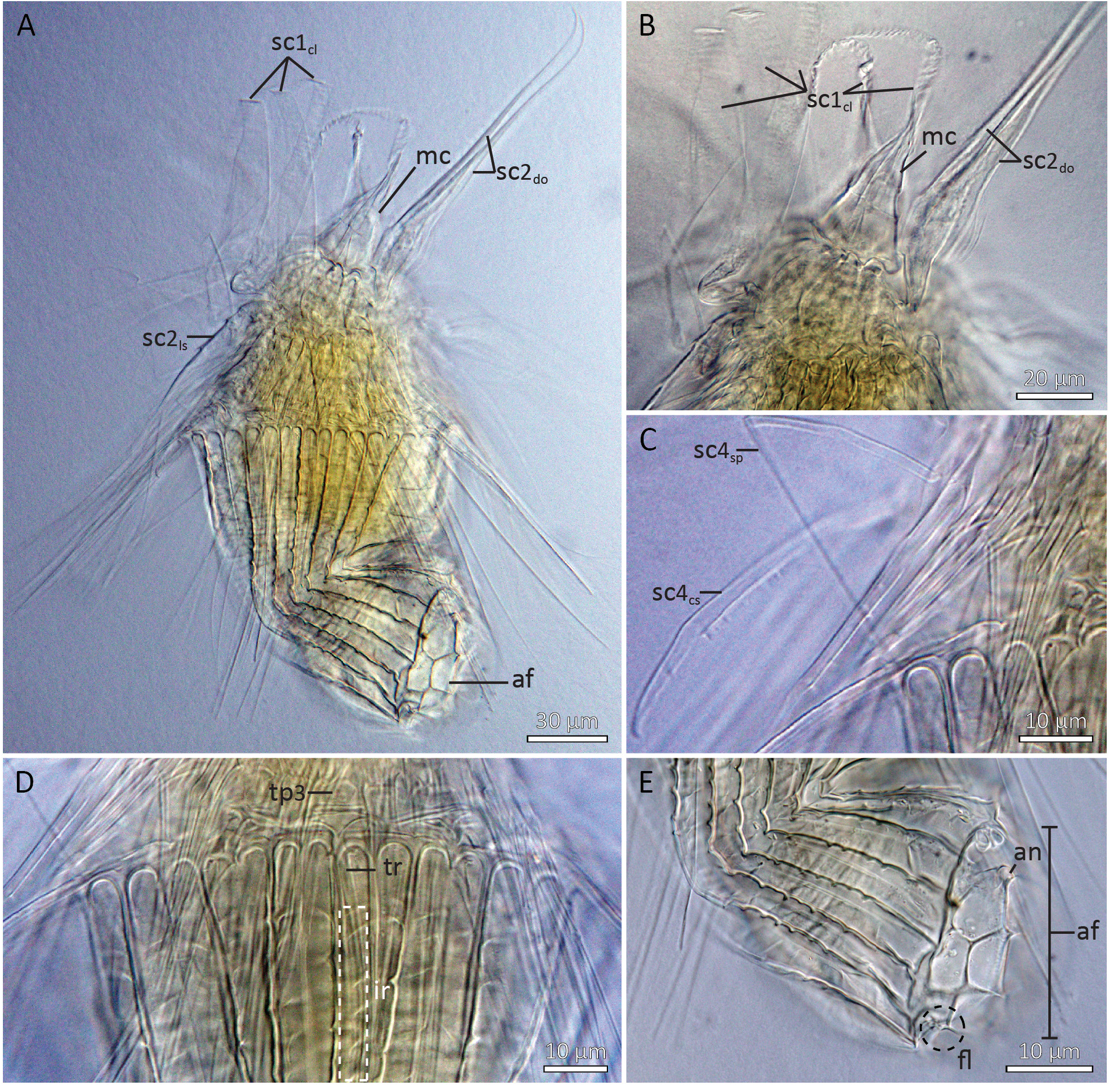
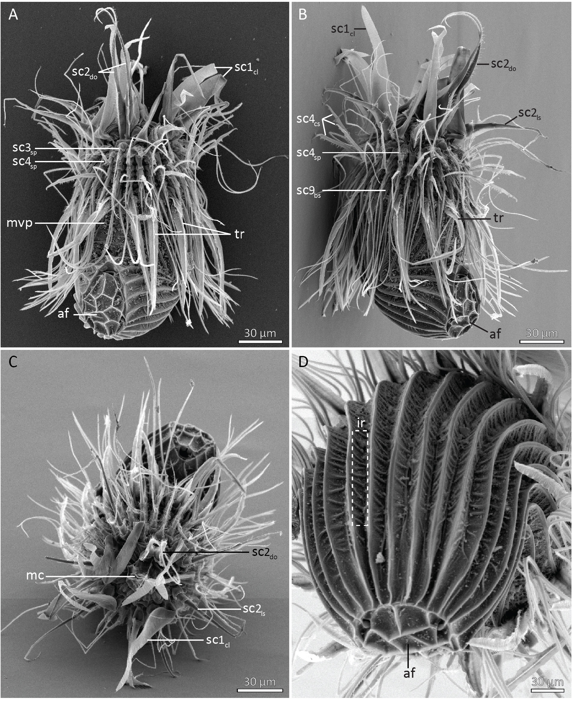
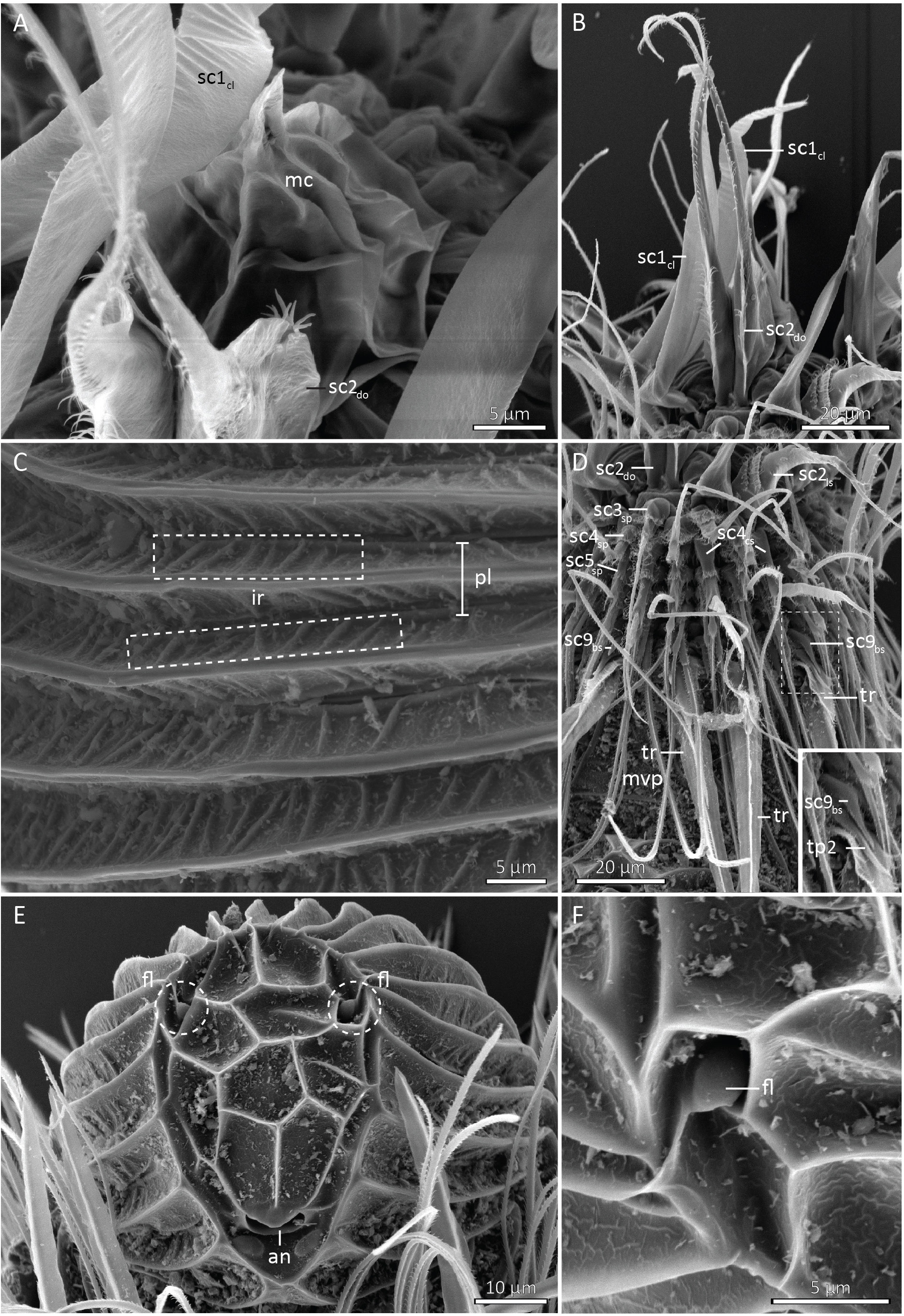
Adult females consist of a head with mouth cone and introvert with nine rows of scalids, a neck region with one row of trichoscalids, a plicated lorica and an anal field with asymmetrically arranged fields ( Figs 2A, C View Fig , 3–6 View Fig View Fig View Fig View Fig , 7A View Fig ). The female holotype measures 240 µm in total length and 100 µm in width at its widest point, i.e., near the anterior lorica margin. The mouth cone measures 68 µm in length, has a narrow basis, but broadens out to reach its maximum width of 25 µm about 1/5 from basis, before gradually tapering again towards the distal tip ( Figs 2A View Fig , 4A–B View Fig , 6A View Fig ). It is slightly retracted in both the holotype and adult paratype, which makes it appear broadest basally. The mouth cone is supported by oral ridges, and a narrow buccal tube is visible inside it. The pharynx does not have any distinct internal structures, and no placoids were observed.
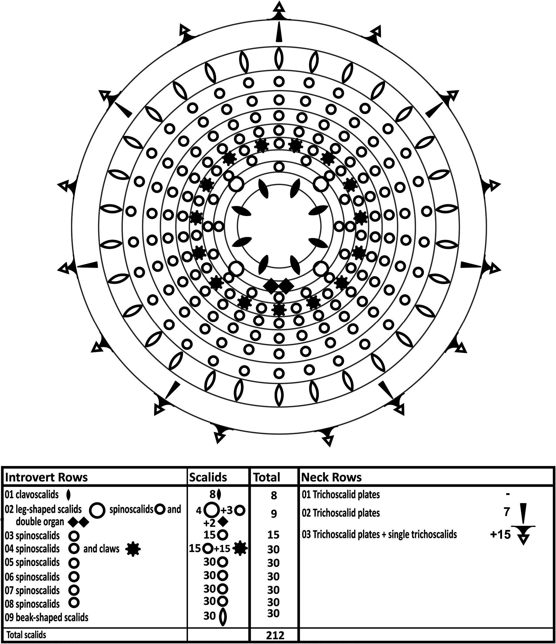
The introvert has nine rows of scalids ( Fig. 3 View Fig ). The anteriormost Row 1 consists of eight club-shaped, 134 µm long clavoscalids. The clavoscalids are covered with extremely fine hairs and consist of a narrow base, a smooth, flattened part representing ⅔ of the total scalid length, and a distal end-piece with about 23–25 annulations; they terminate in small, anteriorly bent hooks ( Figs 2A View Fig , 3 View Fig , 4A–B View Fig , 5A–C View Fig , 6A–B View Fig , 7A View Fig ).
Row 2 consists of four leg-shaped scalids, three regular spinoscalids, and two scalids fused into a ventral double organ. Each leg-shaped scalid (length: 125 µm) consists of a proximal part with three, fringed, articulating units and a spinous end-piece with two partial constrictions about ⅓ from the distal, anteriorly curved tip. The three regular spinoscalids (length: 91 µm) alternate with the leg-shaped scalids and are located middorsally and midlaterally on the introvert. Each spinoscalid is composed of three units: a short, proximal basis with a distal spike, a short cylindrical mid-piece, and a long, acicular end-piece, covered with minute hairs; the end-piece represents more than 4/5 of the total scalid length, and has a distinct bend ⅓ from the distal bending tip. The double organ consists of two appendages (length: 134 µm) with swollen and fused bases, representing ⅓ of the total length; they narrow abruptly into considerably thinner, distally curved end-pieces; the bases have a line of hair along their inferior margins, whereas the end-pieces have hair along both the inferior and superior margins ( Figs 2A View Fig , 3 View Fig , 4A–B View Fig , 5A–C View Fig , 6B, D View Fig , 7A View Fig ).
Row 3 consists of fifteen uniform spinoscalids (length: 58 µm), composed of two short, articulating, ovate bases and a long acicular end-piece, densely covered with minute hairs ( Figs 2A View Fig , 3 View Fig , 5A View Fig , 6D View Fig ).
Row 4 consists of fifteen regular spinoscalids alternating with fifteen claw-like scalids. The spinoscalids (length: 98 µm) are composed of two short, articulating, ovate bases and a long acicular end-piece with a distinct bend ⅓ from its proximal end; end-pieces are covered with minute hairs. Each claw-like scalid (length: 44 µm) is composed of a short basis with hairy, distal margin, a shaft (½ of scalid length) with serration along its inferior margin, and a thinner distal part, with a curved tip; some claw-shaped scalids bend medially, suggesting an articulation between the shaft and the end-piece ( Figs 2A View Fig , 3 View Fig , 4C View Fig , 5A–B View Fig , 6C View Fig , 7A View Fig ).
Rows 5 to 8 with spinoscalids that are very uniform. Each row has thirty spinoscalids (lengths: 68 to 73 µm), each consisting of a short basis composed of two ovate units and a long, smooth acicular end-piece with short hairs along their lateral margins ( Figs 2A View Fig , 3 View Fig , 6D View Fig ).
Row 9 consists of thirty short, beak-shaped scalids (length: 10 µm) ( Figs 2A–B View Fig , 3 View Fig , 5B View Fig , 6D View Fig ).
The neck has fifteen uniform, single trichoscalids ( Figs 2A View Fig , 3 View Fig , 4D View Fig , 5A–B View Fig , 6D View Fig ). The trichoscalids are 79 µm in length, blade-shaped, with a median, longitudinal ridge, and a small, triangular plate covering the proximal attachment point.
Trichoscalid plates are extremely weakly defined. There might, however, be a very weak indication of a row with fifteen trichoscalid plates at the base of each trichoscalid. This indication can only be observed with LM though ( Fig. 4D View Fig ), and not with SEM, which suggests that the plates are mostly intracuticular thickenings, rather than external structures. Interestingly, however, minute (13 µm long) appendages are attached anterior to seven of the fifteen indistinct trichoscalid plates ( Figs 2A View Fig , 3 View Fig , 6D View Fig inset). These appendages are arranged with one in middorsal position anterior to the corresponding trichoscalid, and are otherwise present radially anterior to every second trichoscalid. This pattern corresponds to the arrangement of Row 2 trichoscalid plates in other species of Pliciloricus (see for instance the description of P. apteryx Sørensen et al., 2022 ), and we therefore interpret these short appendages ( Fig. 6D View Fig inset) as Row 2 trichoscalid plates. Accordingly, the weakly defined trichoscalid plates with attached trichoscalid represent Row 3, whereas there are no trichoscalid plates of Row 1 ( Fig. 3 View Fig ).
No particular structures were noted in the thorax region. The lorica is composed of 22 plicae, including a broader midventral plica. Numerous, oblique structures, referred to as ‘interplical radii’, radiate from the primary double ridges of the plicae. The interplical radii are easily observed in LM as well as SEM ( Figs 4D View Fig , 5D View Fig , 6C View Fig ). The anterior lorica margin is straight, and there is no indication of marginal spikes ( Figs 2A View Fig , 4D View Fig ). In both adult specimens, the holotype and the paratype mounted for SEM, the lorica has a highly characteristic, nearly 90° bend on the ventral side ( Figs 4A View Fig , 5 View Fig , 7A View Fig ).
Posteriorly, the lorica forms a large anal field ( Figs 2A, C View Fig , 4A, E View Fig , 5A, D View Fig , 6E–F View Fig ). The anal field is ovoid and limited by a strong ridge, which also marks the posterior limit of the lorical plicae. Numerous cuticular ridges present within the anal field form different pentagonal and hexagonal fields. Whereas the fields closest to the ventral side show a certain level of bilateral symmetry, the shape and arrangement of the remaining fields are more irregular, and overall the anal field ornamentation must be considered as asymmetrical. A single pair of P-flosculi are present in the dorsalmost portion of the anal field; a crescentic anal opening, flanked by a pair of oval thickenings, is present close to the ventral margin of the area.
Internal anatomy was difficult to observe, but two groups of circular muscles are present in the trunk, arranged as an anterior and a posterior group.
Last instar Higgins larva and postlarva
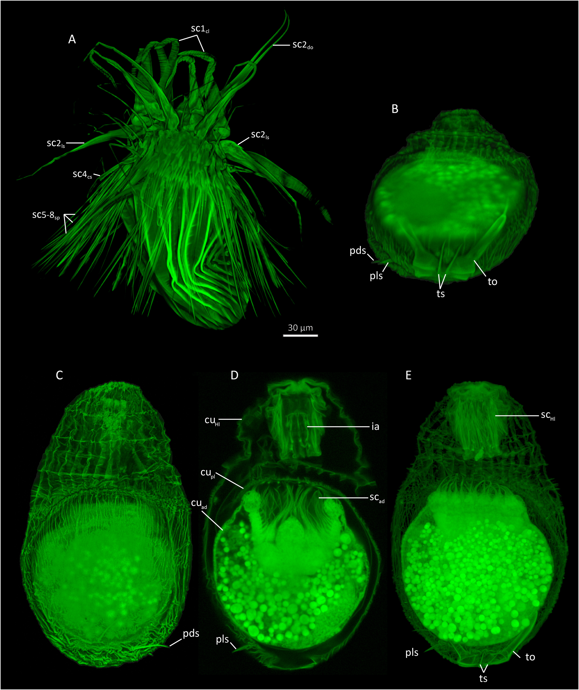
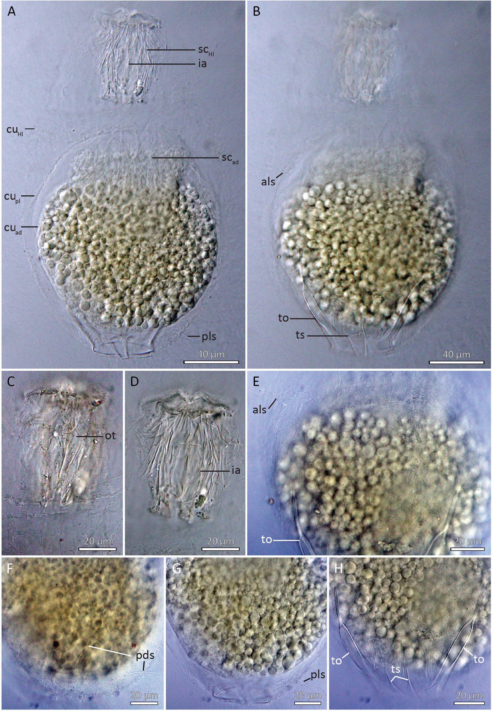
Last instar Higgins larva with head and trunk, the latter divided into a thorax region and a loricated abdomen ( Figs 2B View Fig , 7B–E View Fig , 8 View Fig ). It measures 267 µm in length and 158 µm in width at its broadest point, medially on the abdomen.
The head of the single, paratypic larva was retracted and detailed information on the introvert morphology could not be obtained. Pharyngeal armature consists of oral teeth with tripartite anterior tips and inner armature ( Figs 2B View Fig , 7D View Fig , 8A, C–D View Fig ). The thorax has four transverse folds and numerous (> 22) longitudinal folds. The abdomen has numerous (> 90) indistinct longitudinal lines, which are so weakly developed that they can hardly be referred to as plicae. Two pairs of anterior, simple, unbranched setae are located on the abdomen about ¼ from its anterior margin: anterolateral setae measure 25 µm in length, whereas anteroventral setae are slightly shorter, measuring 22 µm in length. Posterior setae include thin and simple posterodorsal setae (length: 36 µm), swollen and stiff posterolateral setae (length: 18 µm), and the acicular and rigid terminal setae (length: 25 µm) attaching between the toes ( Figs 2B View Fig , 7B–E View Fig , 8A–B, E–H View Fig ).
The toes are long and slender (total toe length: 91 µm) and divided into three portions: relatively broad bases (62 µm), slender mid-pieces each with a central, internal canal (length: 24 µm), and short, abruptly narrowing tips (length: 5 µm) ( Figs 2B View Fig , 7B, E View Fig , 8B, E, H View Fig ).
The Higgins larva contains a postlarva, which is nothing but a cuticle without any further, observable structures. The postlarva is nearly globular and about 152–155 µm in diameter. A developing adult stage of unknown sex is present inside the postlarva, filled with refringent vesicles. The adult scalids of the introvert appear to be well-developed, but internal organs are still not developed, and the lorica is so thin that it appears undifferentiated ( Figs 2B View Fig , 7B–E View Fig , 8A–B View Fig ).

| R |
Departamento de Geologia, Universidad de Chile |
| V |
Royal British Columbia Museum - Herbarium |
No known copyright restrictions apply. See Agosti, D., Egloff, W., 2009. Taxonomic information exchange and copyright: the Plazi approach. BMC Research Notes 2009, 2:53 for further explanation.
|
Kingdom |
|
|
Phylum |
|
|
Order |
|
|
Family |
|
|
Genus |