
Platymessa, Mello-Leitao, 1941
|
publication ID |
https://doi.org/10.11646/zootaxa.4085.1.2 |
|
publication LSID |
lsid:zoobank.org:pub:5093CA8A-2BBC-420D-B7D2-B4B4C0E285D6 |
|
DOI |
https://doi.org/10.5281/zenodo.6078829 |
|
persistent identifier |
https://treatment.plazi.org/id/038487AE-BB4F-4C71-18BE-FA9A8C310C36 |
|
treatment provided by |
Plazi |
|
scientific name |
Platymessa |
| status |
|
Platymessa View in CoL h-inscriptum Mello-Leitão, 1941
Platymessa View in CoL H-inscripta Mello-Leitão, 1941: 167, fig. 2.
Cynorta View in CoL h-inscripta: H. Soares 1970: 325.
Platymessa View in CoL h-inscripta: Kury 2003: 81.
Platymessa View in CoL h-inscriptum: Kury & Alonso-Zarazaga 2011: 51 Platymessa nigrolimbata Mello-Leitão, 1941: 168 View in CoL , fig. 3; B. Soares 1945: 344; Kury 2003: 81. SYN. NOV.
Type data. Platymessa h-inscriptum: COLOMBIA, Santander [and not Tolima as wrongly extrapolated by Kury 2003], Espinal, ♂ holotype ( MNRJ 282 View Materials , examined); Platymessa nigrolimbata : COLOMBIA , Santander, San Gil: ♀ holotype ( MNRJ 463 View Materials , examined) ; Boyacá, La Uvita, 2 ♂ and 1♀ paratypes ( MNRJ 58 View Materials , examined).
Other material examined. COLOMBIA, Boyacá, Tipacoque, Vereda La Calera , (06°23.8’ 91’’N; 072° 43.4’ 10’’W), 2800m, 1-5 April 2013, M. Medrano, 1 ♂ and 11 ♀ (ICN-AO 1186) ; Santander, Zapatoca, Vereda La Cacica, Reserva La Montaña Mágica ( 6°50,044'N; 73°18,241'W), 1964 m, 6 November 2013, C. Perafán and D. Martínez, 1 ♂ and 2 ♀ (ICN-AO 1384). GoogleMaps
Taxonomic background of Platymessa h-inscriptum. By the original description of P. h-inscriptum, Mello- Leitão (1941) used an upper-case H and feminine ending. Those were corrected later to lower-case ( Kury 2003) and neuter gender ( Kury & Alonso-Zarazaga 2011), although the hyphen should be kept. H. Soares (1970) (probably by a lapse) during the description of another species used this species (as a comparison) in combination with Cynorta , thus following the reasoning of Goodnight & Goodnight (1953), although these authors had not mentioned Platymessa in their extensive synonymy.
Remarks. The characters allowing the recognition of P. nigrolimbata as a distinct species as proposed by Mello-Leitão (1941) have not been explicitly stated by him, but by reading the descriptions the differences clearly rely on (1) the pattern of the scutal white blots and (2) tarsomere counts. All of these fall within the range of variation as shown here.
Diagnosis. Scutal area I with a pair, area V with two small blunt tubercles. Ladder-blot with well-marked divergent rails. Tarsus III hexamerous. Length of legs ca. 7/14/9 / 11 mm.
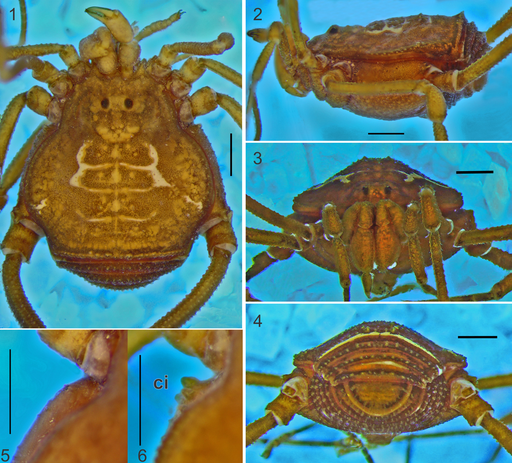
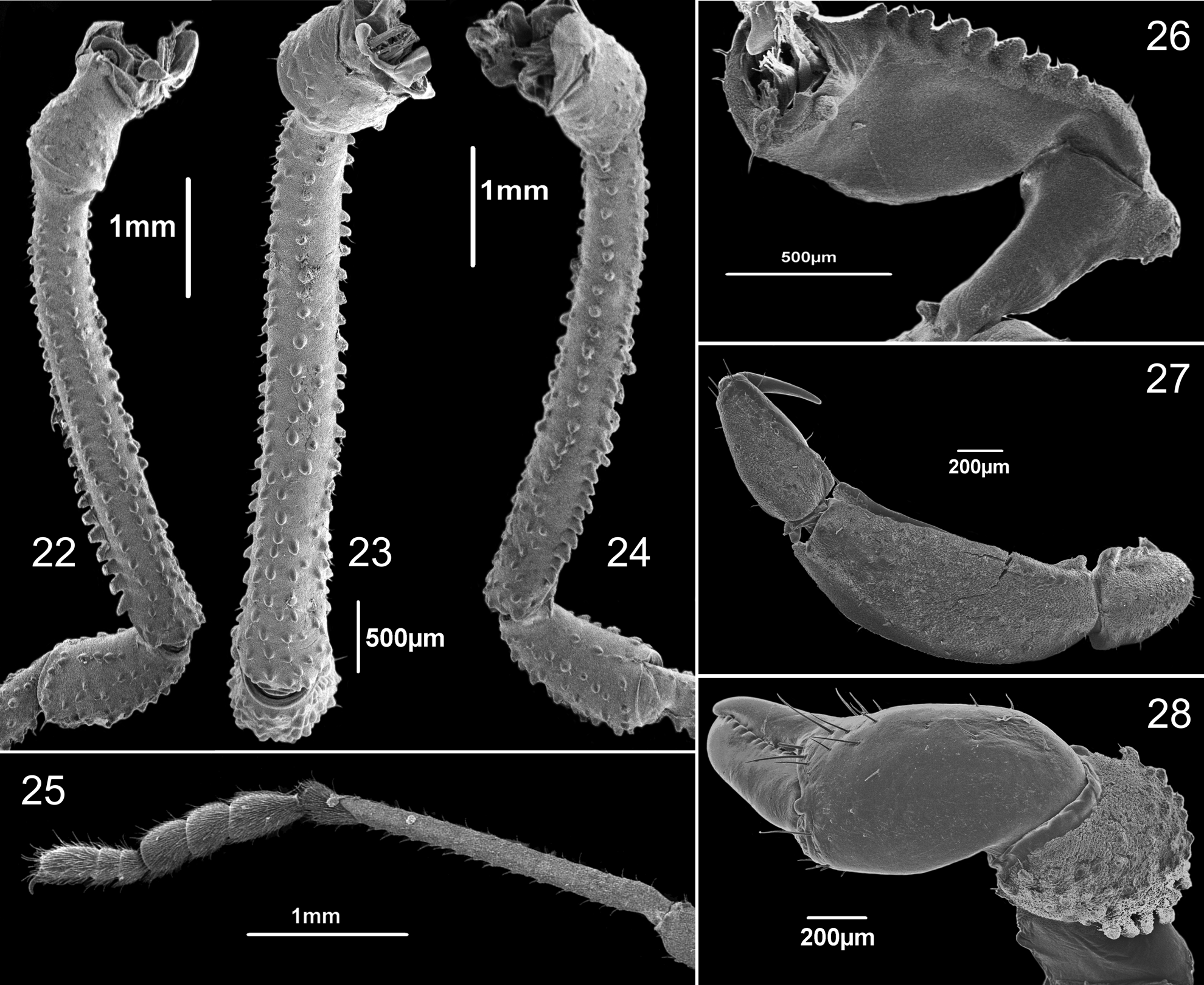
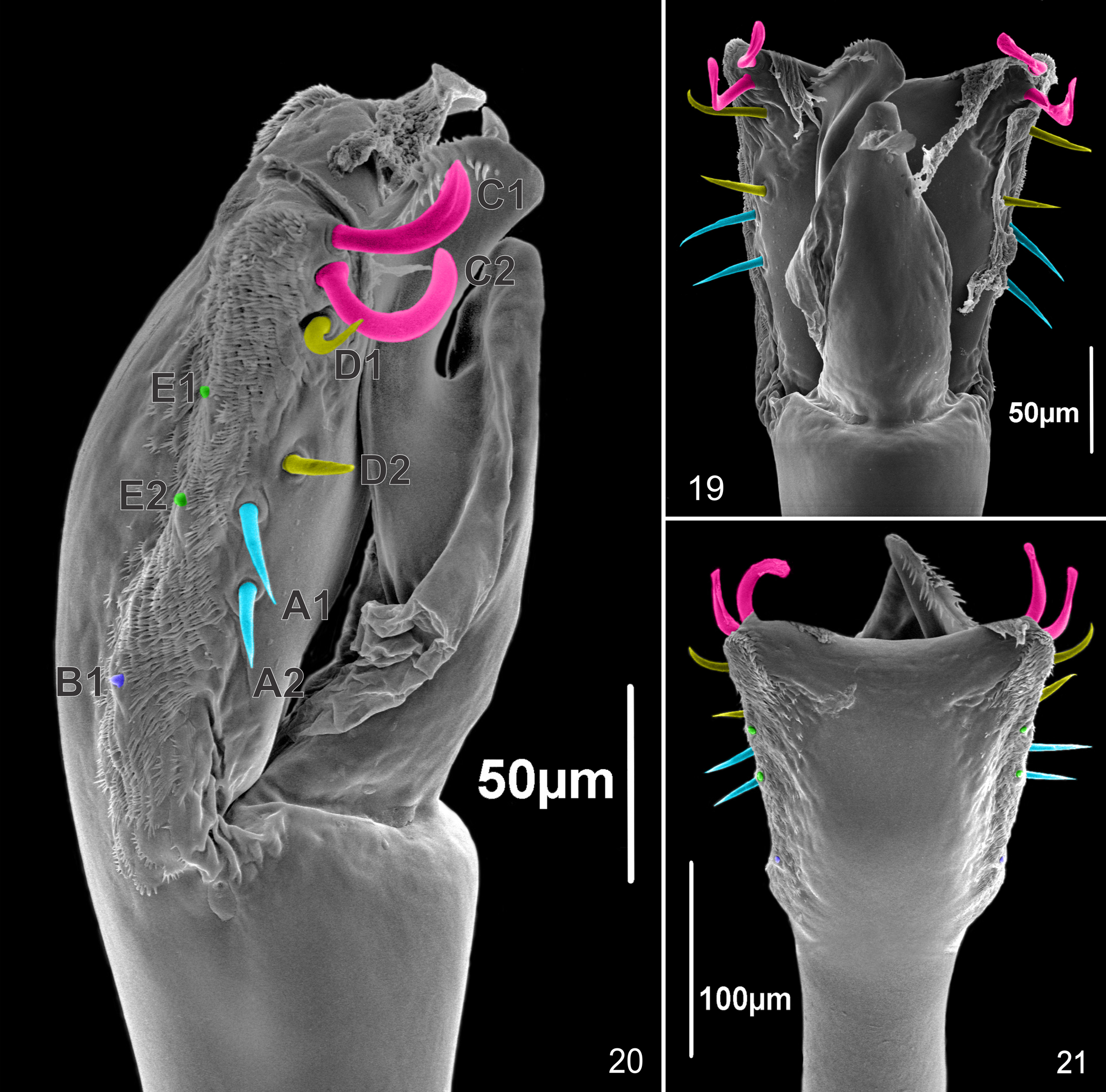
Description of male holotype (with extra figures from other specimens). Measurements. CL = 1.68, AL = 2.77, CW = 2.68, AW = 4.39, Fe IV = 4.13, Ti IV = 3.06. Dorsum ( Figs. 1 View FIGURES 1 – 6 , 7 View FIGURES 7 – 18 ). Dorsal scutum beta-shaped, areas I, III and V with small tubercles and area IV with minute tubercles more separately that the ones of the other areas. Lateral margins with a row of minute tubercles at the bulge. Posterior margin of scutum straight in dorsal view with a row of small tubercles. All tergites and anal operculum finely granular and unarmed. Venter ( Figs 2, 4 View FIGURES 1 – 6 ). Free sternites finely granular, unarmed. Chelicerae ( Fig. 28 View FIGURE 22 - 28 ). Basichelicerite with posterior row of many tubercles, of which six are larger. Pedipalps ( Fig. 26, 27 View FIGURE 22 - 28 ). Trochanter with strong ventral process. Femur with pronounced dorsal keel, with a ventral row of eleven setiferous tubercles and a strong mesodistal process. Shallow slit along tibia mesal surface, separating the dorsal and ventral sides. Legs ( Figs 5, 6 View FIGURES 1 – 6 and 22-24 View FIGURE 22 - 28 ). Coxa IV with groin warts. Trochanter IV with small retro-distal apophysis. Femur IV slightly arched with two longitudinal ventral rows of small tubercles along his entire length, increasing in size apically. Patella IV substraight with small setiferous tubercles. Tarsal counts: 6-6/?- 10/6-6/7 -7. Color. Body and appendages brown. Ladder mask yellowish white. Genitalia ( Figs 19-21 View FIGURE 19 – 21 ). Penis ventral plate subrectangular, narrower basally, with lateral borders parallel and distal border slightly concave; dorsal apophysis of glans subsquare, wattle long. Macrosetae B located in the proximal third of the ventral plate. The shapes and organization of macrosetae are as follows: MS C1-C2 large, curved and flat at the apex; MS D1-D2 large, cylindrical, just curved at the apex, more dorsal than the others; MS D1 as large as C2 and moderately curved, but not flattened, MS D2 more basal and straight (see Discussion for details). MS A1-A2 large, straight, cylindrical and located almost in the middle of the ventral plate; MS B and MS E1-E2 ventral, very small and immersed in the microsetae. The MS B are the most basal MS. Microsetae confined to the lateral margins of the ventral plate.
Variation. Color in 70% ethanol, medium to light brown with a posterior yellowish-white line that can be dissociated in the middle forming two short lines in the extremes, a small spot or nothing. Pattern of yellow spots as in figures 10 to 18. Tubercles of pedipalpal femur vary in number from 9 to 11. Tarsal counts: 5-6; 8-12; 5-6; 6-7. Variation of measurements are given in Table 1.
Males n= 6 Females n=14 Sexual dimorphism. Females subtly larger than males with coda longer and correspondent shorter bulge, making its dorsal scutum alpha-shaped ( Figs. 8, 9 View FIGURES 7 – 18 ). Males with basitarsus thicker but not notably swollen ( Fig. 25 View FIGURE 22 - 28 ), groin warts smaller that in females ( Figs. 5, 6 View FIGURES 1 – 6 ).
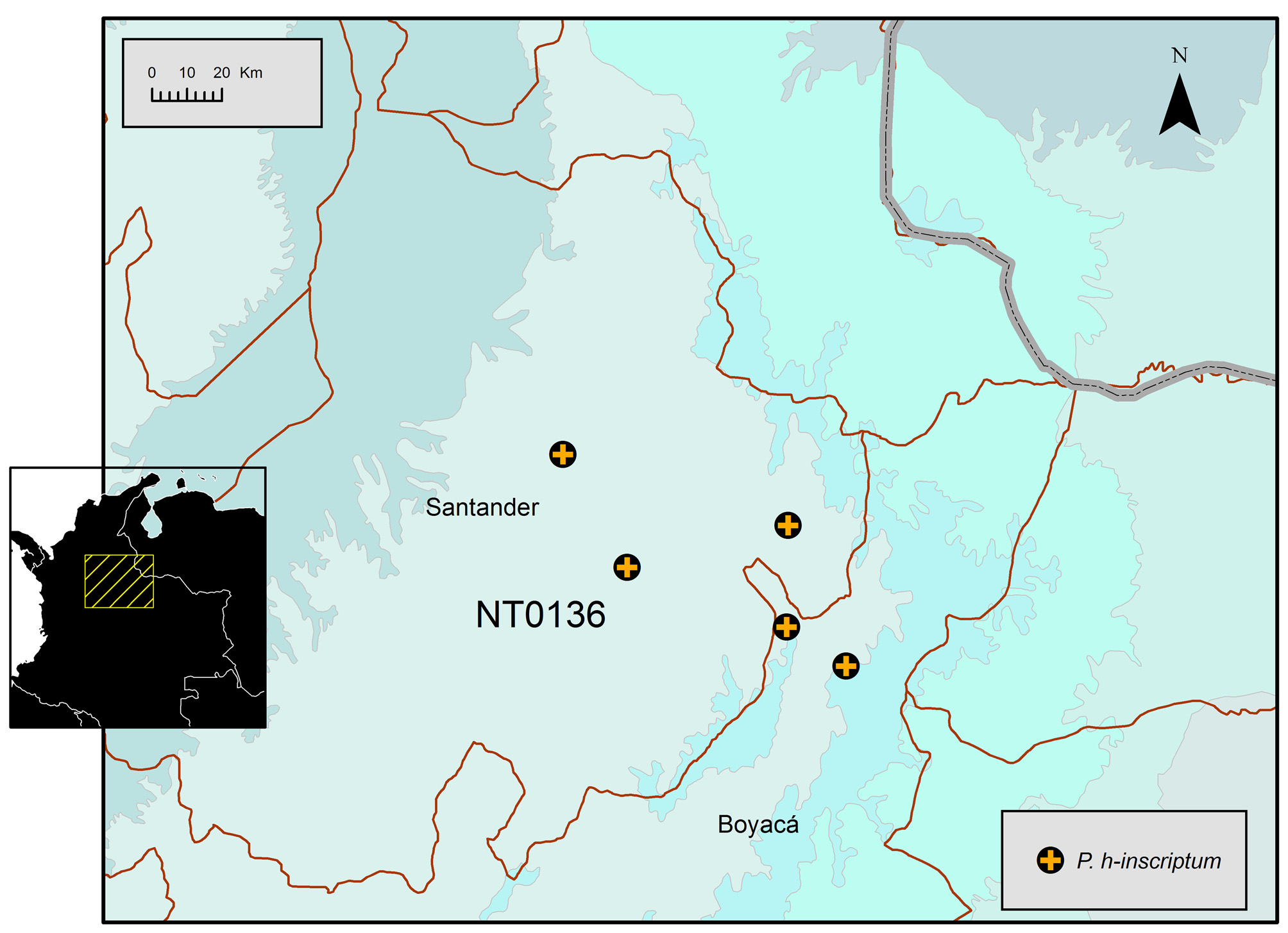
Distribution. P. h-inscriptum was described from the locality “Espinal, Colombia ” by Mello-Leitão (1941), which Kury (2003) wrongly extrapolated as “Espinal, Tolima ”. However, we found a more suitable location for this name, as there is a Espinal in Santander, which corresponds to a place near San Gil, Santander and La Uvita, Boyacá, type locality and record, respectively, of P. nigrolimbata ( Fig. 29 View FIGURE 29 ). All of these occurrences match the WWF ecoregion NT0136 ( Magdalena Valley montane forests).

No known copyright restrictions apply. See Agosti, D., Egloff, W., 2009. Taxonomic information exchange and copyright: the Plazi approach. BMC Research Notes 2009, 2:53 for further explanation.
|
Kingdom |
|
|
Phylum |
|
|
Class |
|
|
Order |
|
|
Family |
Platymessa
| Medrano, Miguel & Kury, Adriano B. 2016 |
Platymessa
| Kury 2011: 51 |
| Kury 2003: 81 |
| Soares 1945: 344 |
| Mello-Leitao 1941: 168 |
Platymessa
| Kury 2003: 81 |
Cynorta
| Soares 1970: 325 |
Platymessa
| Mello-Leitao 1941: 167 |