
Scleromystax reisi, Britto & Fukakusa & Malabarba, 2016
|
publication ID |
https://doi.org/10.1590/1982-0224-20150158 |
|
publication LSID |
lsid:zoobank.org:pub:E96BCCAB-7267-455E-85DE-35096F7CA981 |
|
persistent identifier |
https://treatment.plazi.org/id/C85FB72D-FED6-4F1C-B4D3-B8E2B3C6E5E9 |
|
taxon LSID |
lsid:zoobank.org:act:C85FB72D-FED6-4F1C-B4D3-B8E2B3C6E5E9 |
|
treatment provided by |
Carolina |
|
scientific name |
Scleromystax reisi |
| status |
sp. nov. |
Scleromystax reisi , new species
u r n:lsid:zooba n k.org:pub: E96 BCCA B -7267- 455E -85DE - 35096F7CA981
Figs. 1-7 View Fig View Fig View Fig View Fig View Fig View Fig View Fig
Scleromystax sp. Carvalho et al., 2012 [listed; first record to the laguna dos Patos drainage].
Holotype. MCP 49070, 49.3 mm SL, Brazil, Rio Grande do Sul State, Gravataí, arroio Demétrio, Morungava , 29°47’26”S 50°51’51.55”W, 14 Apr 2014, P. Lehmann. GoogleMaps
Paratypes. Brazil, Rio Grande do Sul State. MCP 48177, 2 View Materials , 13.7-14.4 mm SL, Viradouro, Capela Santana, rio Caí drainage, 29°38’51”S 51°49’12”W, 8 Nov 2013 GoogleMaps , P.
Lehmann. MCP 48178, 2 View Materials , 18.8-46.5 mm SL, same locality of the holotype, 23 Nov 2012, P. Lehmann. MCP 48179, 4 View Materials , 35.8-41.5 mm SL, collected with the holotype. MNRJ 43857 View Materials , 4 View Materials , 2 View Materials cs, 38.3-47.2 mm SL, Amaral Ferrador, stream at Fazenda Ferraria, 30°50’54”S 52°23’19”W, Nov 2006, J. Anza & R. Hirano. UFRGS 8747 View Materials , 1 View Materials , 26.9 mm SL, same data as MNRJ 43857. UFRGS 8754 View Materials , 4 View Materials , 21.4-49.9 mm SL, same data as MNRJ 43857. UFRGS 9228 View Materials , 1 View Materials , 34.8 mm SL, Camaquã, stream at Fazenda Capela Velha, 30°51’36”S 51°53’07”W, Jun 2007, J. Anza & J. Ferrer. UFRGS 18002 View Materials , 1 View Materials , 39.8 mm SL, Mariana Pimentel , stream near Morro Cerro Negro , 30°20’44”S 51°34’08”W, 28 Nov 2012, M. Almeirão. All following lots from Eldorado do Sul , Estação Experimental Agronômica , Universidade Federal do Rio Grande do Sul ( EEA-UFRGS): MNRJ 43858 View Materials , 6 View Materials , 21.6- 35.7 mm SL, small stream tributary to arroio Calombos , 30°06’44.3”S 51°40’44.2”W, 23 Jan 2013, J. Ferrer, C. Fukakusa & J. P. Miranda. UFRGS 17414 View Materials , 6 View Materials , 19.8 View Materials -36.0 mm SL, same data as MNRJ 43858. UFRGS 17416 View Materials , 3 View Materials , 29.7-45.5 mm SL, same locality as UFRGS 17414, 6 Oct 2012, C. K. Fukakusa, J. P. S. Miranda & V. R. Lampert. UFRGS 14964 View Materials , 2 View Materials , 46.8-54.1 mm SL, swamp close to water reservoir, 30°05’56”S 51°40’11”W, 6 May 2011, F. R. Carvalho & C. Fukakusa. UFRGS 14968 View Materials , 1 View Materials , 49.6 mm SL, same data as UFRGS 14964. UFRGS 14965 View Materials , 1 View Materials , 33.5 mm SL, creek below small water reservoir, 30°05’12”S 51°40’57”W, 25 Apr 2011, R. Dala-Corte & C. Fukakusa. UFRGS 16590 View Materials , 1 View Materials , 31.2 mm SL, arroio Calombos , 30°06’03”S 51°41’42”W, 1 Apr 2008, C. Fukakusa. UFRGS 18254 View Materials , 1 View Materials , 47.7 mm SL, 30°05’52.2”S 51°41’26.3”W, 1 Nov 2013, L. R. Malabarba. UFRGS 19188 View Materials , 5 View Materials , 25.4-38.9 mm SL, 30°06’49.10”S 51°41’0.01”W, 20 Sep 2013, C. K. Fukakusa, J. P. Miranda, A. Langoni & L. A. Donin. UFRGS 19189 View Materials , 3 View Materials , 42.1-46.1 mm SL, same data as UFRGS 19188. UFRGS 19191 View Materials , 12 View Materials , 19.2- 44.6 mm SL, unnamed creek, 30°06’49.10”S 51°41’0.01”W, 30 Nov 2013, C. Fukakusa & J. P. S. Miranda. UFRGS 19657 View Materials , 2 View Materials , 23.3-24.3 mm SL, arroio Calombos , 30°06’02”S 51°41’41.2”W, Nov 2009, L. R. Malabarba GoogleMaps .
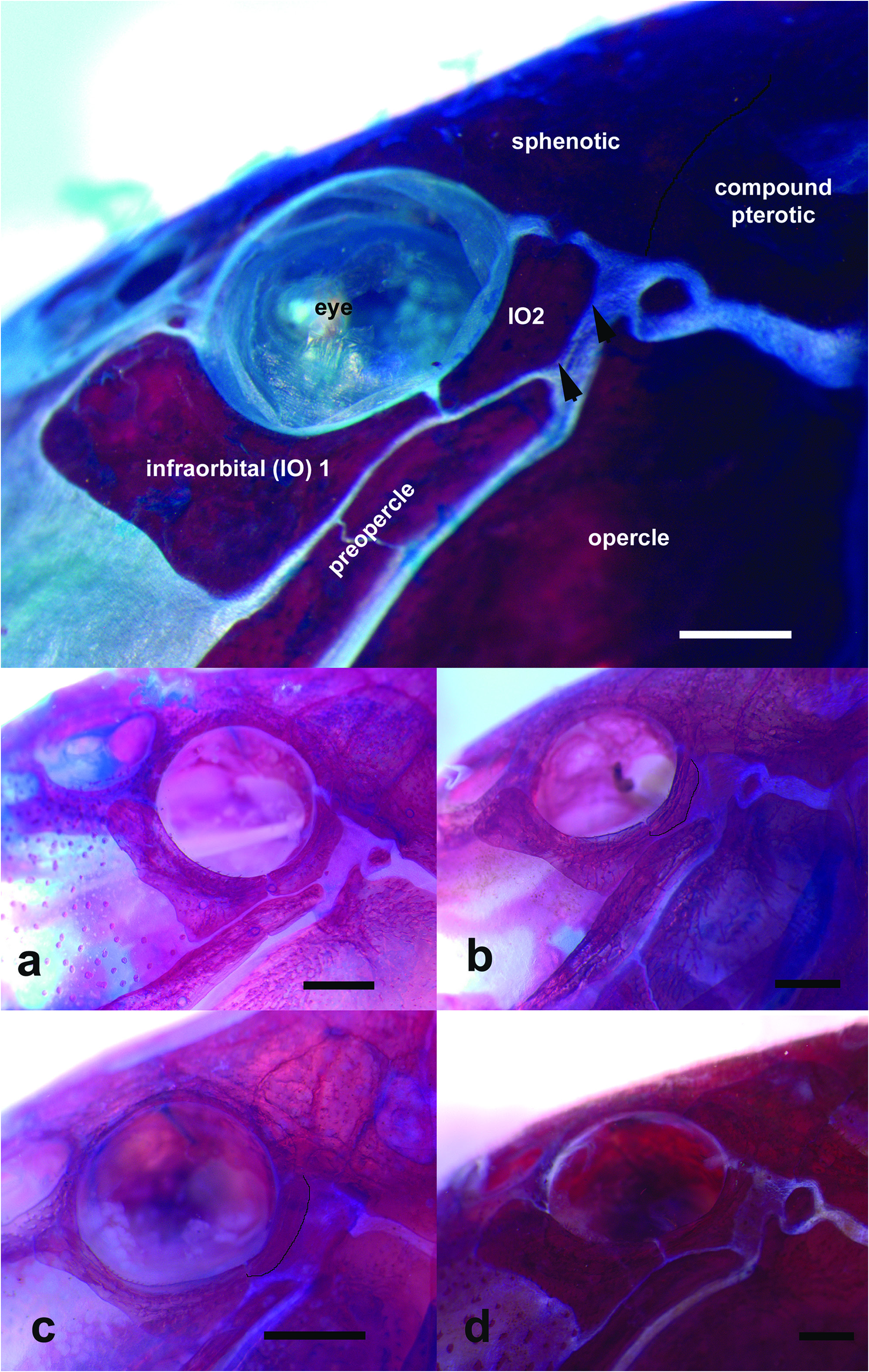
Diagnosis. Scleromystax reisi differs from its congeners by infraorbital 2 expanded ventrally, adjacent to small, almost imperceptible, naked area between sphenotic, compound pterotic, opercle and preopercle ( vs. infraorbital 2 narrow, leaving a somewhat large area between those bones). The new taxon is also distinct from its congeners, except Scleromystax macropterus (Regan) and S. salmacis , by roughly oblique, elongate, dark brown blotches along body ( vs. large, dark brown blotches, often coalesced, mainly on almost all dorsolateral body plates, in S. barbatus ; or ground color yellowish white, with a dark brown stripe along junctions of dorso- and ventrolateral body plates, in S. prionotos (Nijssen & Isbrücker)) . It is also distinguished, except from S. macropterus , by large specimens displaying cranial fontanel completely occluded, leaving just a shallow fossae, delimited by frontal bones, and reaching anterior tip of parieto-supraoccipital ( vs. wide or narrow fontanel between frontals). Scleromystax reisi differs from S. macropterus mainly by the absence of a black dot on the base of median caudal-fin rays ( vs. presence), and sexually dimorphic features inconspicuous, i.e. cheek region similar in males and females, with only minute, scattered odontodes in the former ( vs. males with developed odontodes inserted in fleshy papillae), dorsal and pectoral fins of males slightly longer than in females ( vs. dorsal and pectoral fins of males 2-3 times longer than those fins in females). It is also distinguished from S. salmacis by the tip of snout roughly truncate ( vs. slightly continuous and smoothly tapered).
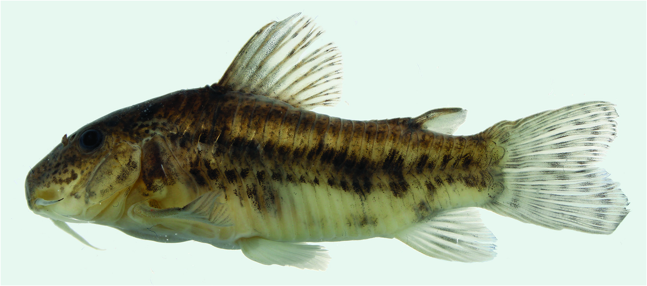
Description. Morphometric data presented in Table 1. Head compressed with slightly convex dorsal profile until posterior tip of parieto-supraoccipital process; roughly triangular in dorsal view. Snout straight and relatively blunt, roughly truncate ( Fig. 2 View Fig ). Profile slightly convex along dorsal-fin base. Postdorsal-fin body profile nearly straight to first preadipose unpaired platelet; slightly convex along preadipose platelets and base of adipose-fin spine. Ventral profile nearly straight from isthmus to anal-fin origin, with pronounced convexity just anterior to pelvic fins. Profile markedly concave from first anal-fin ray to caudal-fin base. Body roughly elliptical in cross section at pectoral girdle, gradually becoming more compressed toward caudal fin.
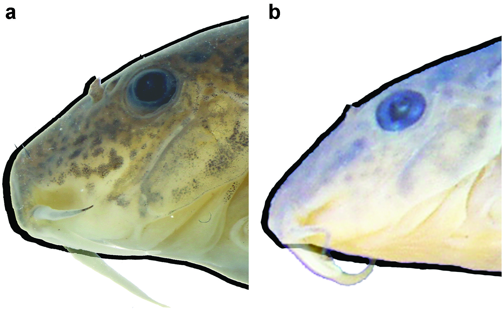
Eye round, located dorso-laterally on head; orbit delimited dorsally by frontal and sphenotic, ventrally by infraorbitals. Anterior and posterior nares close to each other, only separated by flap of skin. Anterior naris tubular. Posterior naris close to anterodorsal margin of orbit, separated from it by distance equal to diameter of naris. Mouth small, subterminal, width slightly larger than bony orbit diameter. Maxillary barbel reaching anteroventral limit of gill membrane in some individuals. Maxillary barbel slightly longer than outer mental barbel, with nearly same length. One specimen (UFRGS 8754, 49.9 mm SL) only with outer mental barbel. Inner mental barbel fleshy, nearly one-third of outer mental barbel length. Small rounded papillae covering entire surface of all barbels, upper and lower lips, and isthmus; more evident on lips and barbels. Minute odontode-bearing platelets scattered on ventral surface, between pectoral and pelvic girdles. Side of snout (cheek, preopercular region just below first infraorbital) conspicuously fleshy; males with several minute, scattered odontodes somewhat larger than those ones over body. Gill membranes united to isthmus. Four branchiostegal rays covered by thick layer of skin; distal two rays united at their tips by branchiostegal cartilage. Teeth on upper pharyngeal tooth plate 32, roughly arranged in two rows, and on fifth ceratobranchial 27, restricted to one row at mesial margin.
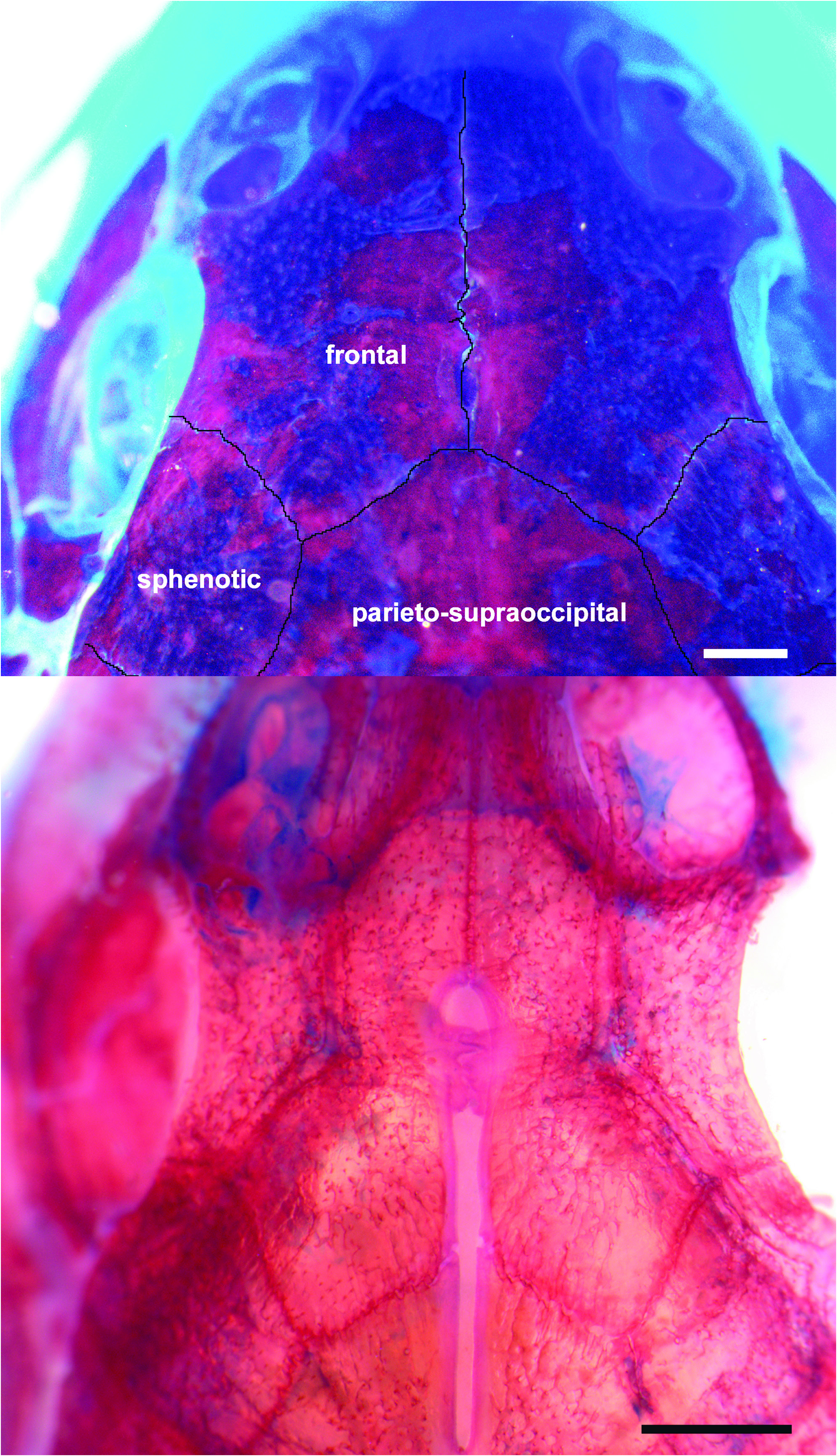
Sphenotic, parieto-supraoccipital, and frontal bones visible externally, all covered by thin layer of skin and bearing minute scattered odontodes. Cranial fontanel completely occluded in large specimens ( Fig. 3 View Fig ), leaving shallow fossae, covered by thin skin, delimited by frontal bones, and reaching anterior tip of parieto-supraoccipital; small specimens (up to 38.3 mm SL) with opened fontanel restricted to region between frontal bones. Sphenotic trapezoid in shape, contacting parieto-supraoccipital dorsally, compound pterotic posteriorly, and second infraorbital ventrally. Parieto-supraoccipital quadrangular with posterior expansion triangular and elongate, but not reaching nuchal plate, leaving naked area with scattered, minute odontode-bearing platelets. Unpaired odontodebearing platelets between parieto-supraoccipital posterior process and nuchal plate.
Two infraorbital bones, externally visible, covered by thin layer of skin; both bearing few minute odontodes. First infraorbital with anterior expansion just surpassing vertical through anterior naris. Second infraorbital expanded ventrally, leaving small, almost absent, naked area between sphenotic, compound pterotic, opercle and preopercle ( Fig. 4 View Fig ). Opercle exposed, ovoid in shape and roughly elongate, with free border variably smooth or angulated. Preopercle externally visible, slender and covered by thin layer of skin. Opercle and preopercle with minute odontodes scattered over their surfaces.
Trunk lateral-line composed of one perforated dorsolateral-body plate and two laterosensory canals, reduced to small ossicles. Lateral-line canal entering neurocranium through compound pterotic, splitting into three branches before entering sphenotic: pterotic, preoperculomandibular, and posterolateral, each with single pore. Posterolateral branch opened in compound pterotic itself, just above upper margin of opercle. Sensory canal continuing through compound pterotic, entering sphenotic as temporal canal, which splits into two branches: one branch giving rise to infraorbital canal, other branch entering frontal through supraorbital canal. Supraorbital canal not branched, running through nasal bone. Epiphyseal pore opening at supraorbital main canal. Nasal canal with single opening at each end. Infraorbital canal running through entire second infraorbital, extending to infraorbital 1 and opening into four pores; one pore at second infraorbital just before articulation point with infraorbital 1, remaining pores opened in first infraorbital. Preoperculomandibular branch not connected to preoperculomandibular canal, which runs through entire preopercle with three openings, leading to pores 3, 4, and 5, respectively.
Body plates with minute, scattered odontodes, larger on posterior margin of plates. Nuchal plate exposed. Cleithrum exposed laterally. Scapulocoracoid not exposed. Dorsolateral body plates not touching counterparts at middorsal line, showing narrow, shallow groove from last dorsal-fin ray to first preadipose platelet.Ventrolateral body plates not touching counterparts at midventral line in all specimens, leaving shallow preanal fleshy skin ridge. Dorsolateral body plates 22(2), 23(20), 24*(13), 25(16) or 26(8); ventrolateral body plates 20(1), 21(16), 22*(28), 23(13) or 24(1); dorsolateral body plates along dorsal-fin base 5(11), 6*(24), or 7(24); dorsolateral body plates from adipose fin to caudal-fin base 5(3), 6(30), 7*(17), 8(7), or 9(1); preadipose platelets 2(2), 4(1), 5(6), 6(24), 7*(20), 8(4), 9(1). Precaudal vertebrae 9; caudal vertebrae 14; six pairs of ribs, first pair conspicuously larger than others.
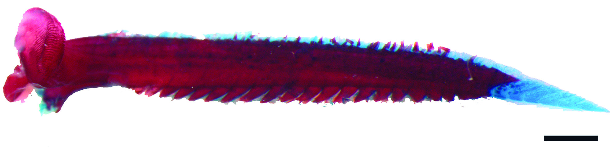
Dorsal fin roughly ovoid; its origin just posterior to third dorsolateral body plate. Dorsal spinelet not embedded in adjacent dorsal spine. Ossified portion of dorsal spine shorter than first five-six branched rays; second branched ray longest. Distal tip of dorsal spine with short, segmented unossified portion. Anterior and posterior border of dorsal spine smooth. Dorsal-fin rays I,7(5), I,8*(54). Adipose fin roughly triangular; its origin separated from base of last dorsal-fin ray by 7(1), 8(19), 9*(23), or 10(11) dorsolateral body plates. Anal fin roughly triangular; its origin located just posterior to 13th ventrolateral body plate, at vertical through posterior margin of penultimate preadipose platelet. Anal-fin rays ii,4(1), ii,5(13), ii,5,i(3), or ii,6*(42). Pectoral fin roughly triangular; its origin located just posterior to gill opening. Ossified portion of pectoral spine shorter than first three branched rays; first branched ray the longest one. Distal tip of spine with short, segmented unossified portion ( Fig. 5 View Fig ). Pectoral spine with 15-20 well-developed proximally-orientated dentations along entire posterior border. Males with thicker pectoral spines, bearing plentiful, randomly scattered odontodes; females with odontodes roughly arranged in longitudinal rows. Small, whiskerlike odontodes on dorsal surface of pectoral spine just adjacent to fin membrane in males. Pectoral-fin rays I,7(11), I,8*(46)or I,9(2).Pelvic fin ellipsoid;its origin just below second ventrolateral body plate, at vertical through base between first and second branched dorsal-fin ray. Pelvic-fin rays i,5. First pelvic-fin ray (unbranched) thickened anteriorly and flattened; its tegument thick, with embedded minute odontodes. Caudal fin bilobed; both lobes equal in size. Principal caudal-fin rays i,5/5,i(1), i,5/6,i(7), i,6/5,i(1) or i,6/6,i*(50); five upper and lower procurrent caudal-fin rays, respectively. All fins with minute odontodes scattered over all rays; pectoral and pelvic fins with odontodes restricted to ventral side.
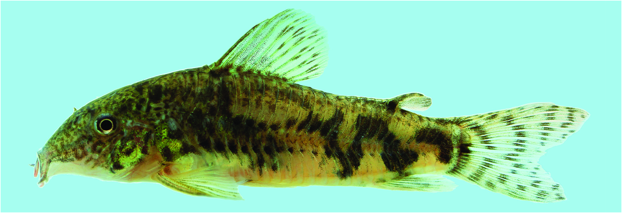
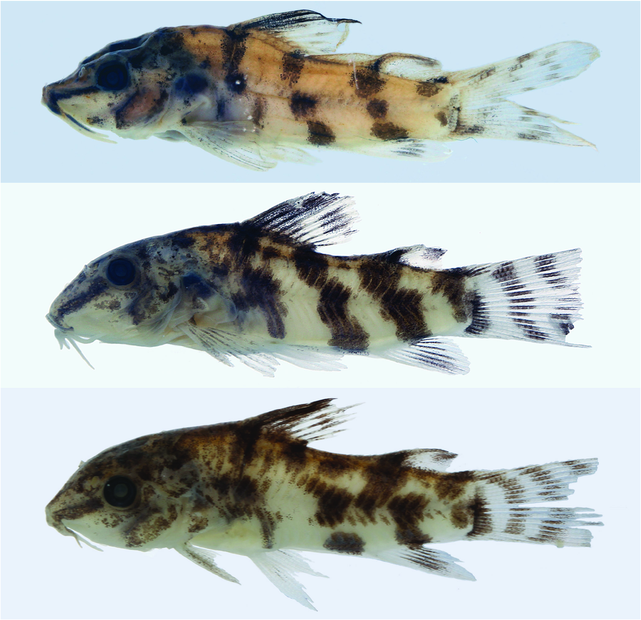
Coloration in alcohol. Ground coloration of head light brown. Several small, irregular, dark brown blotches distributed over dorsal and lateral surface of head, snout, preopercle and opercle. Series of chromatophores forming thin, faint brown ring on skin around orbit. Maxillary barbel yellowish white, with minute, scattered, irregular, small dark brown blotches on base. Inner mental barbel yellowish white, with few chromatophores on tip. Outer mental barbel with light brown coloration. Series of chromatophores forming thin line along profile of parieto-supraoccipital process on both sides, extending to unpaired predorsal platelets. Ventral region of head yellowish white. Ground color of trunk yellowish light brown. Fainted brown coloration along dorsum. Diffuse, dark brown blotch on cleithrum. Two series of small blotches from region just posterior to cleithrum to caudal peduncle, along ventral portion of dorsolateral body plates and dorsal portion of ventrolateral body plates, respectively; nearly close to midlateral plate junctions; more evident in males ( Fig. 1 View Fig ) than in females ( Fig. 6 View Fig ) and absent in juveniles ( Fig. 7 View Fig ). Inner coloration layer on plate junction. Four oblique, trespassing, rough bars on side of trunk, more evident in juveniles with less than 19 mm SL ( Fig. 7 View Fig ). In males, bars variably restricted to dorsolateral body plates, or extending through plate junctions to ventrolateral body plates ( Fig. 1 View Fig ). In females, bars shape variable, usually present on dorsolateral body plates and extending to ventrolateral body plates, sometimes forming a zigzag pattern ( Fig. 6 View Fig ). Ventral surfaces of body yellowish white.
Dorsal spine light brown. Anterior portion of dorsal fin with several chromatophores, coalesced in diffuse, elongated blotch along the membrane between the first and second branched rays and along proximal portion of the membrane between third and fourth branched rays; remaining interradial membranes hyaline. Roughly two series of brown blotches restricted to rays, except by interradial space between first-second, and second-third branched rays ( Figs. 1 View Fig , 6 View Fig ). Juveniles with dorsal spine and two anterior branched rays black pigmented, as well as the membrane between the first and second branched rays and along proximal portion of the membrane between third and fourth branched rays; remaining interradial membranes hyaline; branched dorsal fin rays 2 or 3 to 8 or 9 with small black dots along half-distal length ( Fig. 7 View Fig ).
Ground color of anal-fin rays faintly yellowish white, almost hyaline. Interradial anal-fin membranes in the midlength of anal-fin rays hyaline to light gray with scattered chromatophores on rays and membrane ( Figs. 1 View Fig , 6 View Fig ) and dark gray to black in juveniles (up to 20 mm SL; Fig. 7 View Fig ). Adipose-fin spine brown in adults ( Figs. 1 View Fig , 6 View Fig ) and black in juveniles ( Fig. 7 View Fig ). Adipose-fin membrane with scattered chromatophores; more concentrated on free border. Pectoral spine light brown. Pectoral-fin rays with dark brown chromatophores restricted along first four branched rays; interradial membrane hyaline. Pelvic fin hyaline, with diffuse series of scattered chromatophores on middle region of fin in smaller specimens. Caudal fin with three-four series of small, dark-brown blotches restricted to principal rays.
Coloration in life. General color pattern of black marks similar to that described for specimens in alcohol. Region of opercle and side of scapular girdle iridescent green ( Fig. 6 View Fig ).
Sexual dimorphism. Male representatives of Scleromystax with a well-distinguishable genital papillae display some dimorphic features that help to distinguish them (Britto, 2003). The most useful characteristics are the elongate pectoral and dorsal fins, and the sides of snout (cheek region) bearing pointed, relatively long odontodes embedded in fleshy papillae. Notwithstanding, those features show a variable degree of development within the genus (Britto & Reis, 2005: 486-487), and S. salmacis displays none of the dimorphic conditions in addition to the shape of the genital papillae (Britto & Reis, 2005: figs. 1-2). Scleromystax reisi shared with its congener, S. prionotos , these sexually dimorphic features less evident, i.e. males with only minute, scattered odontodes in the sides of snout (although inserted in fleshy tissue in some specimens of S. prionotos ), and dorsal and pectoral fins slightly longer than in females; conditions even less conspicuous in S. reisi . Males of S. reisi present thicker pectoral spines, with plentiful, randomly scattered odontodes, while females display odontodes roughly arranged in longitudinal rows. Also, males bear small, whiskerlike odontodes on dorsal surface of pectoral spine just adjacent to fin membrane.
Habitat and ecological notes (based on material from EEA-UFRGS). Scleromystax reisi was found mainly in very small ( 0.5-2 m wide) and shallow streams ( 30-60 cm depth), with slow current water and surrounded by relatively preserved riparian vegetation. The bottom was sandy, sometimes covered with a thin layer of mud or fallen leaves. Although there were rocky bottom stretches in the same creeks, the species was never found there. The streams may have a small amount of submerged vegetation. Other species collected along with Scleromystax reisi were Astyanax eigenmanniorum (Cope) , Cheirodon interruptus (Jenyns) , Corydoras paleatus (Jenyns) , Heptapterus mustelinus (Valenciennes) , H. sympterygium (Buckup) , Mimagoniates inequalis (Eigenmann) , Phalloceros caudimaculatus (Hensel) , Pseudobunocephalus iheringii (Boulenger) , Rhamdia sp. and Scleronema sp.
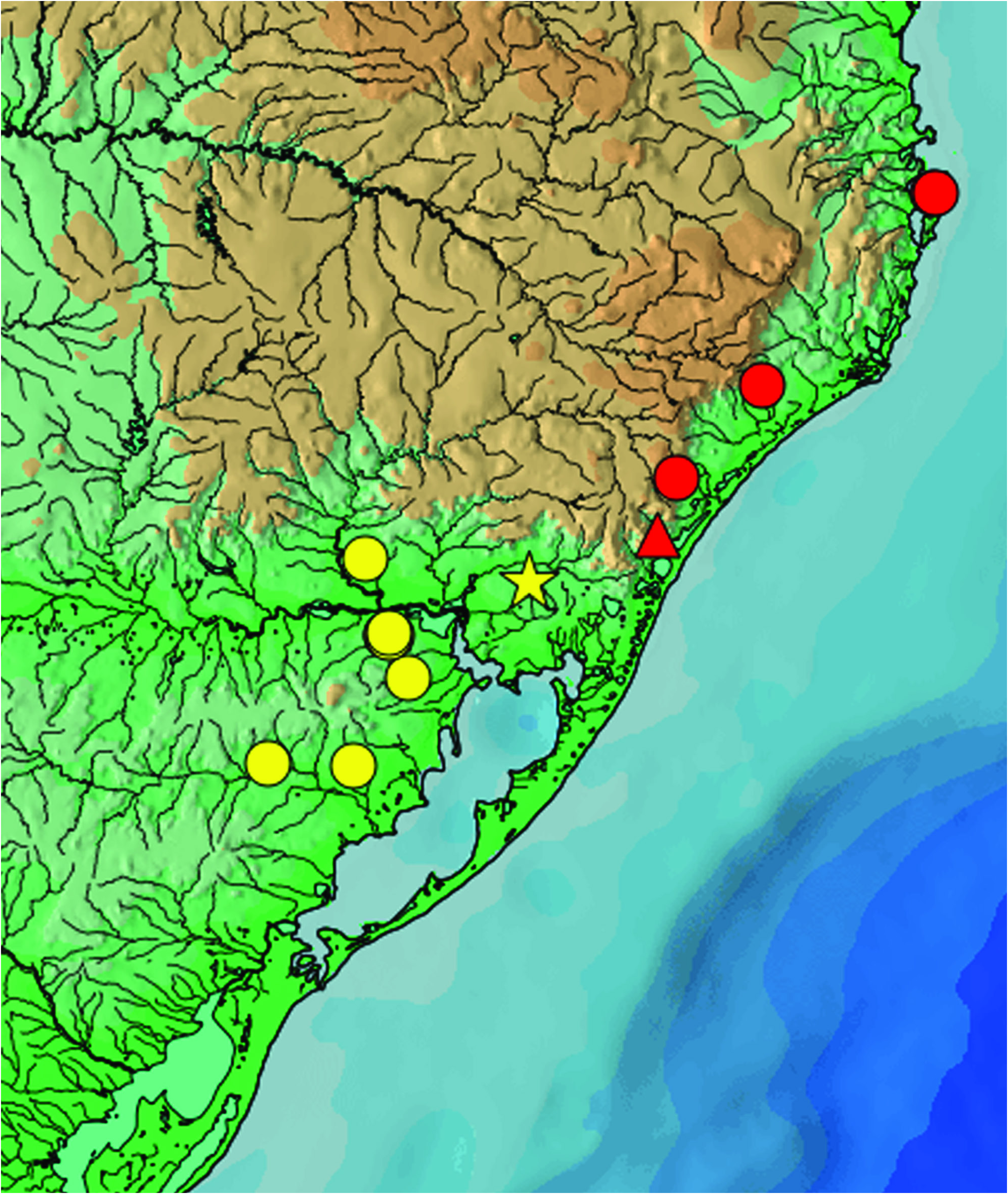
Distribution. Mainly first order streams of tributaries of the rio Jacuí and rio Camaquã, laguna dos Patos drainage, Rio Grande do Sul, Brazil ( Fig. 8 View Fig ).
Etymology. The specific name is given in honor to our colleague Roberto Esser dos Reis, for his many contributions to Neotropical ichthyology, including studies in callichthyid fish. A noun in the genitive case.
Conservation status. Scleromystax reisi is known from an Extent of Occurrence (EOO) of approximately 9,400 km 2 and occurs only in forested streams with preserved riparian vegetation, a habitat naturally fragmented in the southern border of the Atlantic forest. There are no additional treats and it can be categorized as Least Concern (LC) according to IUCN criteria ( IUCN, 2016).
No known copyright restrictions apply. See Agosti, D., Egloff, W., 2009. Taxonomic information exchange and copyright: the Plazi approach. BMC Research Notes 2009, 2:53 for further explanation.
|
Kingdom |
|
|
Phylum |
|
|
Class |
|
|
Order |
|
|
Family |
|
|
Genus |