
Schausiana Viette, 1950a, 1949
|
publication ID |
https://doi.org/ 10.11646/zootaxa.4860.1.3 |
|
publication LSID |
lsid:zoobank.org:pub:38F1E2A5-5DE0-4B95-959A-6347BC593AF0 |
|
DOI |
https://doi.org/10.5281/zenodo.4538724 |
|
persistent identifier |
https://treatment.plazi.org/id/0383282C-7E21-FFE2-4390-FE5341481C07 |
|
treatment provided by |
Plazi |
|
scientific name |
Schausiana Viette, 1950a |
| status |
|
Schausiana Viette, 1950a View in CoL
Type-species: Phassus trojesa Schaus, 1901 View in CoL , by original designation; monotypic.
Viette (1950a: 80).— Viette (1951a: 79).— Paclt (1953: 143).— Edwards & Hopwood (1966: 266).— Nielsen & Robinson (1983: 18).— Robinson & Nielsen (1984: 17).— Nye & Fletcher (1991: 273).— Nielsen et al. (2000: 841).— Grehan (2010: 47; fig. V, appendix).— Mielke & Grehan (2012: 148).— Grehan (2012: 14).— Mielke & Grehan (2015: 113).— Grehan & Rawlins (2016: 47).—Mielke & Grehan (2017: 136).—Grehan et al. (2018: 63).
Diagnosis. Distinguished from all other Hepialidae by unique scales on the FW veins, and by the combination of the following characters: (i) labial palpus unisegmented, sometimes with remnants of segment fusion, (ii) antenna filiform, (iii) ♂ metatibia hypertrophied and androconia present, (iv) ‘hepialine’ venation, (v) ♂ HW 1A incomplete, and (vi) ♀ HW CuP barely complete.
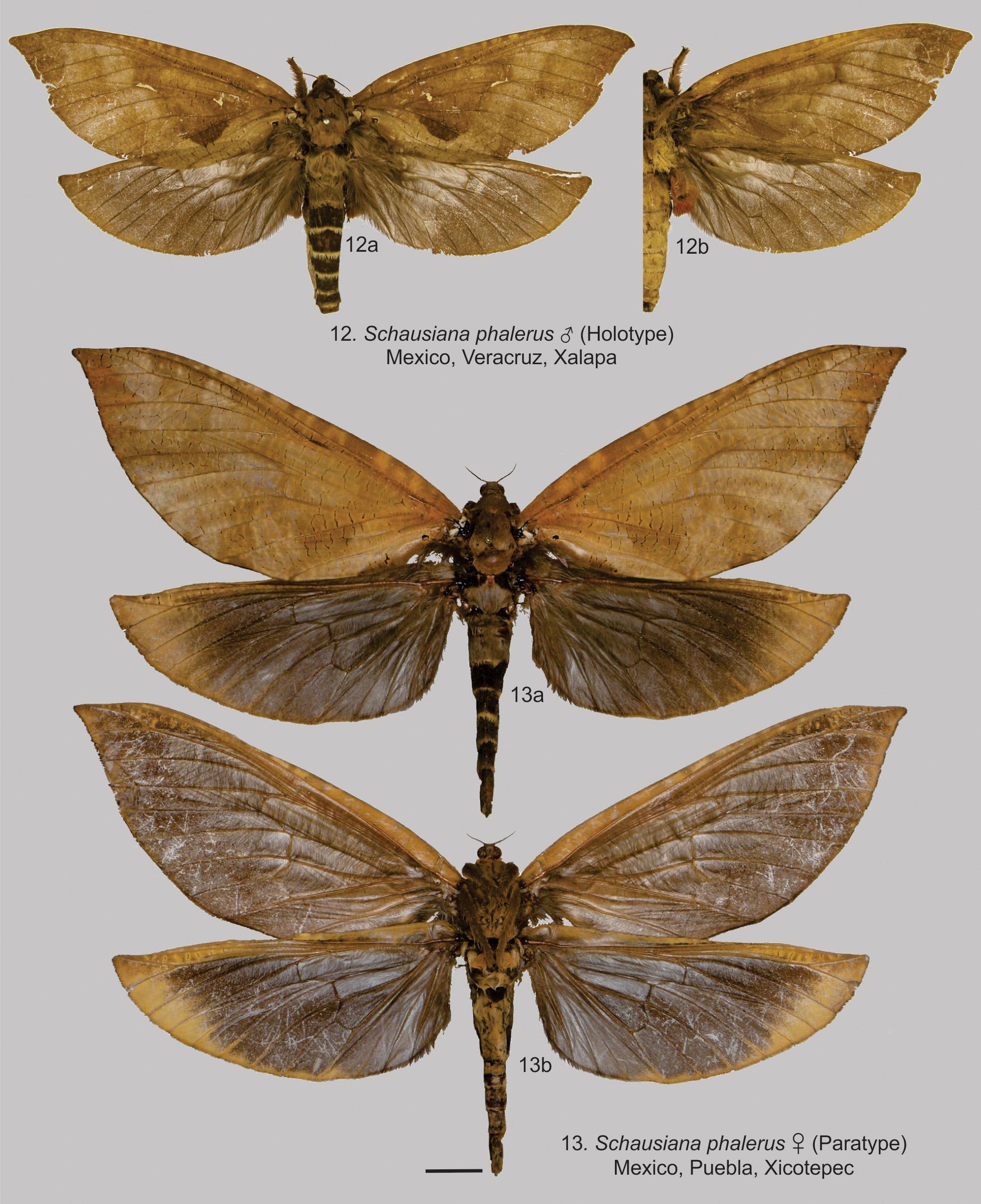
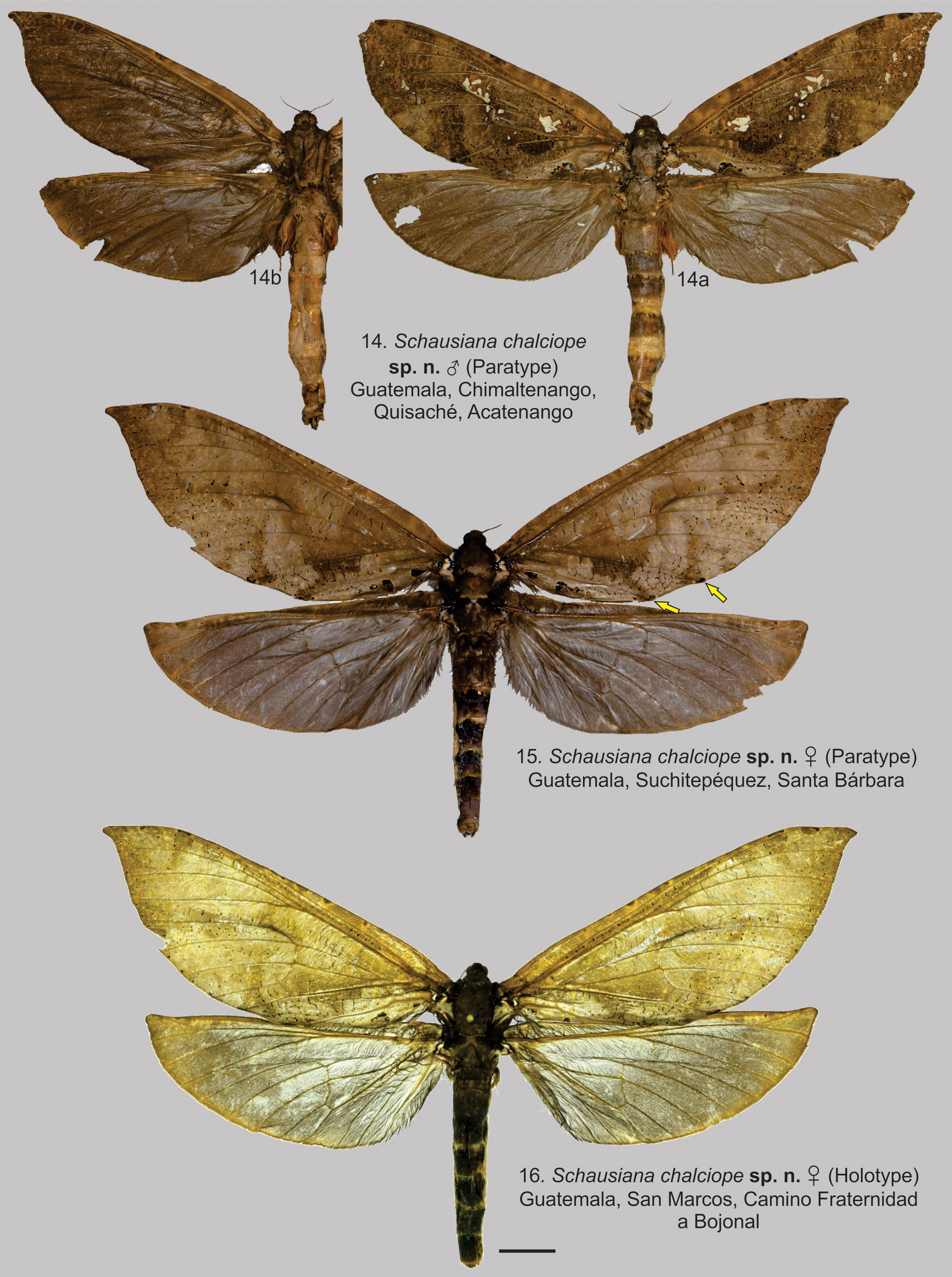
Redescription. Males ( Figs 6–7 View FIGURES 6–8 , 9 View FIGURES 9–11 , 12 View FIGURES 12–13 , 14 View FIGURES 14–16 , 17–18 View FIGURES 17–21 , 23–26 View FIGURES 22–27 , 28, 30, 32 View FIGURES 28–33 , 34–36 View FIGURES 34–36 ).
Head. Clypeus glabrous anteriorly, mesally projected and differentiated from the frons. Frons with piliform and porrect scales. Vertex scales as for the frons, but shorter. Eyes large, occupying 4/5 of the head in anterior view. Labial palps unisegmented ( Fig. 23 View FIGURES 22–27 ) with indentations sometimes indicating fusion of at least two palpomeres. Antenna filiform, segments cylindrical, sensilla caetica and sensilla trichodea present.
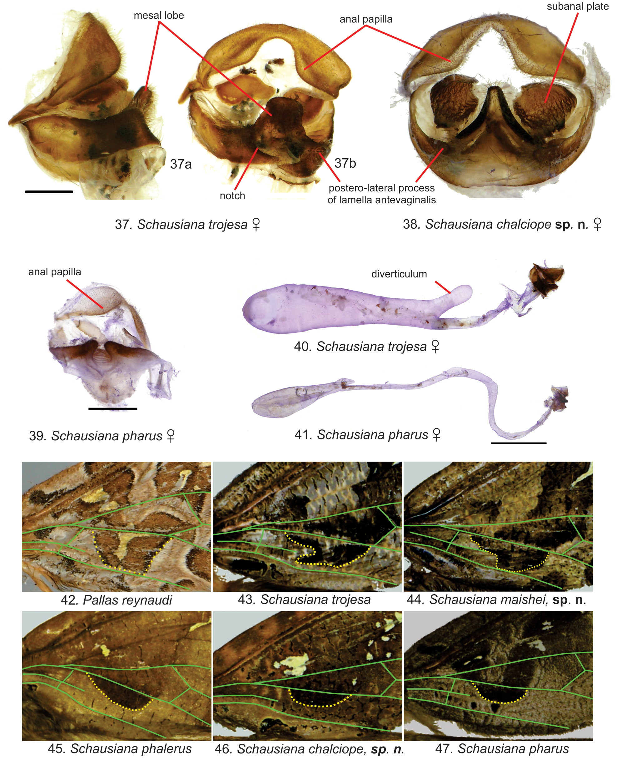
Thorax. Legs ( Figs 24–25 View FIGURES 22–27 ): metatibia hypertrophied (twice as long as wide) with androconia; arolium absent. Venation ( Fig. 26 View FIGURES 22–27 ): FW without Sc1; HW without Sc1, CuP complete, 1A incomplete, and 2A+3A complete. DFW with specialized scales along the veins, long (1–10 mm) and piliform in S. trojesa and S. maishei sp. n., white and shortened spines in S. phalerus (Druce, 1887) comb. n., and S. chalciope sp. n., and thinly piliform, longer than regular porrect scales in S. pharus comb. n. (figured for ♀, Figs 2–5 View FIGURES 1–5 ). Although absent in S. pharus comb. n, all the other species have black ovoid markings in the proximal anal area of the DFW. The FW U-band generally extends posteriorly almost to the A vein.
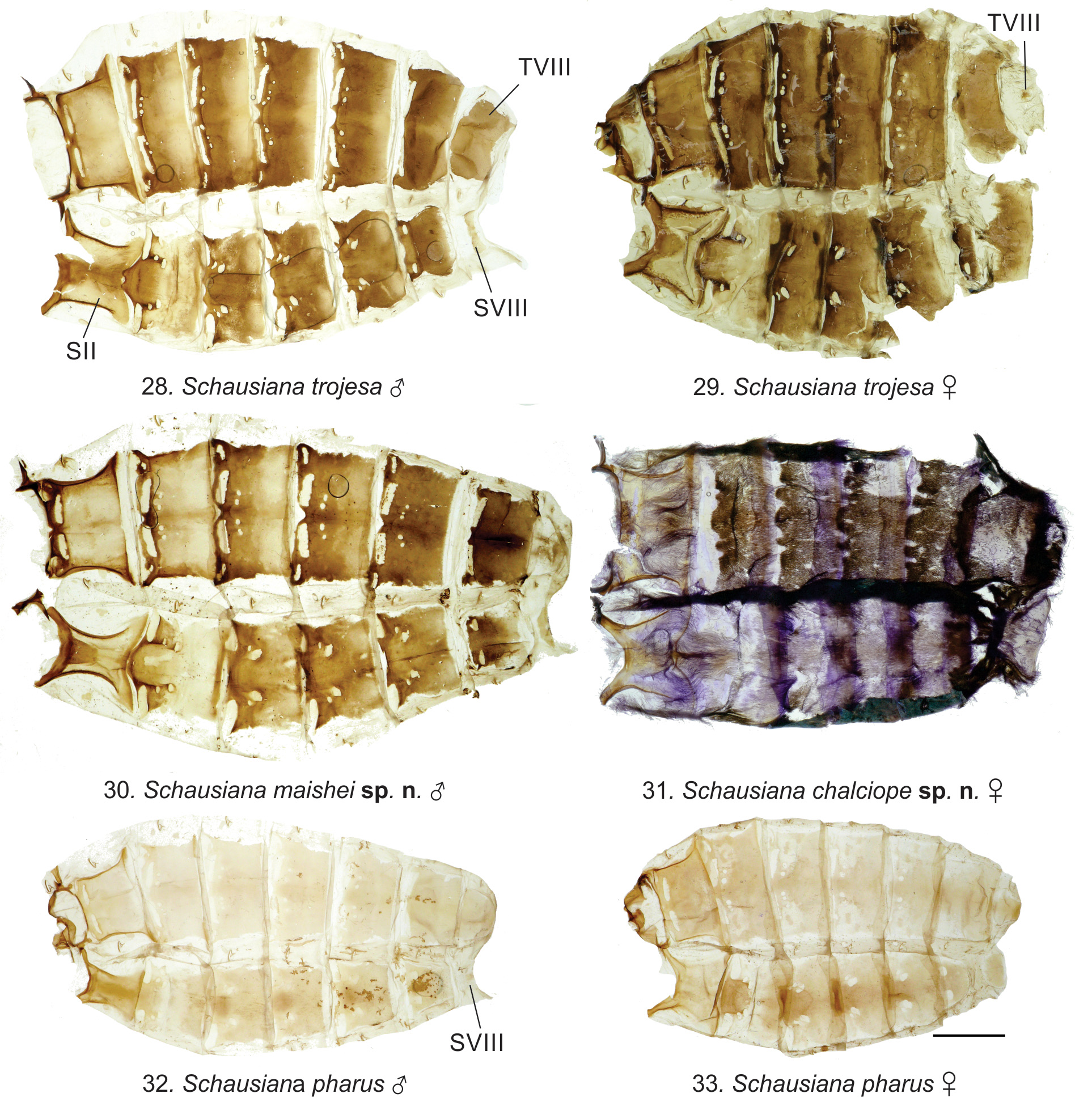
Abdomen ( Figs 28, 30, 32 View FIGURES 28–33 ). Tergosternal sclerite simple, intermediate zone without any protruding knob. Sternite II compressed laterally with shallow concave edges, tergite and sternite VIII sclerotized.
Male genitalia ( Figs 34–36 View FIGURES 34–36 ). Tegumen fused to the pseudotegumen, but distinguished by a less sclerotized fusion line, ventrally articulated with saccus. Saccus U-shaped on posterior and anterior margins. Tergal lobes reduced and fused to the pseudotegumen, but distinctly sclerotized; each dorso-mesally connected by a thick membrane. Pseudotegumen dorso-mesally unfused and ventro-mesally fused. Fultura inferior and fultura superior present. Valva curved with a distinct elbow, and with a basal and densely sclerotized process. Phallus without cornutus.
Females ( Figs 1, 2–5 View FIGURES 1–5 , 8 View FIGURES 6–8 , 10–11 View FIGURES 9–11 , 13 View FIGURES 12–13 , 15–16 View FIGURES 14–16 , 19–22, 27 View FIGURES 17–21 View FIGURES 22–27 , 29, 31, 33 View FIGURES 28–33 , 37–41 View FIGURES 37–47. 37–41 ).
Head. As for the ♂. Labial palps unisegmented ( Fig. 22 View FIGURES 22–27 ).
Thorax. Venation ( Fig. 27 View FIGURES 22–27 ): HW with CuP mesally barely complete, 1A and 2A+3A complete. FW, specialized scales as for the ♂ ( Figs 2–5 View FIGURES 1–5 ).
Abdomen. As for the ♂ ( Figs 29, 31, 33 View FIGURES 28–33 ).
Female genitalia ( Figs 37–41 View FIGURES 37–47. 37–41 ). Corpus bursae with diverticulum for at least S. trojesa and S. pharus comb. n., where is the internal genitalia are preserved.
Geographical distribution. Schausiana is known to occur from Mexico to Costa Rica ( Fig. 48 View FIGURE 48 ).
Etymology. Viette (1950) did not explicitly state the etymology for this genus, but he likely proposed it in honour of William Schaus, a prominent American lepidopterist.
Remarks. Some other potential diagnostic characters have been found only where male and female genitalia have been dissected: (i) pseudotegumen dorso-mesally unfused while ventro-mesally fused, (ii) basal portion of the valve with spiny or hook process (both detected in S. trojesa , S. maishei sp. n., and S. pharus (Druce, 1887) comb. n), and (iii) corpus bursae with diverticulum (detected in S. trojesa and S. pharus comb. n.). For comparison with other genera, see the discussion section.


No known copyright restrictions apply. See Agosti, D., Egloff, W., 2009. Taxonomic information exchange and copyright: the Plazi approach. BMC Research Notes 2009, 2:53 for further explanation.
|
Kingdom |
|
|
Phylum |
|
|
Class |
|
|
Order |
|
|
Family |