
Trichomycterus igobi, Wosiacki & Pinna, 2008
|
publication ID |
https://doi.org/ 10.1590/S1679-62252008000100003 |
|
persistent identifier |
https://treatment.plazi.org/id/03826B04-7106-FFEA-FC47-FC58FF02FE2D |
|
treatment provided by |
Carolina |
|
scientific name |
Trichomycterus igobi |
| status |
sp. nov. |
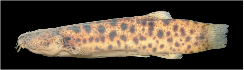
Trichomycterus igobi View in CoL , new species
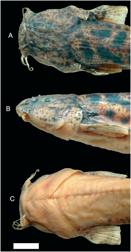
Figs. 1 View Fig and 2 View Fig
Holotype. MPEG 13352 View Materials (90.1 mm SL), Brazil, Estado do Paraná, Município de Candói, rio Jordão above the hydroelectric dam of Santa Clara, Apr 2005, NUP team col.
Paratypes. NUP 4007 , 2 , 62.0- 66.3 mm SL, Brazil, Estado do Paraná, Município de Candói, rio do Sobradinho (tributary to rio Jordão ), 8 Apr 2005 , NUP team col; MPEG 13353 View Materials , 2 View Materials , 72.4-82.7 mm SL, collected with NUP 4007 ; MZUSP 94842 View Materials , 2 View Materials , 72.1-86.6 mm SL, collected with NUP 4007 ; MZUSP 94843 View Materials , 3 View Materials , 63.0- 89.8 mm SL, Brazil, Estado do Paraná, Município de Candói, rio Jordão , no date , NUP team col; MPEG 13354 View Materials , 1 View Materials c&s, 70.9 mm SL, collected with MZUSP 94843 View Materials .
Diagnosis. Trichomycterus igobi is distinguishable from all other species currently in Trichomycterus by its large head (23.8-26.8% SL), which is proportionally the largest head in any Trichomycteridae . Other diagnostic features that distinguish the new species from most or all of its congeners include an almost entirely cartilaginous second hypobranchial (with only vestigial ossification); a mesially expanded palatine ossification; a narrow cleithrum, falciform in shape; and the lack of a proximal posterior concavity on the third ceratobranchial. The rigid spine-like morphology of individual procurrent rays of the caudal fin, the extension of the dorsal caudal-fin procurrent ray series (extending for ten neural spine tips), and the presence of ten or eleven branchiostegal rays each distinguish T. igobi from all congeners except T. stawiarski and T. sp. C (see Discussion). Other characters shared with various other species of Trichomycterus yet useful for identification include a dorsal fin located on a concavity on dorsal profile of trunk; the short caudal peduncle (15.4- 19.7% SL); and the first anal-fin ray base posterior to the vertical through the base of the last dorsal-fin ray.
W. B. Wosiacki & M. de Pinna 19
Description. Morphometric data for holotype and paratypes given in Table 1.
Body short, rounded at level of pectoral girdle and gradually more compressed posteriorly. Dorsal profile of trunk straight or slightly convex, ventral profile gently convex. Dorsal and ventral profiles of caudal peduncle slightly convex. Integument thick and opaque, especially over dorsal-, anal- and pectoral-fin bases. Small papillae visible under stereomicroscope over surface of body; papillae larger and more densely concentrated over oral surface of lips. Head large, longer than wide, depressed. Head width larger than maximum body width. Snout broad, convex in dorsal view. Dorsal profile of head straight in lateral view; ventral profile slightly convex. Interorbital region flat. Lateral portion of head swollen by well-developed jaw muscles. Eyes round, dorso-laterally oriented, slightly converging anteriorly towards midline. Orbital rim not free. Skin covering eye thin and transparent. Anterior nostrils smaller than eye, separated by space approximately equal to interorbital, surrounded by fleshy flap of integument posterolaterally continuous with nasal barbel. Posterior nostrils smaller and more closely positioned to each other than anterior ones, partially surrounded anteriorly, laterally and medially by thin flap of skin. Branchial membranes thick, narrowly united to isthmus anteriorly. Gill opening wide, not constricted. Ten or eleven (modally ten) branchiostegal rays, most of which clearly visible externally.
Mouth wide, subterminal, with corners laterally oriented. Upper and lower lips fleshy, similar-sized. Lower lip with large fleshy lobes located posteromedially to origin of rictal barbels.
Barbels short and thin, gradually narrowing to fine tips. Nasal barbels flat and narrow, posteriorly reaching middle of eyes. Origin of nasal barbels on posterolateral portion of integument flap around anterior nostril. Maxillary and rictal barbels thin and moderately long, both reaching vertical through posterior margin of eyes.
Pectoral fin narrow, with rounded margin. Pectoral-fin rays i,7, first one longest, unbranched, not prolonged as filament. Dorsal fin round, with i,6-7 rays (modally i,7, including holotype), second and third longest. Anal fin shorter and narrower than dorsal fin. Base of first anal-fin ray located posterior to vertical through base of last dorsal-fin ray. Anal-fin 20 A new trichomycterid catfish from the rio Iguaçu drainage rays i-ii,5, second ray longest. Pelvic fin with i,4 rays, second one longest. Pelvic-fin base anterior to dorsal-fin origin; inner rays of contralateral pelvic-fin not overlapping when in repose; distal part of fin covering urogenital opening. Caudal fin truncate with straight corners; distal margin slightly deeper than fin base; 6+7 principal rays.
Opercular patch of odontodes very small, with 9-12 short, thin, straight odontodes with thick tips. Interopercular patch of odontodes with nine short, thick, conic odontodes arranged in a single main series, with additional odontodes posteriorly.
Cephalic sensory canals including complete supraorbital canal and incomplete infraorbital canal. Infraorbital restricted to posterior region corresponding to pores i10 and i11. Supraorbital pores S1, S3 and S6. Two paired pores S6. Laterosensory canal reduced on trunk, comprising two pores at vertical through middle of pectoral fin. Vertebra 37, ribs 13 pairs, first thickest, 6th and 7th longest, last rib rudimentary. Dorsal-fin pterygiophores 8, first one inserting anterior to neural spine of 17th free vertebra. Anal pterygiophores 6, first one anterior to hemal spine of 23rd free vertebra. Two hypural ossifications on upper hypural plate, parahypural and hypurals one and two fused on lower plate. Procurrent caudal-fin rays 24 dorsally and 13 ventrally.
Color in preservative. Body pigmentation arranged in at least two different layers of integument. Deep layer composed of relatively large irregular dark spots of variable size and shape, larger and more concentrated on dorsum of trunk, gradually becoming more scattered and smaller on sides, entering dorsal surface of head only posteriorly and completely absent on ventral surface of trunk and head. A more superficially-located pattern of fine spots overlays large marking with relatively uniform freckle, densest on dorsum. Smaller superficial markings extend onto entire dorsal and lateral surfaces of head, as fine spots smaller than those on body, and form main component of dark pigmentation at that region. Sides of head slightly less heavily pigmented than dorsal region. Opercular patch of odontodes with pigmentation continuous with that on remainder of head. Odontode-bearing area of interopercle almost devoid of dark pigment, in contrast to area of head immediately dorsal to it. Upper lip with small spots similar to those on rest of head. Lower lip with small dark fields close to its anterior margin. Dorsal fin with irregular dark markings along its basal portion and anterior edge, with additional spots along rays on rest of fin, in some specimens aligned so as to form a poorlydefined stripe across the distal third of fin. Anal fin with dark pigmentation similar to that of dorsal fin, but generally fainter. Caudal fin covered with small irregular spots, often aligned to form one or more rough vertical lines across fin. Dorsal surface of pectoral fin with small dark markings similar to those on rest of flanks, fading distally to transparent margin. Pelvic fins mostly white, with only few dark chromatophores at base. Nasal barbels with irregular dark fields along entire length, on both surfaces. Maxillary and rictal barbels with dark markings mostly on dorsal surface.
Distribution. Lower rio Jordão, close to its mouth on rio Iguaçu, rio Paraná drainage, Southern Brazil ( Fig. 3 View Fig )
Etymology. The species name igobi is based on a mythological character of the Tupi-Guarani Indian legend about the origin of the Iguaçu waterfalls.
Ecological data. Specimens of Trichomycterus igobi were collected in the same general locality as T. sp. C in the rio
W. B. Wosiacki & M. de Pinna 21
Jordão, and presumably occupy the same kind of fast water, rocky-substrate environment reported for that species (cf. Wosiacki & de Pinna, in press). The details of its microhabitat, however, are as yet unknown. The stomach of the cleared and stained specimen contained larvae of Diptera (Simulidae), Ephemeroptera, and Trichoptera, indicating benthic feeding habits.

No known copyright restrictions apply. See Agosti, D., Egloff, W., 2009. Taxonomic information exchange and copyright: the Plazi approach. BMC Research Notes 2009, 2:53 for further explanation.