
Lepraliella contigua ( Smitt, 1868 )
|
publication ID |
https://doi.org/ 10.11646/zootaxa.5125.2.4 |
|
publication LSID |
lsid:zoobank.org:pub:6DDA279A-FA5F-4993-98DD-FC40133292BB |
|
DOI |
https://doi.org/10.5281/zenodo.6425469 |
|
persistent identifier |
https://treatment.plazi.org/id/03825422-2A3E-444B-8A9F-FE6D2B2CFCB6 |
|
treatment provided by |
Plazi |
|
scientific name |
Lepraliella contigua ( Smitt, 1868 ) |
| status |
|
Lepraliella contigua ( Smitt, 1868)
( Fig. 5 View FIGURE 5 ; Table 4 View TABLE 4 )
Cellepora ramulosa forma contigua Smitt, 1868: 31 , figs 198–201.
Lepraliella contigua: Levinsen 1917: 467 .
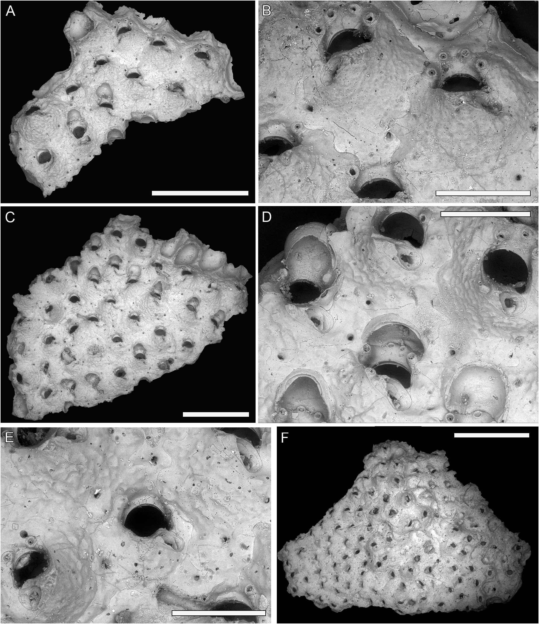
Material examined. Lectotype (designated here) SMNH-Type-1695a ( Fig. 5C–E View FIGURE 5 ), and paralectotypes SMNH-Type-1695b ( Fig. 5A, B View FIGURE 5 ) and SMNH-Type-1695c ( Fig. 5F View FIGURE 5 ); three colony fragments; North Atlantic Ocean , Hammerfest, Norway, depth 73–110 m. Leg. S. Lovén 1837.
Description. Colony encrusting, multiserial, uni- to multilaminar.
Autozooids quincuncially arranged ( Fig. 5A, C View FIGURE 5 ) or irregularly arranged if in multilayered colonies ( Fig. 5F View FIGURE 5 ); boundaries ill-defined, sometimes marked by marginal areolar pores and undulate grooves.
Frontal shield slightly convex centrally, nodular, imperforate apart from circular, marginal areolar pores, usually two per zooid, one located on the proximal corner and the other on the lateral corner ( Fig. 5B, D View FIGURE 5 ) either side, 15–18 µm in diameter, from which secondary calcification obliterating the zooidal boundaries seems to spread.
Primary orifice semicircular, hidden proximally by the raised suboral avicularium, with two robust, rounded lateral condyles and smooth anter ( Fig. 5D View FIGURE 5 ); secondary orifice reduced and eye-shaped ( Fig. 5E View FIGURE 5 ); 3–5 oral spines (more commonly 4), equidistantly spaced, the base 25–45 µm in diameter, in both ovicellate and non-ovicellate zooids; all spines or only the most distal pair often obliterated because of the spreading of secondary calcification ( Fig. 5D, E View FIGURE 5 ).
Adventitious avicularium suboral, placed on one side of the orifice, elliptical to slightly spatulate, raised distally, sloping proximally, directed proximo-laterally, seemingly with two triangular condyles often broken.
Ovicells subimmersed with a widely arched opening; ooecium globular, resting on the next distal zooid, smooth, covered distally by secondary calcification ( Fig. 5D View FIGURE 5 ).
Remarks. Lepraliella contigua is recorded from the Arctic, the northern Atlantic and the northern Pacific ( Bock 2022). Smitt (1868) referred to the specimens from Hammerfest given to him by Prof. Lovén when first introducing this species, initially considering them as a variety of Omalosecosa ramulosa ( Linnaeus, 1767) (previously Cellepora ). Unfortunately, it is not possible to match the specimens with Smitt’s drawings (1868, figs 198–201).
Levinsen (1917) when introducing the genus Lepraliella referred to two of Smitt’s species, L. contigua and L. hippopus , without specifying a type, but with the latter subsequently used as the type species of the new genus Hippoporella by Canu (1917). Canu & Bassler (1920, p. 509) formally selected Lepraliella contigua as the type species of the genus Lepraliella . Levinsen’s description and drawings of L. contigua were based on some specimens collected from rocks off north-east Greenland during the Denmark Expedition 1906–1908, at depths shallower (10–15 and 20–40 m) than those of the syntypes. Levinsen’s drawings (1917, figs 2–14) agree with the features shown by the syntypes studied here ( Fig. 5 View FIGURE 5 ).
Levinsen (1917) placed his new genus in the family Reteporidae (now Phidoloporidae ), based especially on the size and shape of the ooecia. Canu & Bassler (1920), Bassler (1935), and Osburn (1952) followed Levinsen (1917), while Gordon (1993) placed Lepraliella in the family Lepraliellidae Vigneaux, 1949 . Gordon (1993) based his decision on the observation in Lepraliella of some features typical of the family Celleporariidae Harmer, 1957 . These features included the smooth frontal shield, asymmetrical suboral mucro equipped with avicularium, widely open ovicell, ancestrula encircled with small spines, but not the lepralioid frontal shield. In addition, Lepraliella lacked the typical beaded anter of phidoloporids. Celleporariids were then considered allied to lepraliellids and the family Celleporariidae substituted with Lepraliellidae , this latter family name having priority.
Here, Lepraliella is returned to the Phidoloporidae based on the latest molecular phylogeny of cheilostome bryozoan available (Orr et al. in press), which shows Lepraliella contigua (specimen from the White Sea) wellnested within the monophyletic Phidoloporidae ( Orr et al. 2021, in press), as sister clade to Plesiocleidochasma Soule, Soule & Chaney, 1991 , another phidoloporid genus having a smooth anter. Lepraliella shows also a close similarity in general appearance with other phidoloporid genera such as Pleuromucrum Vigneaux, 1949 and Fodinella Tilbrook, Hayward & Gordon, 2001 . The main difference with Pleuromucrum is in ovicell formation: in Lepraliella the ovicells are formed solely by the distal zooid, in Pleuromucrum they are kenozooidal (Di Martino & Taylor 2017), while Fodinella has a denticulate anter ( Tilbrook et al. 2001; Di Martino & Taylor 2017). Recent molecular phylogenies ( Orr et al. 2021, in press) have shown that, with a few exceptions, cheilostome families are arbitrary and need to be rethought.
SEM images of L. contigua were first provided by Gordon (1993, figs 1–5), who figured a specimen from Svalbard, part of A.M. Norman’s collection at the NHMUK, to illustrate the umbonuloid frontal shield.
Five additional species have been included in the genus Lepraliella , three fossil (Miocene of France and Pleistocene of Japan) and two Recent (Hawaii and Philippines). The unusual distribution of the genus in time and space was first noticed by Canu & Bassler (1929, p. 375) when describing a new species from the Philippines. Although originally recorded from the northern seas, these authors considered the discovery of the first fossil species of Lepraliella from the Aquitanian of France by Duvergier (1921) as a proof of a plausible much larger geographical distribution of the genus. However, Lepraliella strophiae Duvergier, 1921 is described and figured with a pseudoporous frontal shield that contrasts with the imperforate, umbonuloid frontal of the type species. Unfortunately, the low quality of the images provided in the original publications prevent to confirm or reject the generic attribution of the other species, calling for urging revision of the type specimens.
No known copyright restrictions apply. See Agosti, D., Egloff, W., 2009. Taxonomic information exchange and copyright: the Plazi approach. BMC Research Notes 2009, 2:53 for further explanation.
|
Kingdom |
|
|
Phylum |
|
|
Class |
|
|
Order |
|
|
Family |
|
|
Genus |
Lepraliella contigua ( Smitt, 1868 )
| Martino, Emanuela Di 2022 |
Lepraliella contigua: Levinsen 1917: 467
| Levinsen, G. M. R. 1917: 467 |
Cellepora ramulosa forma contigua Smitt, 1868: 31
| Smitt, F. A. 1868: 31 |