
Exogonia, Melichar, 1926
|
publication ID |
https://doi.org/ 10.11646/zootaxa.2046.1.3 |
|
publication LSID |
lsid:zoobank.org:pub:C0AA096C-FF8F-4F15-AB5F-A6F8451BBF1A |
|
DOI |
https://doi.org/10.5281/zenodo.5333994 |
|
persistent identifier |
https://treatment.plazi.org/id/035F87B3-1616-FFAE-FF2B-FF32FCD2FEBE |
|
treatment provided by |
Felipe |
|
scientific name |
Exogonia |
| status |
|
Key to species of Exogonia View in CoL
1. Dorsum with pair of pale yellow stripes extending from crown to apical cells of forewings ( Figs 28, 29 View FIGURES 28–35 , 66, 68 View FIGURES 66–75 ) .... 2
- Dorsum without such stripes......................................................................................................................................... 3
2. Crown with stripes extending anteriorly beyond frontogenal sutures and without maculae on median portion ( Fig. 29 View FIGURES 28–35 ); forewings with distinct longitudinal pale stripe on costal area ( Fig. 28 View FIGURES 28–35 ); subgenital plates gradually narrowed toward apex ( Fig. 31 View FIGURES 28–35 ); styles without preapical lobe on inner margin ( Fig. 32 View FIGURES 28–35 ); connective with narrow stalk ( Fig. 32 View FIGURES 28–35 ); aedeagus with dorsal margin smooth and concave preapically ( Fig. 33 View FIGURES 28–35 ); paraphyses with posterior rami divergent at basal half ( Fig. 35 View FIGURES 28–35 ); female sternite VII broader than long ( Fig. 36 View FIGURES 36–38 ).................................................................. ................................................................................................................ E. luteovittata sp. nov. (Rio de Janeiro State)
- Crown with stripes not extending anteriorly beyond frontogenal sutures and with small pale yellow maculae on median portion ( Fig. 68 View FIGURES 66–75 ); forewings with small aligned pale maculae on costal area ( Fig. 66 View FIGURES 66–75 ); subgenital plates abruptly narrowed toward apex on basal half ( Fig. 71 View FIGURES 66–75 ); styles with preapical lobe on inner margin ( Fig. 72 View FIGURES 66–75 ); connective with broad stalk ( Fig. 72 View FIGURES 66–75 ); aedeagus with dorsal margin serrate and approximately rectilinear ( Fig. 73 View FIGURES 66–75 ); paraphyses, in dorsal view, with posterior rami approximately parallel at basal half ( Fig. 75 View FIGURES 66–75 ); female sternite VII longer than broad ( Figs 76, 77 View FIGURES 76–80 ).......................................................................................................................................................... ................. E. hyalinosparsa (Melichar, 1932) View in CoL , in part (Rio de Janeiro, São Paulo, Paraná, and Santa Catarina states)
3. Dorsum with very small pale maculae ( Figs 9, 10 View FIGURES 9–16 , 20, 21 View FIGURES 20–27 , 39, 40 View FIGURES 39–46 , 69 View FIGURES 66–75 ). ....................................................................... 4
- Dorsum marked differently, small maculae when present always occurring with larger ones ( Fig. 67 View FIGURES 66–75 )...................... 7
4. Subgenital plates abruptly narrowed toward apex on basal half ( Fig. 71 View FIGURES 66–75 ); connective stalk broad ( Fig. 72 View FIGURES 66–75 ). ............... .............................................................. E. hyalinosparsa (Melichar, 1932) View in CoL , in part (Paraná and Espírito Santo states)
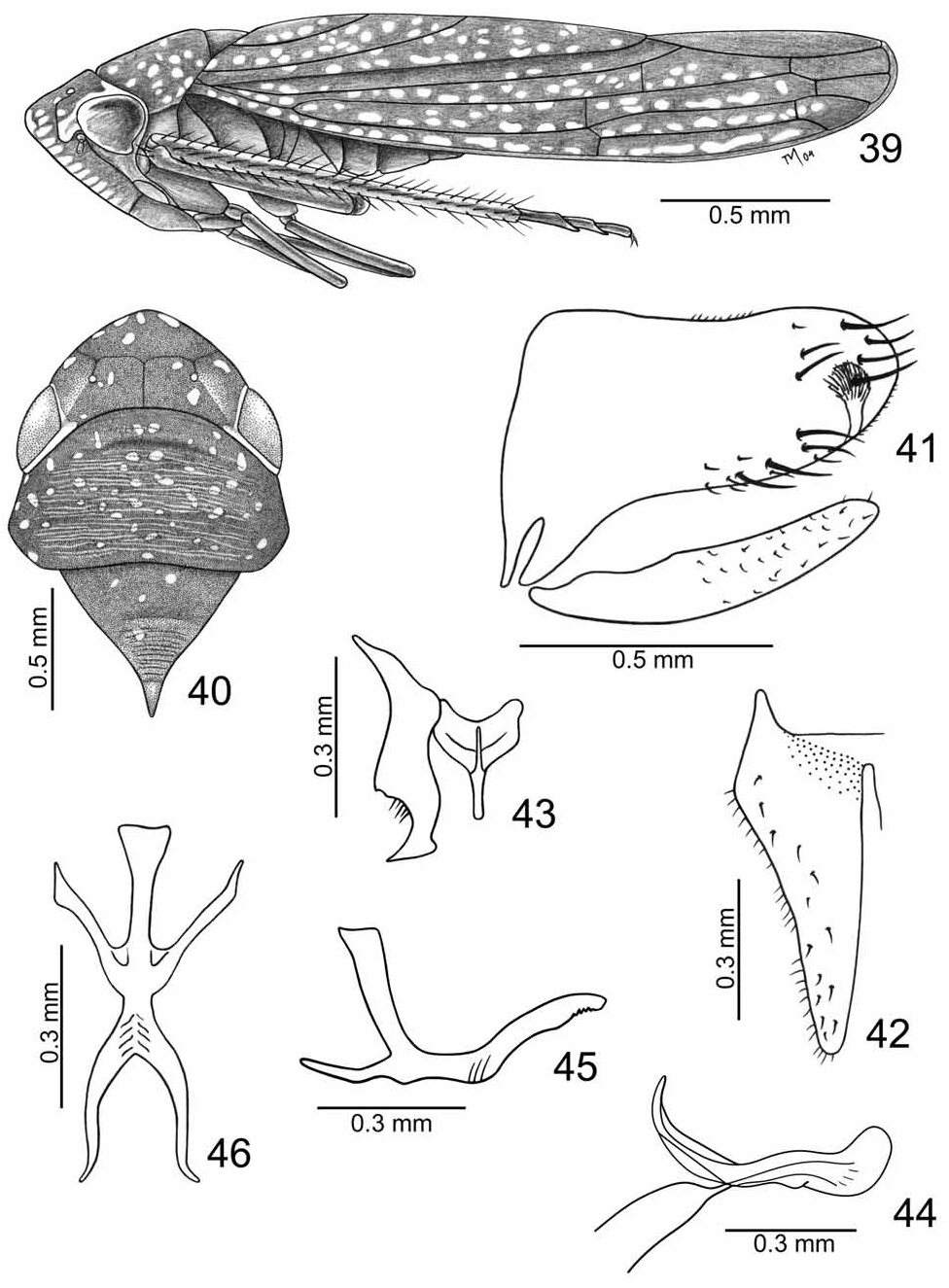
- Subgenital plates gradually narrowed toward apex ( Figs 12 View FIGURES 9–16 , 23 View FIGURES 20–27 , 42 View FIGURES 39–46 ); connective stalk narrow ( Figs 13 View FIGURES 9–16 , 24 View FIGURES 20–27 , 43 View FIGURES 39–46 )....... 5
5. Aedeagus with small dorsoapical process ( Fig. 25 View FIGURES 20–27 ). .................................. E. longinqua sp. nov. (Mato Grosso State)
- Aedeagus without small dorsoapical process ( Figs 14 View FIGURES 9–16 , 44 View FIGURES 39–46 ).......................................................................................... 6
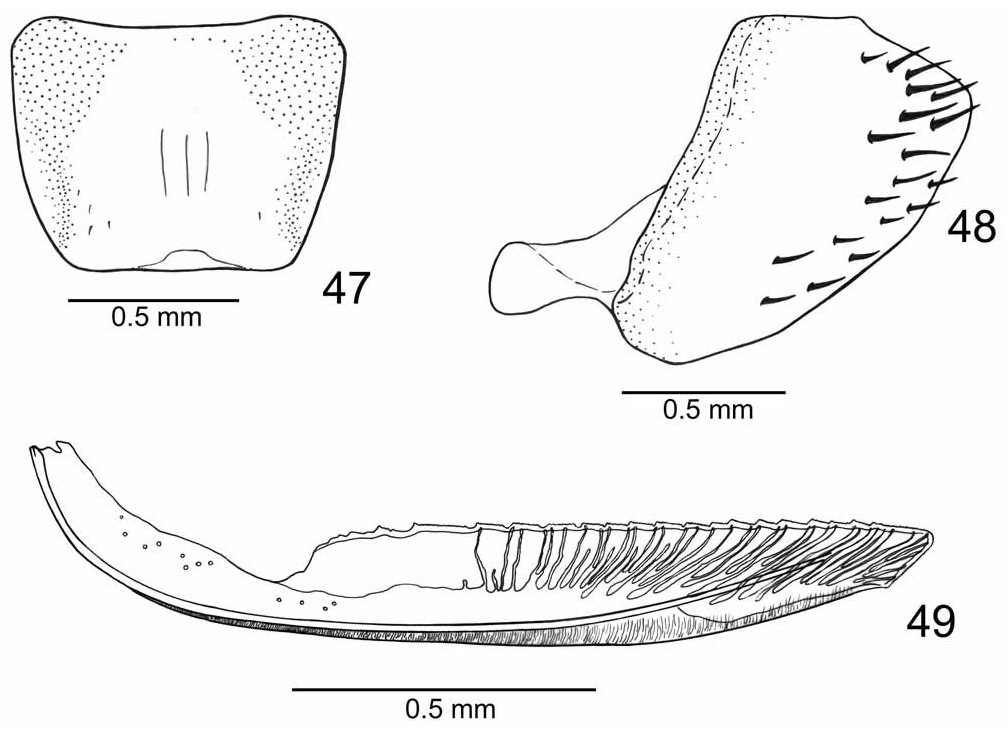
6. Connective shallowly concave between arms ( Fig. 43 View FIGURES 39–46 ); aedeagus ( Fig. 44 View FIGURES 39–46 ), in lateral view, without dorsal projection on basal half; paraphyses with posterior rami narrower toward apex ( Figs 45, 46 View FIGURES 39–46 ); female sternite VII with posterior margin approximately rectilinear ( Fig. 47 View FIGURES 47–49 ); second pair of valvulae with ventral margin distinctly convex on apical third up to preapical prominence, basal teeth (except first three) large and rectangular, and becoming irregularly smaller and subtriangular apically ( Fig. 49 View FIGURES 47–49 ). ..................................................... E. paranaensis sp. nov. (Paraná State)
- Connective deeply concave between arms ( Fig. 13 View FIGURES 9–16 ); aedeagus ( Fig. 14 View FIGURES 9–16 ), in lateral view, with dorsal projection on basal half; paraphyses with posterior rami broader toward apex ( Figs 15, 16 View FIGURES 9–16 ); female sternite VII with posterior margin concave ( Fig. 17 View FIGURES 17–19 ); second pair of valvulae with ventral margin uniform, without distinct convexity before preapical prominence, basal teeth large and rectangular, and becoming gradually smaller and quadrate apically ( Fig. 19 View FIGURES 17–19 ). .. ............................................................................................................ E. flavomaculata sp. nov. (Espírito Santo State)
7. Ground color of forewings yellowish-orange................................................................................................................. .................................................. E. semivitta (Walker, 1851) View in CoL ( Young 1977: fig. 184. Rio de Janeiro and Bahia states)
- Ground color of forewings brown to dark brown......................................................................................................... 8
8. Frons dark brown to black with pale yellow macula mostly on median portion.......................................................... 9
- Frons yellow with brown marks mostly on muscle impressions................................................................................ 12
9. Forewings with two yellow maculae ( Figs 50 View FIGURES 50–57 , 58 View FIGURES 58–65 )..................................................................................................... 10
- Forewings with three or more ivory or yellowish-white maculae.............................................................................. 11
10. Pronotum with transverse yellow macula not extending to lateral pronotal lobes ( Figs 58, 59 View FIGURES 58–65 ); forewings with claval transcommissural macula and macula on corium not reaching costal margin ( Fig. 58 View FIGURES 58–65 )................................................. .......................................................................................................................................... E. una sp. nov. (Bahia State)
- Pronotum with transverse yellow macula extending to lateral pronotal lobes ( Figs 50, 51 View FIGURES 50–57 ); forewings with claval macula not transcommissural and macula on corium transverse and reaching costal margin ( Fig. 50 View FIGURES 50–57 )......................... ............................................................................................................................ E. paulista sp. nov. (São Paulo State)
11. Paraphyses with one pair of rami.................................................................................................................................... ............. E. leucampix ( Signoret, 1853) View in CoL ( Young 1977: fig. 182. Santa Catarina, São Paulo, and Rio de Janeiro states)
- Paraphyses with two pairs of rami.................................................................................................................................. .... E. assimilis ( Signoret, 1853) View in CoL ( Young 1977: fig. 181. Santa Catarina, São Paulo, Rio de Janeiro, and Bahia states)
12. Subgenital plates abruptly narrowed toward apex on basal half ( Fig. 71 View FIGURES 66–75 ); connective stalk broad ( Fig. 72 View FIGURES 66–75 ). ............... .............................................................. E. hyalinosparsa (Melichar, 1932) View in CoL , in part (Paraná and Espírito Santo states)
- Subgenital plates gradually narrowed toward apex ( Fig. 4 View FIGURES 1–8 ); connective stalk narrow ( Fig. 5 View FIGURES 1–8 )...................................... .................................................................................................................... E. capixaba sp. nov. (Espírito Santo State)
Exogonia capixaba Mauro-Barr, Carvalho et Felix sp. nov.
( Figs 1–8 View FIGURES 1–8 )
Length: Male holotype, 10.0 mm.
Male holotype. Head ( Figs 1, 2 View FIGURES 1–8 ), in dorsal view, moderately produced, anterior margin broadly rounded; crown with median length 3/5 interocular width and 2/5 transocular width; epicranial suture distinct; temporal sutures partially visible; frontogenal sutures distinct. Pronotum ( Figs 1, 2 View FIGURES 1–8 ), in dorsal view, with width approximately equal to transocular width. Forewings ( Fig. 1 View FIGURES 1–8 ) with membrane slightly developed on apical portion of apical cells.
Ground color of dorsum ( Figs 1, 2 View FIGURES 1–8 ) brown. Crown with anterior margin yellow; median portion with dark brown areas delimiting three brown maculae, one anteriorly and pair posteriorly. Pronotum with irregularshaped yellow maculae on anterior two-thirds. Forewings ( Fig. 1 View FIGURES 1–8 ) with five yellow maculae; one on basal portion of clavus and four on corium, one on basal portion of discal cells, two near apex of clavus and one on apex of first apical cell and base of second apical cell. Frons yellow with pair of narrow brown stripes on dorsal half; clypeus with brownish-yellow macula on median portion. Lateral and ventral portions of thorax yellow except for brown to dark brown areas on anepisternum and lateral portions of mesosternum; legs yellow.
Pygofer ( Fig. 3 View FIGURES 1–8 ), in lateral view, with macrosetae on apical portion and extending anteriorly along ventral margin; apical portion with inner process bearing numerous setae. Subgenital plates ( Fig. 4 View FIGURES 1–8 ), in ventral view, gradually tapered toward apex; ventral surface with macrosetae near outer margin. Styles ( Fig. 5 View FIGURES 1–8 ), in dorsal view, extending posteriorly beyond apex of connective; with distinct external median lobe; apical portion beyond lobe curved, with few setae; apex truncate. Connective ( Fig. 5 View FIGURES 1–8 ), in dorsal view, Y-shaped. Aedeagus ( Fig. 6 View FIGURES 1–8 ) with shaft, in lateral view, curved ventrally, with distinct dorsoapical expanded lobe. Paraphyses ( Figs 7, 8 View FIGURES 1–8 ) with anterior pair of rami directed anteroventrally in lateral view; posterior pair slightly shorter than anterior one, directed posterodorsally in lateral view; stalk approximately as long as anterior rami. Anal tube (segment X), in lateral view, with pair of small processes on ventroapical margin bearing numerous setae similar to those of pygofer.
Female genitalia. Unknown.
Etymology. The specific epithet, capixaba , is from the indigenous language Tupi-Guarani and refers to the natives from Espírito Santo State, Brazil.
Type-material. Brazil, Espírito Santo State. Holotype: male, “Rib.[Ribeirão] do Engano E.S.\ Vale do Itauna \ Trav.[Travassos] e Santos-9-10-42” ( MNRJ).


| MNRJ |
Museu Nacional/Universidade Federal de Rio de Janeiro |
No known copyright restrictions apply. See Agosti, D., Egloff, W., 2009. Taxonomic information exchange and copyright: the Plazi approach. BMC Research Notes 2009, 2:53 for further explanation.