
Marma wesolowskae, Salgado & Ruiz, 2020
|
publication ID |
https://doi.org/10.11646/zootaxa.4899.1.16 |
|
publication LSID |
lsid:zoobank.org:pub:27E67BBB-DFD0-4A96-8269-9E1CB6153B83 |
|
DOI |
https://doi.org/10.5281/zenodo.4456881 |
|
persistent identifier |
https://treatment.plazi.org/id/03403F11-FFBE-FFA3-538B-FF73082DF8EE |
|
treatment provided by |
Plazi (2020-12-30 11:02:11, last updated 2024-11-27 00:17:40) |
|
scientific name |
Marma wesolowskae |
| status |
sp. nov. |
Marma wesolowskae View in CoL sp. nov.
Figures 1E View FIGURE 1 , 29–33 View FIGURE 29 View FIGURE 30 View FIGURE 31 View FIGURE 32 View FIGURE 33 , 57D View FIGURE 57 , 58F View FIGURE 58
Etymology. The epithet commemorates the career and life of Polish Professor Wanda Wesołowska. During her career, she has authored/co-authored the description of hundreds of jumping spider species.
Diagnosis. Among all Marma species, Marma wesolowskae sp. nov. shares most similarities with M. nigritarsis and M. pipa sp. nov. The males of these species have an embolic disc with a straight prolateral border and curved retrolateral border ( Figs 25C View FIGURE 25 , 27A View FIGURE 27 , 30C View FIGURE 30 , 32A View FIGURE 32 , 36C View FIGURE 36 , 37A View FIGURE 37 , 57 View FIGURE 57 C–E). Besides, in the females, the copulatory ducts do not touch each other and they encircle the primary spermathecae ( Figs 26 View FIGURE 26 C–D, 27F–G, 31C–D, 32F–G, 35H–I, 37H–I, 58E–G). The males of M. wesolowskae sp. nov. differ from the males of these species by having a narrower embolic disc ( Figs 30C View FIGURE 30 , 32A View FIGURE 32 , 57 View FIGURE 57 C–E), and the females differ by having the posterior border of the epigynal plate projected and the copulatory openings placed more posteriorly (posterior to the distal region of the primary spermathecae) ( Figs 31 View FIGURE 31 C–D, 32G, 58E–G).
Description. Male (Holotype, MPEG 34355). Total length: 3.38. Carapace 1.96 long, 1.31 wide, 0.97 high. Ocular quadrangle 0.78 long. Anterior eye row 1.18 wide, posterior 0.97 wide. Legs 3412. Length of legs: I 3.53 (1.13 + 1.32 + 1.08); II 3.17 (1.02 + 1.15 + 1.00); III 4.09 (1.39 + 1.41 + 1.29); IV 3.94 (1.25 + 1.30 + 1.39).
Leg macrosetae: Femur I–III d1-1-1, p1di, r1di; IV d1-1-1, p0, r1di. Patella I–II 0; III–IV p0, r1. Tibia I p0 (or p0-1-0), r0, v1r-2-2; II p0-1-0, r0-1-0, v1r-1r-2; III p0-1-1-0, r1-1-1-0 (or r0-1-1-0), v1p-0-0-2; IV p0-1-1-0, r1-1- 1-0, v1p-0-0-2. Metatarsus I p1-1, r0-1, v1r-2 (or v2-2); II p1-1, r1di, v2-2; III d1p-0-0, p1-0-2, r1-0-2, v2-0-2; IV p1-1-2, r1-1-2, v1p-0-2.
Color in alcohol ( Figs 30 View FIGURE 30 A–B): carapace covered entirely by scales (homogenous distribution); abdomen with thick dark brown stripe and pale lateral borders; legs: femur I–II with dark dorsal spot distally, III with distal and proximal regions with incomplete dark ring (distal ring: not colored ventrally; proximal ring: not colored dorsally), IV with dark distal ring; patella I–IV with dark proximal spot; tibia I–II with proximal region with dark dorsal spot, III–IV proximal and distal regions with dark dorsal spot; tarsus I with dark tip, II without dark marks, III–IV with dark proximal ring.
Palp: RTA finger-shaped ( Figs 30D View FIGURE 30 , 32B View FIGURE 32 , 33E View FIGURE 33 ); embolic disc narrow, with straight prolateral edge and curved retrolateral edge ( Figs 30C View FIGURE 30 , 32A View FIGURE 32 , 33A View FIGURE 33 ); PED slightly longer than exposed portion of embolic disc and emerging from the prodistal part of embolic disc ( Figs 32A, E View FIGURE 32 , 33A View FIGURE 33 ); tip of embolus ending beyond tip of PED ( Figs 30C View FIGURE 30 , 32A, E View FIGURE 32 ).
Female (Paratype, MPEG 34354). Total length: 4.01. Carapace 1.90 long, 1.43 wide, 1.11 high. Ocular quadrangle 0.97 long. Anterior eye row 1.27 wide, posterior 1.07 wide. Legs 3421. Length of legs: I 2.97 (0.93 + 1.17 + 0.87); II 3.00 (0.95 + 1.16 + 0.89); III 4.17 (1.37 + 1.45 + 1.35); IV 4.09 (1.27 + 1.37 + 1.45).
Leg macrosetae: Femur I d1-1-1, p1di, r0; II d0 (d1-1-1), p0 (p1di), r0 (r1di); III d1-1-1, p1di, r1di; IV d1-1-1, p0, r0 (r1di). Patella I–II 0; III–IV p0, r1. Tibia I p0-1-0, r0, v1r-2-2; II p1di (p1-1-1), r0, v0 (v1r-2-0); III p0-1-0- 0 (or p0-1-1-0), r1-1-1-0 (or r0-1-1-0), v1p-0-0-1-2; IV p1-1-1-0 (or p0-1-1-0), r1-1-1-0, v0-1p-0-2. Metatarsus I p1-1, r1di, v2-2; II p1di (p1-1), r1di, v2di (v2-2); III d1p-0-0, p1-0-2, r1-0-2, v2-2; IV p1-1-2, r1-1-2, v0-1p-2 (or v1p-1p-2).
Color in alcohol ( Figs 31 View FIGURE 31 A–B): carapace as in male; abdomen ventrally pale; legs: femur I–II with distal region with dark spot, III with dark distal ring and proximal region with dark proventral spot, IV with dark distal ring; patella I–II with dark proximal spot; tibia I–II with dark proximal ring, III–IV with proximal and distal dark ring; tarsus I–II without dark marks, III–IV with dark proximal ring.
Epigyne ( Figs 31 View FIGURE 31 C–D, 32F–G, 33J): copulatory openings very close to each other and placed more posteriorly than end region of primary spermathecae; copulatory ducts long, encircling primary spermathecae; proximal copulatory duct shorter than distal section; primary spermathecae with homogenous diameter, narrow distal end and anteriorly projected.
Type material. Holotype ♁: BRAZIL: Pernambuco: Buíque, Paraíso Selvagem camping area, 08°35’08.8”S, 37°14’28.4”W, leg. A. Salgado, 21.VII.2019 ( MPEG 34355 View Materials ). GoogleMaps
Paratypes: Same data as holotype, 1♁ ( MPEG 37130 View Materials ), 3♀ ( MPEG 34354 View Materials , 37131 View Materials , IBSP 267943 View Materials ) GoogleMaps . BRAZIL: Pernambuco: Venturosa, Pedra Furada , 08°34’12.6”S, 36°49’34.4”W, leg. A. Salgado, 21.VII.2019, 1♁ ( IBSP 267944 View Materials ) GoogleMaps .
Other material examined. Same data as holotype, 2 GoogleMaps ♁ 1♀ ( MPEG 37132 View Materials ) , 1♁ ( MPEG 37133 View Materials ) , 1♁ ( MPEG 37134 View Materials ) , 1♀ ( MPEG 37135 View Materials ) . BRAZIL: Pernambuco: Venturosa, Pedra Furada , 8°34’12.6”S, 36°49’34.4”W, leg. A. Salgado, 21.VII.2019, 1♁ ( MPEG 37136 View Materials ) GoogleMaps .
Distribution. Known only from the Brazilian state of Pernambuco ( Fig. 59B View FIGURE 59 ).
Natural History. The specimens were collected from sandstone ( Figs 1E View FIGURE 1 , 29 View FIGURE 29 ).

FIGURE 1. Marma Simon, 1902. A–E live specimens in different habitats [A M. nigritarsis from Belém/Pará/Brazil, on urban construction (Photo credit: César Favacho); B M. linae sp. nov. from São Geraldo do Araguaia/Pará/Brazil, on tree trunk; C M. rosea from São Geraldo do Araguaia/Pará/Brazil, on a wall of an urban construction; D M. nigritarsis from Belém/Pará/Brazil, on a substrate with moss; E M. wesolowskae sp. nov. from Buíque/Pernambuco/Brazil, on rocks].
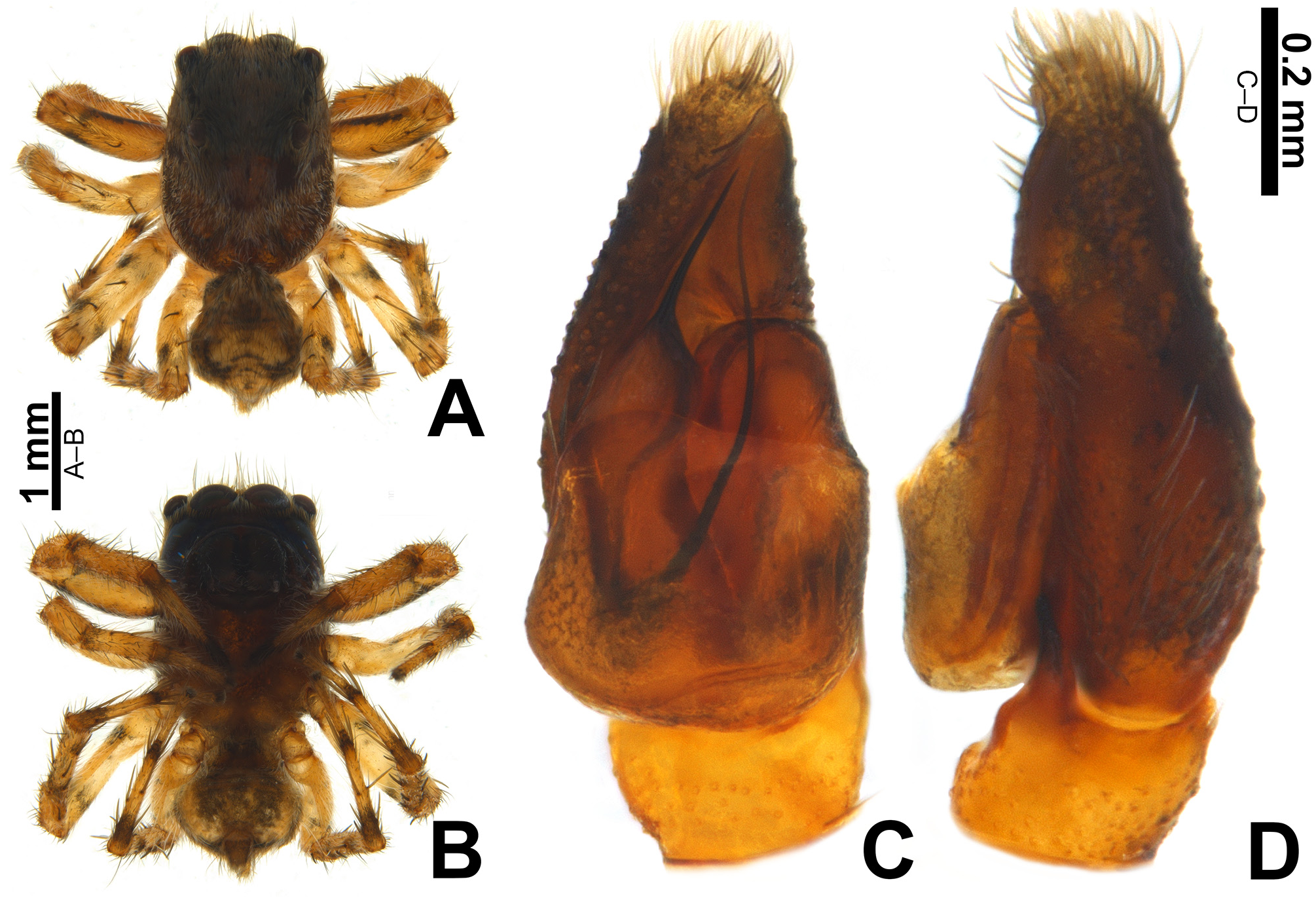
FIGURE 25. Male holotype of Marma pipa sp. nov. (MPEG 34352). A–B habitus (A dorsal, B ventral); C–D left palp (C ventral; D retrolateral).
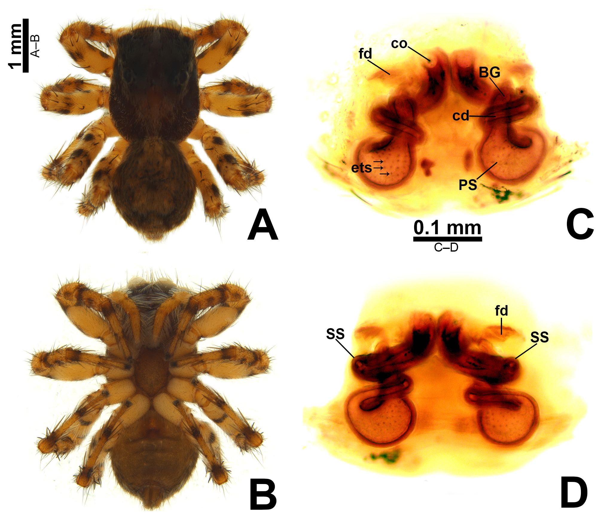
FIGURE 26. Female paratype of Marma pipa sp. nov (MPEG 34349). A–B habitus (A dorsal, B ventral); C–D cleared epigyne/ vulva (C ventral, D dorsal). Abbreviations: BG—Bennett’s gland; cd—copulatory duct; co—copulatory opening; ets—endothe- cal spikes; fd—fertilization duct; PS—Primary spermatheca; SS—secondary spermatheca.
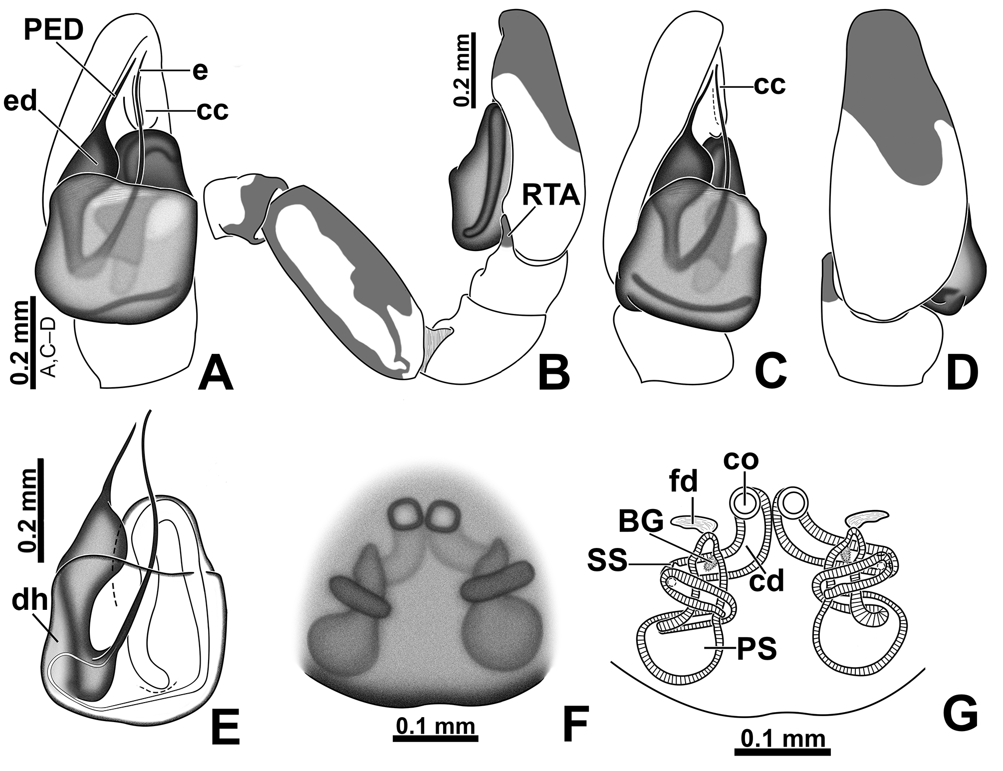
FIGURE 27. Marma pipa sp. nov. A–E left male palp (A ventral; B retrolateral; C proventral; D dorsal; E cleared bulb, ventral); F–G epigyne/vulva (F ventral; G cleared, ventral). Abbreviations: BG—Bennett’s gland; cc—cymbial conductor; cd—copu- latory duct; co—copulatory opening; dh—distal hematodocha; e—embolus shaft; ed—embolic disc; fd—fertilization duct; PED—process on embolic disc; PS—primary spermatheca; RTA—retrolateral tibial apophysis; SS—secondary spermatheca.

FIGURE 29. Marma wesolowskae sp. nov. live specimens from Buíque/Pernambuco/Brazil on sandrock. A–C male (A–B dorsolateral, C dorsal); D–F female (D–E dorsolateral, F dorsal).
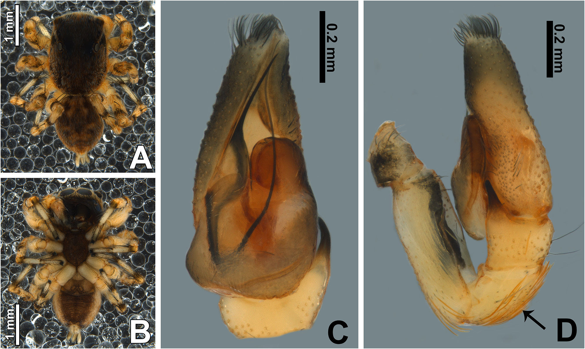
FIGURE 30. Male holotype of Marma wesolowskae sp. nov. (MPEG 34355). A–B habitus (A dorsal, B ventral); C–D left palp (C ventral; D retrolateral).
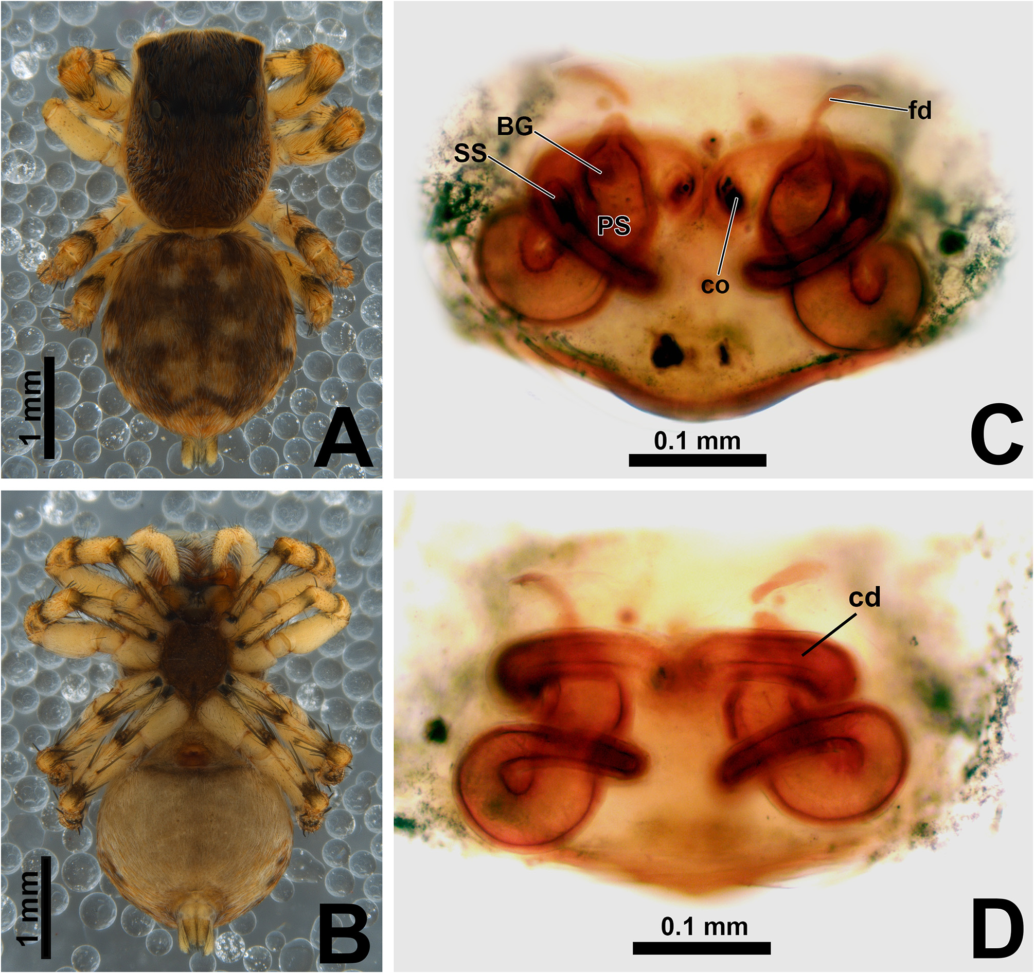
FIGURE 31. Female paratypes of Marma wesolowskae sp. nov. A–B habitus, MPEG 34354 (A dorsal, B ventral); C–D epi- gyne/vulva, MPEG 37131 (C ventral, D dorsal). Abbreviations: BG—Bennett’s gland; cd—copulatory duct; co—copulatory opening; fd—fertilization duct; PS—Primary spermatheca; SS—secondary spermatheca.

FIGURE 32. Marma wesolowskae sp. nov. A–E left male palp (A ventral; B retrolateral; C proventral; D dorsal; E cleared bulb, ventral); F–G epigyne/vulva (F ventral; G cleared, ventral).Abbreviations: BG—Bennett’s gland; cc—cymbial conductor; cdcopulatory duct; co—copulatory opening; dh—distal hematodocha; e—embolus shaft; ed—embolic disc; fd—fertilization duct; PED—process on embolic disc; PS—primary spermatheca; RTA—retrolateral tibial apophysis; SS—secondary spermatheca.
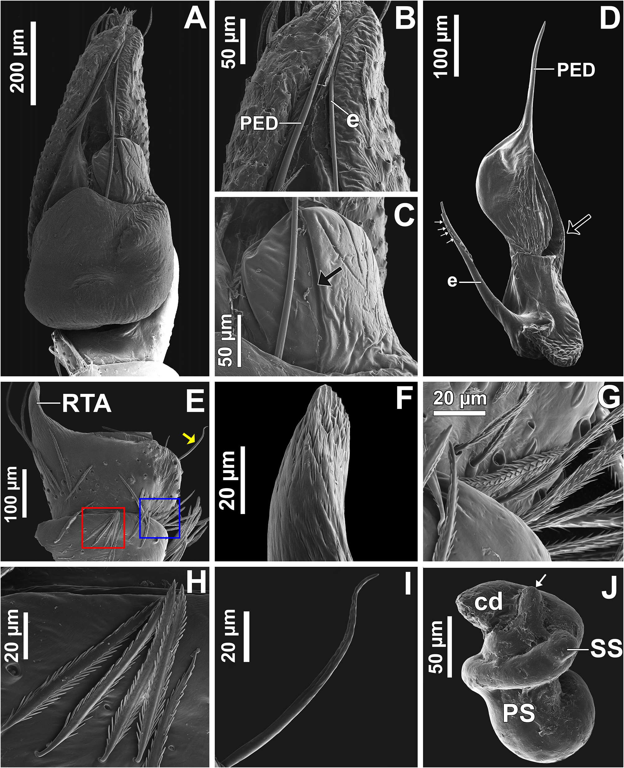
FIGURE 33. Marma wesolowskae sp. nov. A–I left male palp [A ventral; B apical portion of palp, ventral; C tegular shoulder, ventral; D dissected embolic disc and embolus, dorsal; E patella and tibia, retrolateral; F tip of RTA, retrolateral; G detail of patella and tibia setae (blue rectangle in 33E); H detail of patella setae (red rectangle in 33E); I detail of smooth scale of tibia (yellow arrow in 33E]. J left spermatheca and copulatory duct, ventral. Abbreviations: cd—copulatory duct; e—embolus shaft; PED—process on embolic disc; PS—primary spermatheca; RTA—retrolateral tibial apophysis; SS—secondary spermatheca. Black arrow in C shows depression of tegular shoulder; in D shows groove; white arrows in D show spikes of embolus shaft; in J shows end region of the primary spermatheca (broken fertilization duct).
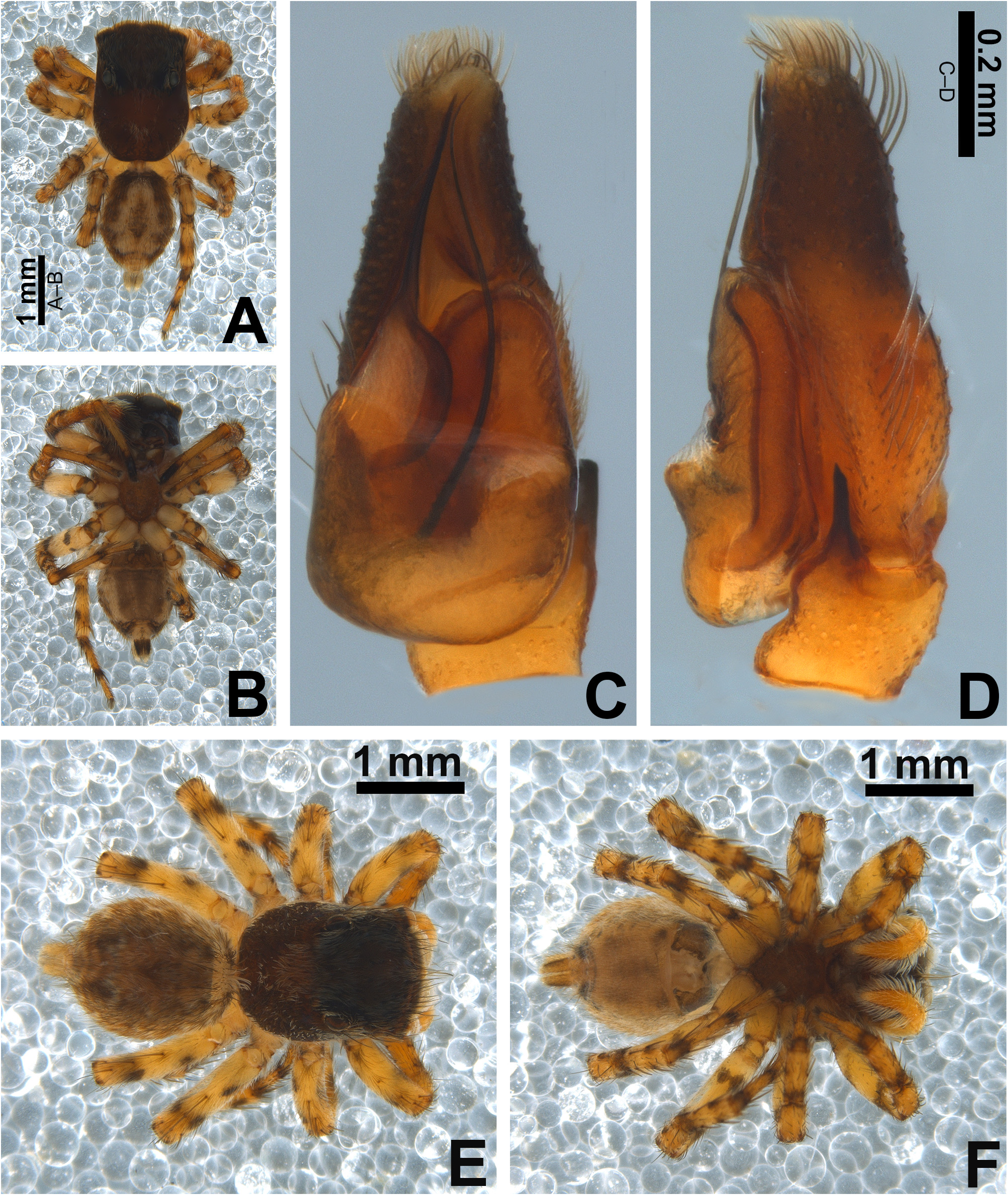
FIGURE 36. Marma nigritarsis (Simon, 1900), specimens from Belém/Pará/Brasil. A–D male, MPEG 34357 (A habitus, dorsal; B ventral; C left palp, ventral; D retrolateral). E–F female, MPEG 34358 (E habitus, dorsal; F ventral).

FIGURE 37. Marma nigritarsis (Simon, 1900). A–E, G specimen from Belém/Pará/Brazil. F holotype illustrated by Galiano (1962), reflected.A–G left male palp (A ventral, B retrolateral, C proventral, D dorsal, E–F retroventral; G cleared bulb, ventral). H–I female holotype of M. trifidocarinata Caporiacco, 1947, epigyne/vulva (H ventral; I cleared, ventral).Abbreviations: BG— Bennett’s gland; cc—cymbial conductor; cd—copulatory duct; co—copulatory opening; dh—distal hematodocha; e—embolus shaft; ed—embolic disc; fd—fertilization duct; PED—process on embolic disc; PS—primary spermatheca; RTA—retrolateral tibial apophysis; SS—secondary spermatheca.

FIGURE 57. Left male palp, ventral [A Marma baeri Simon, 1902; B M. linae sp. nov.; C M. pipa sp. nov.; D M. wesolowskae sp. nov.; E M. nigritarsis (Simon, 1900); F M. sinuosa sp. nov., G M. argentina (Mello-Leit„o, 1941), H M. rosea (Mello- Leit„o, 1941), I M. spelunca sp. nov.]. Colors represent homologous structures.

FIGURE 58. Epigyne/vulva, A–C, E–K ventral, D dorsal [A Marma baeri Simon, 1902; B M. linae sp. nov.; C M. abaira sp. nov.; D M. femella (Caporiacco, 1955); E M. pipa sp. nov.; F M. wesolowskae sp. nov.; G M. nigritarsis (Simon, 1900); H M. sinuosa sp. nov.; I M. argentina (Mello-Leitão, 1941); J M. rosea (Mello-Leitão, 1941); K M. spelunca sp. nov.]. Colors represent homologous structures; arrow in D shows hypothetical position of secondary spermatheca.

No known copyright restrictions apply. See Agosti, D., Egloff, W., 2009. Taxonomic information exchange and copyright: the Plazi approach. BMC Research Notes 2009, 2:53 for further explanation.
|
Kingdom |
|
|
Phylum |
|
|
Class |
|
|
Order |
|
|
Family |
|
|
SubFamily |
Salticinae |
|
Tribe |
Euophryini |
|
Genus |
1 (by plazi, 2020-12-30 11:02:11)
2 (by diego, 2021-01-20 16:56:11)
3 (by ExternalLinkService, 2021-01-20 17:06:48)
4 (by diego, 2021-01-22 10:52:40)
5 (by ExternalLinkService, 2021-01-22 11:05:27)
6 (by ExternalLinkService, 2021-09-19 03:48:30)
7 (by plazi, 2023-11-01 01:15:53)