
Coelostoma (Lachnocoelostoma) jaechi, Jia, Fenglong, Lin, Renchao, Chan, Eric, Skale, Andre & Fikáček, Martin, 2017
|
publication ID |
https://doi.org/ 10.11646/zootaxa.4232.1.8 |
|
publication LSID |
lsid:zoobank.org:pub:B304D4FA-FA87-45C6-9B90-82DE4A71AB50 |
|
DOI |
https://doi.org/10.5281/zenodo.6038242 |
|
persistent identifier |
https://treatment.plazi.org/id/025B394E-543F-FFE3-FF1C-5C2EFD40F94B |
|
treatment provided by |
Plazi |
|
scientific name |
Coelostoma (Lachnocoelostoma) jaechi |
| status |
sp. nov. |
Coelostoma (Lachnocoelostoma) jaechi View in CoL sp. nov.
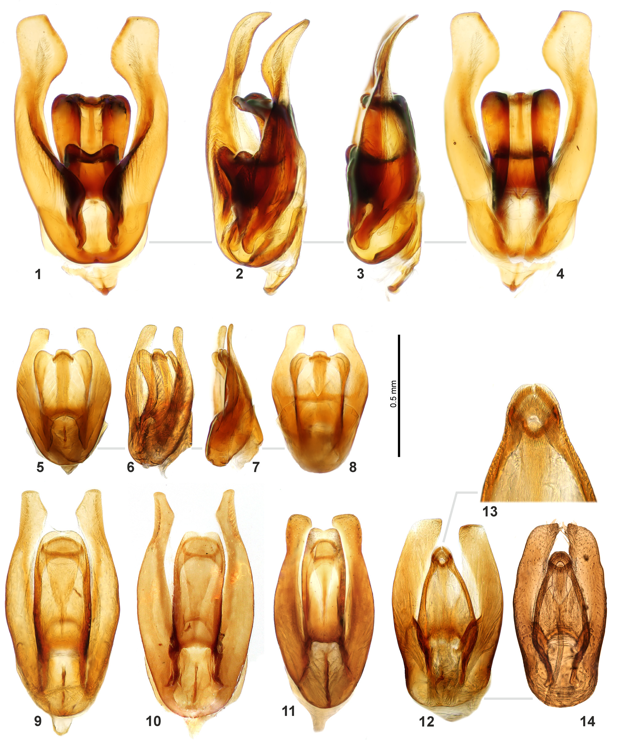
( Fig. 11 View FIGURES 1 – 14 )
Type material. Holotype: male ( SYSU): Hong Kong, Sha Lo Tung [translation; labeled in Chinese], 22°28.555′N, 114°10.909′, 185m, in light, Fenglong Jia, Weicai Xie, Alex lgt . Paratypes (18): 5 males, 12 females: same data as holotype ; 1 male: Hong Kong Cuk Keng, 24.ix.2013, Fenglong Jia, Yingming Li & Eric Chen lgt. ( SYSU, AFCD, NMPC, NMW) .
Diagnosis. Body length 4.3–4.9 mm. Prosternum moderately carinate throughout medial portion and forming an anteromedian spine. Pronotum with punctures slightly finer than on head and elytra; elytra with lateral portions more strongly punctate, without traces of series of punctures laterally. Mesofemora densely pubescent except at extreme apex. First abdominal ventrite with a sharp carina reaching beyond basal half; last abdominal ventrite emarginate and with a row of stout setae apically. Aedeagus ( Fig. 11 View FIGURES 1 – 14 ) 0.8 mm long. Median lobe broad, subparallel throughout, only slightly widened subapically, slightly emarginate at apex, gonopore situated subapically, wider than long; parameres longer than median lobe, outer face sinuate at anterior third, truncate at apex with round outer corner and rectangular inner corner.
This species is similar to C. coomani Orchymont, 1932 by the weakly emarginate apex of the median lobe, and to C. martensi Hebauer, 2002 and C. huangi Jia, Aston & Fikáček, 2014 by the subapically sinuate outer margin of the paramere. It is easy to distinguish it from C. coomani (Fig. 24 in Jia et al. 2014) by the median lobe being widest in apical third (widest at midlength in C. coomani ), the gonopore situated subapically (situated at midlength in C. coomani ) and parameres sinuate on outer margin subapically (without subapical sinuation in C. coomani ). It differs from C. martensi (Fig. 28 in Jia et al. 2014) by generally broader median lobe widest in apical third (narrower and widest ca. at midlength in C. martensi ) and parameres wider in apical portion (narrower in C. martensi , Fig. 28 in Jia et al. 2014). It differs from C. huangi by the gonopore situated subapically (situated at apex in C. huangi : Fig. 9 View FIGURES 1 – 14 in this paper, Fig. 27 in Jia et al. 2014) and relatively shorter basal apodemes of the median lobe (as long as ca. one fourth of total length of the median lobe in C. jaechi , as long as nearly half of total length of the median lobe in C. huangi ).
Description. Form and Color. Body length 4.3–4.9 mm, width 3.0– 3.1 mm. Body oval, strongly convex. Dorsum black. Labrum, maxillary palpi and labial palpi yellowish to reddish brown, antennae yellowish to reddish brown with dark club. Ventral surface black with reddish pubescence. Femora and tibiae dark reddish brown, tarsi pale reddish.
Head. Dorsal surface with dense and moderately strong punctures. Intervals between punctures smooth, but with clear shagreen and transverse microsculpture on posterior margin of head (this part sometimes covered by pronotum). Clypeus truncate anteriorly. Eyes moderately sized, separated by ca. 5× the width of one eye, not emarginate anteriorly. Mentum with transverse microsculpture posteriorly and moderately strong punctures laterally, strongly emarginate anteriorly and depressed in anterior half. Antennae with 9 antennomeres, antennal club loosely segmented. Maxillary palpomere 2 strongly swollen, palpomere 4 almost symmetrical, rather longer than palpomere 3. Gula very narrow, glabrous.
Thorax. Pronotum with slightly finer punctures than on head; anterior margin strongly bisinuate; posterior margin slightly bisinuate; lateral marginal bead reaching posterior corner, not continuing to posterior margin; posterior corner almost rectangular. Prosternum moderately carinate throughout medial portion, bearing strong anteromedian spine. Mesoventrite with raised, arrowhead-shaped process, surface pubescent. Metaventrite with strongly raised median portion broadly projecting anteriorly between mesocoxae and abutted to mesoventral process; lateral portions of metaventrite densely pubescent, middle portion more shining, only sparsely pubescent. Metepisterna ca. 5× as long as wide, parallel-sided. Scutellar shield slightly longer than wide, with punctation as on pronotum. Elytra with slightly coarser punctures than on pronotum, punctures on lateral and posterior portions somewhat coarser than those on disc; elytra without traces of punctural series; sutural stria reaching basal half of elytra. Femora with deep tibial groove posteriorly. Mesofemora pubescent except at extreme apex. Metafemora sparsely punctate, with dense microsculpture. Tarsi with long dorsal setae and gold ventral setae; metatarsi with fifth tarsomere almost as long as third and fourth combined. Claws moderately curved, with a pair of long setae beneath.
Abdomen. Abdominal ventrites densely pubescent. First abdominal ventrite with a sharp carina present in slightly more than basal half; last ventrite emarginate and with a row of stout setae apically.
Aedeagus ( Fig. 11 View FIGURES 1 – 14 ). 0.8 mm long. Median lobe broad, subparallel throughout, only slightly widened subapically, slightly emarginate at apex; gonopore situated subapically, wider than long; parameres longer than median lobe, outer face sinuate at anterior third, truncate at apex with round outer corner, inner margin truncate apically.
Etymology. The species is named after Dr. Manfred Jäch, a specialist on aquatic beetles from the Natural History Museum, Vienna, Austria, who gave us the opportunity to study the hydrophilid beetles deposited in NMW collection.
Biology. Unknown; examined specimens were collected at light.
Distribution. Only known from type locality.
No known copyright restrictions apply. See Agosti, D., Egloff, W., 2009. Taxonomic information exchange and copyright: the Plazi approach. BMC Research Notes 2009, 2:53 for further explanation.
|
Kingdom |
|
|
Phylum |
|
|
Class |
|
|
Order |
|
|
Family |
|
|
SubFamily |
Sphaeridiinae |
|
Tribe |
Coelostomatini |
|
Genus |