
Pseudorogneda shenda Wang & Xie, 2020
|
publication ID |
https://doi.org/ 10.11646/zootaxa.4808.3.13 |
|
publication LSID |
lsid:zoobank.org:pub:3A84CCF5-CD1D-4893-8A0A-DD59207DB59F |
|
DOI |
https://doi.org/10.5281/zenodo.4327949 |
|
persistent identifier |
https://treatment.plazi.org/id/B15BF1D8-55FC-411F-A143-7E09C83906F0 |
|
taxon LSID |
lsid:zoobank.org:act:B15BF1D8-55FC-411F-A143-7E09C83906F0 |
|
treatment provided by |
Plazi |
|
scientific name |
Pseudorogneda shenda Wang & Xie |
| status |
sp. nov. |
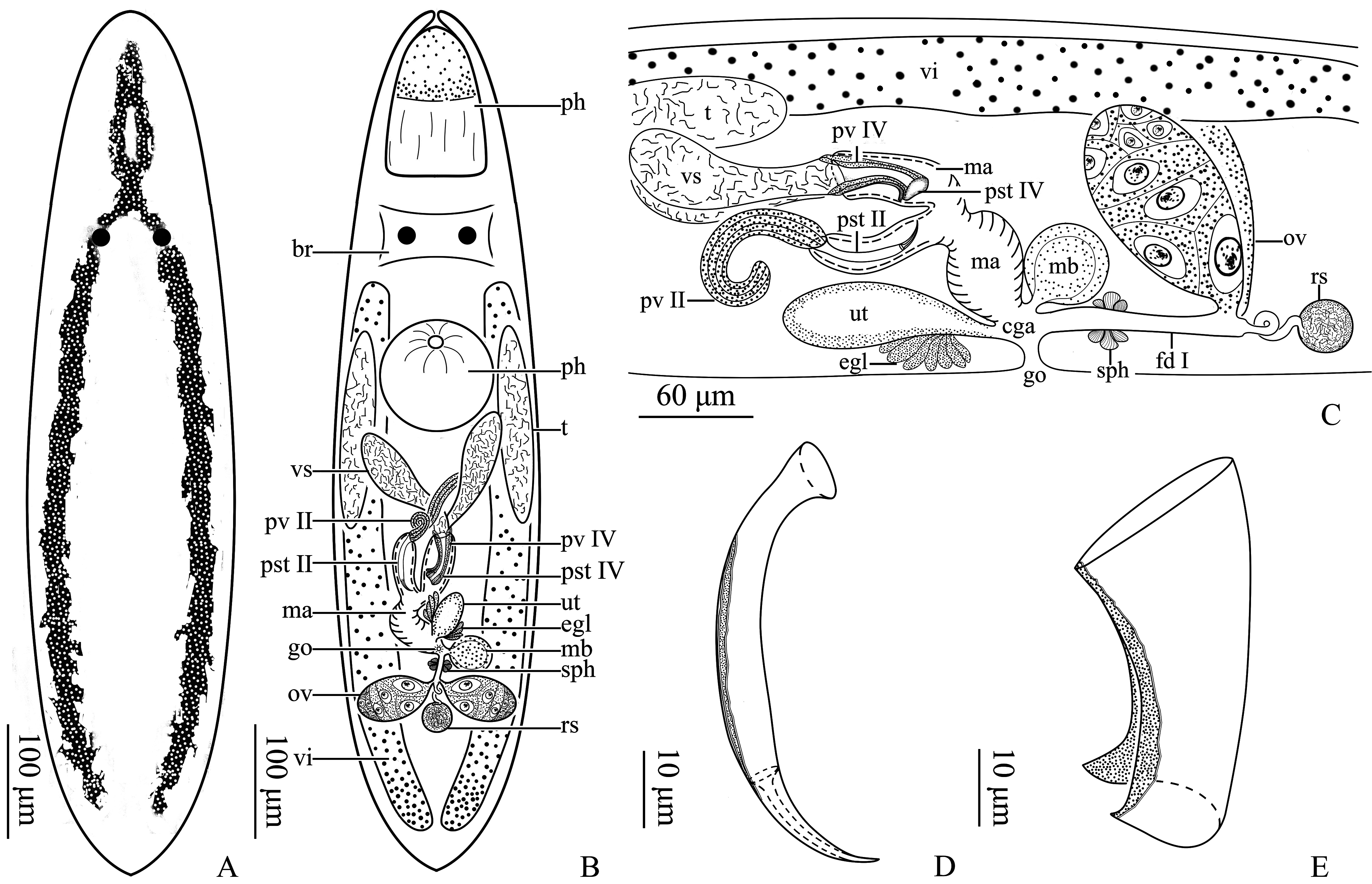
Pseudorogneda shenda Wang & Xie n. sp.
( Figs. 3-4 View FIGURE 3 View FIGURE 4 )
Locality. Specimens collected at Shekou Port (water temperature: 18–20℃; salinity: 17–20‰), Shenzhen City, Guangdong Province, China (22°28′N, 113°54′E), by Yaohang Xie in March, 2018, where the species inhabits the muddy surface of the submersed stones GoogleMaps .
Material examined. Holotype: PLA-Po121, one whole-mounted specimen. Paratypes: PLA-Po122-123, two whole-mounted specimens; PLA-Po124-126, three serially sectioned (sagittal) specimens; PLA-Po127-129, three mounted prostate stylets.
Etymology. The new species was named as “shenda”, the Pinyin spelling of abbreviated Chinese name of Shenzhen University.
Description. The animal measures 0.8–1.0 mm long and 130–160 μm wide (n=3) ( Fig. 3A View FIGURE 3 ). Mouth opens at anterior 3/5 ventral side of the body at level of the eyes. Pharynx is spherical and 115–120 μm in diameter (n=3) ( Fig. 3 View FIGURE 3 A–B, 4B). Inverted-Y-shaped brownish-yellow stripes are present dorsally, one anterior around eyes and two extending from pharynx to cauda tip. Proboscis is 125–135 μm (n=3) in length. Paired circular black eyes are situated at anterior 1/4 position of the body, 15–20 μm apart (n=3) ( Fig. 3 View FIGURE 3 A–B, 4A–B). The brain lies ventral to the eyes ( Fig. 4B View FIGURE 4 ).
Paired cylindrical testes, 250–300 μm long (n=3), are located at the posterior end of pharynx. The two seminal vesicles lie at midpoint of the body, measuring 220–235 μm long (n=3) ( Fig. 3 View FIGURE 3 B–D, 4B–C). They fuse at their pos- terior ends to form the ejaculatory duct which enters prostate stylet type IV through prostate vesicle type IV. Prostate stylet type IV is tubular, measuring 50–60 μm in length and 20–35 μm in basal diameter (n=5). It bears a slit and a distal fold ( Fig. 3 View FIGURE 3 C–G, 4B–C, E). Prostate vesicle type II (135–150 μm, n=3) lies between the two seminal vesicles with its distal end entering prostate stylet type II ( Fig. 3 View FIGURE 3 C–D, 4B–C). Prostate stylet type II is double-walled (120– 130 μm in length, n=5) and crescent-shaped (10–15 μm in diameter, n=5), with a sharp-pointed distal end where an elongate funnel-shaped inner wall can be observed ( Fig. 3 View FIGURE 3 C–D, H–J, 4B–D). Male atrium lies at posterior 1/4 of the body and is surrounded by strong muscular layer. It opens to the common genital atrium at the point where the spherical male bursa (75–100 μm in diameter, n=3) ( Fig. 3D View FIGURE 3 , 4 View FIGURE 4 B–C) connects to that common atrium.
Paired vitellaria lie dorsal and extend from pharynx to the cauda tip of the body ( Fig. 4 View FIGURE 4 B–C). The paired ellipsoidal ovaries lie in the posterior 1/3 of the body, measuring 75–90 μm long and 40–60 μm wide (n=3) ( Fig. 3B & D View FIGURE 3 , 4 View FIGURE 4 B–C). Female duct type I is connected to two oviducts and bears a sphincter ( Fig. 4 View FIGURE 4 B–C). The seminal receptacle is spherical and sperm-filled ( Fig. 4 View FIGURE 4 B–C); it measures 60–70 μm in diameter (n=3), and connects to the female duct via a coiled duct ( Fig. 3D View FIGURE 3 , 4 View FIGURE 4 B–C).
The gonopore is located ventrally at 1/4 body length from the posterior tip. The uterus bears a bundle of eosinophilic glands ( Fig. 4 View FIGURE 4 B–C) and connects to the anterior end of the common genital atrium. A mature egg is spherical in shape and encloses two embryos ( Fig. 3K View FIGURE 3 ).
Phylogenetic analysis. We compared sequence similarity of 18S rDNA and 28S rDNA using BLAST (BLAST N) of NCBI. The two new species are most similar to Rogneda Uljanin, 1870 . Specifically the 18S and 28S rDNA sequences of P. sinensis n. sp. are most similar to those of Rogneda reticulata Brunet, 1969 (>95%, n=2) and Rogneda sp. 2 (>91%, n=2), respectively; those of P. shenda n. sp. are most near Rogneda sp. 1 (>95%, n=3) and Rogneda sp. 2 (>89%, n=3).
The phylogenetic trees generated from concatenated 18S rDNA and 28Sr DNA sequence by Bayesian-inference (BI) and Maximum Likelihood (ML) methods with Pogaina sp. and Baicalellia beauchampi (Ax, 1956) Stephenson, Van Steenkiste & Leander, 2018 ( Provorticidae of the Dalytyphloplanida ) as outgroups are shown in Figs. 5 View FIGURE 5 , and the two trees agree with each other. The tree is divided into two main clades. The new species form a well-supported clade and share sister-group relationship with Rogneda Uljanin, 1870 , Alcha Marcus, 1949 and Paraustrorhynchus Karling & Schockaert, 1977 . They themselves form two well-supported clades, with both Bootstrap and Bayesian posterior probability indicating 100%, supporting the establishment of two new species.


No known copyright restrictions apply. See Agosti, D., Egloff, W., 2009. Taxonomic information exchange and copyright: the Plazi approach. BMC Research Notes 2009, 2:53 for further explanation.
|
Kingdom |
|
|
Phylum |
|
|
Class |
|
|
Order |
|
|
Family |
|
|
SubFamily |
Polycystidinae |
|
Genus |